INTRODUCTION
Aesthetic makeovers, as well as rehabilitation of worn dentitions, are procedures that have always been considered extremely expensive, complicated, and lengthy and as having many risks. Rehabilitations are surrounded by many myths, which in fact can make them more complicated and potentially damaging to the oral health of the patient. Examples of these myths include:
• The use of long-term provisionals is required as a needed “test” to see if one should proceed with the definitive treatment. While it’s emotionally reassuring to routinely prescribe long-term provisionals or temporaries and then wait and see how they adapt, this approach has disadvantages. Historically, this approach has required the use of full-crown preparations to allow for more robust provisionals as very thin partial coverage temporaries are weaker and will likely not last many months without the need for repairs. Also, using direct composite as an alternative for long-term provisional restorations adds additional cost and complications. It is important to note that it doesn’t take months to know if a patient will adapt to changes in aesthetics, phonetics, and function. This process usually takes a few days or a couple of weeks at most for the patient and the clinician to know if there are any issues. Proper diagnosis and protocol can negate the need for the wait-and-see approach.
• Crowns are more predictable. It is often argued that the use of more than 2.0 mm of unsupported porcelain on the incisal edges of anterior veneers will lead to more failures1 or that—in patients with severe wear—bonded, minimally invasive onlays are less predictable,2 and thus full-coverage crowns are recommended.
• Occlusal issues are very complicated and hard to understand. This kind of thinking may become a barrier to clinicians who need to seek more knowledge about occlusion.
If one can set aside the above myth-based approach to care, rehabilitations using supragingival minimally invasive dentistry, and the simple and evidence-based Occlusal Disease Management System3 with the 3 Golden Rules, can prove to be less complicated, healthier, and less expensive, as demonstrated herein. This case report will show how, in previous treatment of this patient, a combination of the above myths caused severe pain; unnecessary stress; and, ultimately, failure of what should have been a simple partial rehabilitation. Using supragingival adhesive techniques, combined with the Occlusal Disease Management System, made resolving the challenges that presented simpler and with less complications.
CASE REPORT
Diagnosis and Treatment Planning
A 57-year-old female patient presented with provisional crowns on teeth Nos. 6 to 11 (right maxillary to left maxillary canine). She reported that porcelain veneers that had been placed only one year ago had failed due to chipping, so her dentist then decided to replace the veneers with stronger, full-coverage zirconia crowns. At first, the patient reported being satisfied with the new crowns, but within a couple of days after the delivery of the new crowns, she started to get headaches. She also felt her jaw was locked and that her muscles extending into her neck were tight all the time. She reported these symptoms to her dentist, and he made several attempts to adjust the bite on the crowns. After a few months passed, the decision was made to remove the zirconia crowns because her symptoms were not resolving. The treating clinician told the patient that he was going to place her in long-term laboratory provisionals to allow her to “adapt.” After placement of the provisionals, the patient continued to have symptoms of pain and muscle tiredness. After 6 months and numerous occlusal adjustments, she finally started to feel relieved of the muscle pain, but her teeth started to become sensitive upon biting and to cold and sweets. The provisionals broke a few times, which prompted the patient to want final restorations, but the treating dentist continued to encourage waiting. Finally, the patient lost confidence in the treating dentist and decided to seek care elsewhere.
 |
 |
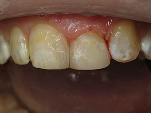
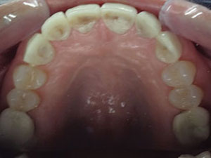
| Figure 1. Long-term provisionals were paper-thin and perforated on the lingual surfaces. | Figure 2. Crown preparations on teeth Nos. 6 to 11 (as done by the previous dentist). |
 |
 |
| Figure 3. Evaluation of wax-up. | Figure 4. Observe the minimal preparation for the premolars compared to the adjacent, previous crown preparations. |
 |
 |
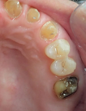
| Figure 5. Occlusal view of the same minimally invasive preparations for the premolars compared to the adjacent crown preparations. | Figure 6. A 10-second enamel etching (SureEtch [Zest Dental Solutions]). |
 |
 |
| Figure 7. Application of the bonding adhesive (CLEARFIL SE Protect [Kuraray America]). | Figure 8. Occlusal view of the finished restorations (IPS e.max Press [Ivoclar Vivadent]). (The restoration for tooth No. 14 was cemented later.) |
 |
| Figure 9. Lateral view of the final restorations, showing nice aesthetics and good gingival health. |
Upon examination, it was evident that the provisionals on the lingual side were paper-thin and fully perforated in multiple places with her lower teeth in full contact on the lingual surfaces (Figure 1). It was also evident that the patient suffered from bruxism.
This case exemplifies the downfalls of the myths described above and how the combination of them created a failing outcome along with the associated stress and unhappiness of all concerned. While lacking all of the information from the beginning of the case, one can still deduce some of the factors involved in the outcome. We can assume that lack of confidence in adhesive dentistry prompted the treating dentist to reverse his initial conservative approach with veneers and to revert to the old myth that full crowns are more predictable. The correct management, once it was reported that the newly placed veneers were chipping, would have been to re-evaluate the patient’s occlusion and not to replace the veneers with crowns.
Cutting the teeth down for crowns (Figure 2) led to lingual reduction, a lack of space, and a violation of the envelope of function. When handled properly, zirconia will not fracture, but the high strength of a material should never be the sole focus of a material choice in cases like this; it was the diagnosis and management of the occlusion that had been sorely missing. In addition, the decision to use long-term provisionals further complicated the case. During the 6 months in provisionals, and after many adjustments, the patient finally started to feel better. However, because of the lingual wear, the lower anterior teeth were allowed to slowly extrude, thus further decreasing the available restorative space.
The approach taught at the Los Angeles Institute of Clinical Dentistry requires taking proper records and the establishment of a full diagnosis before any partial or full rehabilitation is ever started. Patient records include accurate study casts using a quality hydrophilic alginate substitute (such as Silginat [Kettenbach LP]). A face-bow transfer and centric relation (CR) bite (Futar Fast [Kettenbach LP]) were taken to properly mount the study casts. This was done to permit a clinical occlusal analysis, allow for proper occlusal diagnosis, and determine the stability and health of the TMJ. Aesthetic cases require creating a smile design using the Dento-Facial Esthetic Diagnosis System.4
 |
| Figure 10. Side-by-side before-and-after images showing improved aesthetics. |
In this case, after proper diagnosis, the patient was informed that there was insufficient lingual space to allow for restorative material thickness and envelope of function space. Further tooth reduction would involve pulpal exposure(s). Options to regain space included orthodontics or opening the vertical dimension of occlusion (VDO). The patient chose opening the VDO because it was faster and less expensive.
 |
| Figure 11. Side-by-side case images of before, showing heavy wear, and after, a 15-year postoperative view of functioning porcelain veneers (from a different case example). |
The CR-mounted casts were sent to the laboratory team (Burbank Dental Lab (Burbank, Calif) for a diagnostic wax-up. The instructions to the lab team included opening the vertical dimension just enough to allow for the 1.5 mm of porcelain thickness required on the lingual of the anterior teeth, and to establish a 2.0-mm overjet (Figure 3). Aesthetic changes were requested based on the smile diagnosis. After approval of the wax-up, a matrix for the intraoral fabrication of the provisionals was created using a silicone putty (Panasil [Kettenbach LP]).
Clinical Protocol
Preparation day was very simple as all anterior teeth had already been prepped for crowns and were in provisionals. No additional tooth reduction was necessary as space would be achieved by opening the VDO. Additionally, both first molars had old crowns, which were removed, and the preparations were cleaned up, avoiding too much additional tooth removal. The 4 maxillary bicuspids were virgin teeth and were very lightly prepped for veneer/onlay restorations using the LA Institute Bur Kit (Brasseler USA) to prepare a 0.4-mm cervical, a 0.5-mm facial, and a 0.5-mm occlusal reduction. Knowing the bite had been opened 1.0 mm, it was expected to have a total occlusal restorative thickness of 1.5 mm (Figures 4 and 5) for the planned lithium disilicate restorations (IPS e.max [Ivoclar Vivadent]). The manufacturer recommends a minimum of 1.0 thickness for this material, however since the patient had a history of bruxism, greater material strength from using a thicker restoration was warranted. Bonded onlay/veneer preparations using supra-gingival preparation techniques has been discussed in previous articles.5,6 All the new preparations were left supragingival, with the exception of crown teeth, which had already been prepped with subgingival margins. Supragingival margins are always preferable as they are more predictable, with no bleeding cementation, as well as healthier for the patient.
The new provisionals were fabricated intraorally in 3 segments using the silicon matrix and a strong bis-acryl material (VISALYS TEMP [Kettenbach LP]). Next, the bite was adjusted on the uncemented temporaries using bimanual manipulation to ensure all teeth touched evenly in CR. After the bite was adjusted on the provisionals, the posterior portion was removed, leaving only the already adjusted anterior portion. Using Futar Fast, the bite was taken from the first bicuspid and back, again using bimanual manipulation to ensure the patient was in CR. (Alternatively, a leaf gauge technique can be used.)
The final upper impression was taken using a light/heavy-body-technique vinyl polysiloxane (Panasil light body [Kettenbach LP]) in a prefabricated full-impression tray. Panasil was chosen due to its excellent hydrophilicity, accuracy, and tear strength. The author’s experience is that physical impressions are the optimal choice in cases like this because, while digital scanning can achieve excellent accuracy, model printing and duplication has not achieved the same degree of accuracy. Furthermore, having been trained as a dental laboratory technician, I know that technicians prefer stone dies for minimally invasive preparations with minor contact separation and very shallow, slightly supragingival chamfers. At this point in time, margin definition is less crisp and defined with printed casts and dies. Currently, digital impressions and dies are best suited for traditional crown and bridge dentistry with deeper chamfers and more aggressive interproximal cavosurface margin separation as well as model-less single crowns. The author prefers to not do larger cases that are not best suited, or practical, with digital or model-less fabrication.
Next, the 3 provisional segments were luted into place using a non-eugenol temporary cement (Temrex TNE [Temrex]). Then the bite was adjusted to ensure it fulfilled the 3 Golden Rules of Occlusion7: (1) equal contact on all teeth in CR, (2) posterior disocclusion, and (3) a free and unobstructed envelope of function. Remember, the improper management of this patient’s occlusion had been identified as the main cause of her problems. Final aesthetic touches were provided, and the patient was dismissed. The patient remarked upon dismissal that the visit had been the easiest she ever had since her whole ordeal started.
At the one-week aesthetic and occlusion check, the patient reported no pain in her teeth nor discomfort and/or tension in her muscles. She said her bite did not feel constricted, which was to be expected after providing appropriate space for the envelope of function. She was very satisfied with the aesthetics, which was also to be expected with proper diagnosis and planning. An impression (Silginat) and bite (Futar Fast) were taken of the now-approved provisionals. These were sent to the laboratory team to be cross-mounted and used, along with a silicon matrix, to fabricate final restorations that would closely resemble what the patient had approved. The patient was dismissed, and an appointment was made for delivery of the final restorations.
IPS e.max Press (Ivoclar Vivadent) was the chosen material for all anterior and posterior restorations. Burbank Lab sent the waxed restorations for approval and later provided excellent final restorations. Bonded e-max is approximately 70% as strong as zirconia.8 On heavy grinders, it is wise to request non-layered e-max and to make them a bit thicker to maximize strength, as mentioned above, rather than the minimum thickness of 1.0 mm. While layered e-max is prettier, it is also weaker, especially on the incisal edges.
At the final delivery appointment, the provisionals were removed, and the preparations were cleaned using a flour pumice paste containing chlorhexidine (such as Consepsis Scrub [Ultradent Products], Preppies Plus [WhipMix], or Surpass [Apex]). Bonded cementation was very easy on the teeth with supragingival margins as no bleeding was present (Figure 5). The enamel was then etched (SureEtch [Zest Dental Solutions]) for 10 seconds (Figure 6), washed, and dried. Next, bonding adhesive (CLEARFIL SE Protect [Kuraray America]) was applied over the dentin and enamel, agitating it on dentin using the Microbrush (Microbrush Int) for 25 seconds (Figure 7). Careful removal of the solvent was achieved using a gentle stream of dry, oil-free air on the tooth until the primer stopped moving. A subsequent thin layer of bond (second component) was applied and not cured. For all anterior restorations, RelyX Veneer Cement A1 (3M) was used, and for all the posterior restorations, Panavia V5 Universal (Kuraray America) was used. All light curing was done using a VALO (Ultradent Products) cordless LED curing light. Finally, the bite was adjusted, fulfilling the 3 Golden Rules of Occlusion, and the patient was dismissed.
At the 2-week postoperative visit, a night guard was delivered, the bite was again checked, and any minor adjustments were made. The patient was extremely satisfied. She stated that her bite “felt great” and that her husband also liked the enhanced smile (Figures 8 to 10). Longevity is expected for this patient per the author’s extensive experience when employing a thorough diagnosis and treatment plan, the 3 Golden Rules of Occlusion, and proven restorative techniques (Figure 11).
References
- Castelnuovo J, Tjan AH, Phillips K, et al. Fracture load and mode of failure of ceramic veneers with different preparations. J Prosthet Dent. 2000;83:171-180.
- Kois DE, Chaiyabutr Y, Kois JC. Comparison of load-fatigue performance of posterior ceramic onlay restorations under different preparation designs. Compend Contin Educ Dent. 2012;33(special issue 2):2-9.
- Ruiz JL, Coleman TA. Occlusal disease management system: the diagnosis process. Compend Contin Educ Dent. 2008;29:148-152.
- Ruiz JL. Achieving predictable, beautiful smiles using a dento-facial esthetic diagnosis system. Compend Contin Educ Dent. 2007;28:50-55.
- Ruiz JL, Christensen GJ, Sameni A, et al. Clinical performance of bonded ceramic and resin-based composite inlays and onlays using a self-etch bonding system: a 51-month report. Inside Dentistry. 2007;3:62-65.
- Ruiz JL. Avoiding subgingival margins for healthier dentistry: using a supragingival preparation protocol. Dent Today. 2015;34:82-86.
- Ruiz JL. The three golden rules occlusion. Dent Today. 2010;29:92-93.
- Ma L, Guess PC, Zhang Y. Load-bearing properties of minimal-invasive monolithic lithium disilicate and zirconia occlusal onlays: finite element and theoretical analyses. Dent Mater. 2013;29:742-751.
Dr. Ruiz practices in Los Angeles, and he is the owner and director of the Los Angeles Institute of Clinical Dentistry and of many continuing education courses at the University of Southern California. He is an associate instructor at Dr. Gordon Christensen’s Practical Clinical Courses in Provo, Utah, and an independent product evaluator for CR Foundation, also in Provo. He can be reached at (818) 558-4332 or at ruiz@drruiz.com.
Disclosure: Dr. Ruiz reports no disclosures.
Related Articles
Health and Biocompatibility With Supragingival Dentistry
Two Crucial Techniques for Healthier Restorations
Focus On: Minimally Invasive Dentistry