INTRODUCTION
In recent decades, dental professionals have increasingly advocated and explained the importance of, and proper technique for, daily oral hygiene habits for children and adults. Simultaneously, dental hygienists are sharing more information regarding the negative oral health consequences of excessively consuming acidic and sugary foods and beverages. Unfortunately, a variety of factors may affect an individual’s compliance with accepted oral hygiene recommendations, ultimately contributing to tooth demineralization (eg, active caries lesions and/or white spot lesions) and/or periodontal disease.
White spot lesions are clinically detectable areas of demineralized enamel that indicate the initiation of the caries process.1 Characterized by a milky white appearance of varying opacities on smooth tooth surfaces, white spot lesions develop from subsurface porosities caused by the loss of calcium, phosphate, and other minerals.1-3 As a result, white spot lesions exhibit 2 distinct areas: dense interprismatic surface enamel and prismatic porous enamel below the surface.4
Traditionally, teeth exhibiting white spot lesions have been treated with direct composite restorations and/or veneers. Unfortunately, significant tooth reduction is typically required in order to accommodate sufficient material thickness, mask discolored tooth structure, and establish aesthetic morphology. Today, however, many white spot lesions—including those with complete demineralization (eg, cavities)—can be treated and remineralized chemically and topically without the need to permanently sacrifice tooth structure.5
Fluoride has historically been applied to teeth as a means of preventing and reducing caries development (eg, fluoride varnishes), as well as for remineralizing tooth structure. However, fluoride treatments are only effective in the presence of available and sufficient salivary calcium and phosphate ions, which bind to the salivary protein Statherin.6 If saliva quantity or quality is compromised, acid-producing biofilms exist on smooth surfaces, or teeth are constantly challenged by acid (eg, erosion), then significantly more calcium and phosphate are required to enhance fluoride’s effectiveness. Therefore, calcium phosphate technologies—including amorphous calcium phosphate (ACP) and casein phosphopeptide-ACP (CPP-ACP)—are increasingly becoming preferred therapies for arresting the caries process and remineralizing and strengthening tooth enamel.1,7
Implications of CPP-ACP for Remineralization
CPP-ACP are complex molecules derived from milk proteins that react in a manner similar to statherin. Like statherin, the combination of CPP-ACP (eg, RECALDENT) delivers bio-available calcium and phosphate to tooth structure in addition to neutralizing acid challenges.8-10 As a result, CPP-ACP promotes remineralization while simultaneously inhibiting demineralization.7
 |
 |
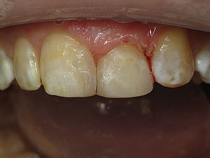
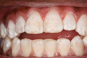
| Figure 1. Pretreatment photo of the patient with a natural smile. Initial white spots and cavities in her anterior teeth were obvious. | Figure 2. The hygienist performed a thorough prophylaxis using a micro-abrasive slurry, prophy angles, and prophy cups. |
 |
 |
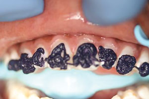
| Figure 3. The teeth were etched for 30 seconds using a phosphoric acid etchant. | Figure 4. View of the further-demineralized teeth after acid etching. |
Although the limiting factor for the effectiveness of remineralizing therapies is calcium and phosphate availability, products containing CPP-ACP have shown greater potential to create fluorapatite based on its availability of calcium and phosphate.10 In fact, research has demonstrated that CPP-ACP has a significant remineralizing effect on early caries lesions in both in vivo and in vitro studies.11 Additionally, enamel surfaces treated with CPP-ACP paste have exhibited the less lesion depths than enamel surfaces treated with fluoridated toothpaste.12
What’s more, pastes containing CPP-ACP can reverse the visible appearance of white spot lesions as well as rebuild other tooth structures that are water-rich, demineralized, or otherwise defective. In cases of incipient carious lesions, the application of CPP-ACP converts subsurface water back into enamel by diffusing neutral-ion species through the porous surfaces. When the consequently formed hydroxyapatite reacts with the water, it regenerates in subsurface spaces. Once 80% to 85% regeneration has occurred, tooth enamel appears optically normal (ie, white spot lesions disappear).
Currently the only professional-use and dentist-dispensed products containing CPP-ACP are MI Paste, MI Paste Plus (contains fluoride), and MI Paste ONE (contains fluoride) (GC America). MI Paste and MI Paste Plus can be used safely several times daily or as directed by the dentist and/or hygienist. MI Paste ONE is a dentifrice that can replace a patient’s current toothpaste yet provide the same preventive and reparative benefits as the other MI Paste products in a single application. The CPP-ACP in MI Paste products is naturally occurring and derived from milk casein.
Each MI Paste product containing CPP-ACP has specific indications. MI Paste is indicated for general caries prevention, preventing sensitivity prior to and during whitening treatments, relieving general tooth hypersensitivity, protecting exposed root surfaces, and sensitivity prevention during and after periodontal care. It is ideal for pregnant and nursing women and for children younger than 6 years of age. MI Paste Plus and MI Paste ONE are also appropriate for those indications; however, because these products contain fluoride, they are also indicated for remineralization and cavity prevention, patients suffering from xerostomia, the treatment of white spot lesions, and pH buffering to neutralize oral pH levels.
Treating White Spot Lesions With CPP-ACP
According to the manufacturer, white spot lesions can be treated successfully using MI Paste Plus and MI Paste ONE twice daily for 8 to 12 weeks or longer, as required. Arrested white spot lesions should be etched for no more than 15 seconds using a low-concentration phosphoric acid prior to applying either MI Paste Plus or MI Paste ONE in order to make the surface permeable. Active white spot lesions do not require pretreatment etching.
While MI Paste (non-fluoridated) is not indicated by the manufacturer for treating either active or arrested white spot lesions, the author and his hygienist have used it successfully for this purpose for more than 10 years. The following case presentation outlines the protocol developed by the author and describes the long-term results that were achieved.
CASE REPORT
Diagnosis and Treatment Plan
In 2010, a then-13-year-old girl presented with varying degrees of cavitated and noncavitated white spot lesions on her anterior maxillary (ie, central incisors and cuspids) and mandibular teeth that produced different aesthetically detrimental effects, including white streaks and holes in her teeth (Figure 1). Mercilessly teased in school due to the appearance of her teeth, she said she was frequently called “Swiss Cheese Mouth.”
A thorough examination was performed, and the patient was asked about her dietary habits. She admitted to consuming 2 sodas per day and a great deal of candy and junk food, as well as practicing poor oral hygiene habits. A resident of the metropolitan New York City area, the patient was exposed to fluoridated water, but fluorosis was not likely the cause of the white spots.
 |
 |
| Figure 5. Close-up retracted view after 8 weeks of treatment. Note that the previously visible cavities in the patient’s central incisors were now closed. | Figure 6. Photo of her natural smile after 8 weeks of treatment. Further time for remineralizing the canines was required. |
 |
 |
| Figure 7. Tooth whitening was accomplished after 10 weeks of treatment. | Figure 8. Close-up retracted view 2 years after treatment, confirming the longevity of the treatment results achieved for the patient’s anterior teeth. |
 |
 |
| Figure 9. Retracted view 2 years after treatment, confirming remineralization of the patient’s maxillary and mandibular teeth. | Figure 10. Post-treatment view at 8 years. The patient, now 21 years old, with her teeth still looking healthy. |
The patient’s father requested composite fillings for her teeth, but it was explained to the patient and her father that her diet and poor oral hygiene habits were likely contributing to many of her dental problems. It was further explained that with the required behavior modifications, her teeth could be treated without restorations. In particular, the patient’s white spot lesions would be treated according to a strict demineralization/remineralization protocol that would combine weekly in-office enamel etching and daily at-home application of a CPP-ACP topical cream (eg, MI Paste). The paste would be protected against the teeth using a custom tray (eg, bleaching tray) for at least 30 minutes a day.
Treatment Protocol (As Developed by the Author)
Step 1. To initiate treatment, the hygienist first performed a thorough prophylaxis using prophy angles, prophy cups, and a micro-abrasive slurry (Figure 2).
Step 2. After rinsing, an alginate impression of the patient’s teeth was made, and models were poured for use in making custom trays; these would be used to hold the MI Paste in close proximity to the teeth while the patient applied it at home as well as for tooth whitening later in the treatment process.
Step 3. While the dental assistant poured the impressions and made the models, the teeth were etched for 15 seconds with a 6.6% hydrochloric acid slurry (eg, Opalustre [Ultradent Products]) using a prophy cup or angle and light pressure, followed by a water rinse. This opened up the enamel pores to allow further penetration of the CPP-ACP paste (eg, MI Paste), in addition to facilitating white spot removal.
Step 4. Next, 37% phosphoric acid gel was applied to all affected tooth areas for 30 seconds once a day (Figure 3) and then rinsed off (Figure 4).
Step 5. The CPP-ACP paste (eg, MI Paste) was loaded into the custom-made bleaching trays, which were then seated into the patient’s mouth. The patient was instructed to wear the trays with MI Paste for one application of at least 30 minutes each day.
The patient was also specifically instructed to eliminate carbonated beverages and sugary foods and to brush and floss properly. The importance of her compliance was not overstated to the patient and her father, who both committed to the required weekly follow-up appointments.
Follow-up Appointment Protocol
Thereafter, for a period of 10 weeks, the patient was seen weekly. At each appointment, the teeth were re-etched using only phosphoric acid and then rinsed. The CPP-ACP was applied to the teeth for 5 minutes with the custom tray, after which the patient was dismissed until the next weekly follow-up appointment. After 8 weeks of treatment, the patient’s teeth showed visible improvement as a result of reportedly applying/wearing the CPP-ACP between 1 and 2 hours per day (Figures 5 and 6).
After 10 weeks of treatment, approximately 50% to 60% remineralization had been achieved, which enabled the incorporation of an at-home tooth whitening component to even out the overall tooth color. In this case, a 15% hydrogen peroxide whitening gel (Opalescence [Ultradent Products]) was used with the custom tray for between 20 and 30 minutes a day (Figure 7). The patient also continued her daily CPP-ACP cream application and weekly appointments.
CLOSING COMMENTS
Treating white spot lesions in this manner is a time-consuming process, one that requires patient commitment and compliance. Not all white spot lesion cases are the same; some require fewer weekly appointments, while others could require 6 months. However, it’s important to note that aesthetic, remineralized smiles are possible without removing any tooth structure when a strict protocol that incorporates CPP-ACP (eg, MI Paste) is followed. In this case, the patient’s ideal outcome was achieved after 14 weeks of treatment, and those results have endured for more than 9 years (Figures 8 to 10).
References
- AlShehri A, Kwon SR. Etiology and management of white spot lesions. Decisions in Dentistry. January 1, 2016. https://decisionsindentistry.com/article/lesions-0116/. Accessed February 27, 2020.
- Cochrane NJ, Cai F, Huq NL, et al. New approaches to enhanced remineralization of tooth enamel. J Dent Res. 2010;89:1187-1197.
- Mizrahi E. Enamel demineralization following orthodontic treatment. Am J Orthod. 1982;82:62-67.
- Takahashi N, Nyvad B. Caries ecology revisited: microbial dynamics and the caries process. Caries Res. 2008;42:409-418.
- Kugel G, Arsenault P, Papas A. Treatment modalities for caries management, including a new resin infiltration system. Compend Contin Educ Dent. 2009;30(special issue 3):1-10.
- Shen P, Manton DJ, Cochrane NJ, et al. Effect of added calcium phosphate on enamel remineralization by fluoride in a randomized controlled in situ trial. J Dent. 2011;39:518-525.
- Kwon SR, Kolker J. Implement a minimally invasive approach. Dimens Dent Hyg. 2014;12:22-25.
- Reynolds EC. Anticariogenic complexes of amorphous calcium phosphate stabilized by casein phosphopeptides: a review. Spec Care Dentist. 1998;18:8-16.
- Schüpbach P, Neeser JR, Golliard M, et al. Incorporation of caseinoglycomacropeptide and caseinophosphopeptide into the salivary pellicle inhibits adherence of mutans streptococci. J Dent Res. 1996;75:1779-1788.
- Rose RK. Effects of an anticariogenic casein phosphopeptide on calcium diffusion in streptococcal model dental plaques. Arch Oral Biol. 2000;45:569-575.
- Li J, Xie X, Wang Y, et al. Long-term remineralizing effect of casein phosphopeptide-amorphous calcium phosphate (CPP-ACP) on early caries lesions in vivo: a systematic review. J Dent. 2014;42:769-777.
- Somasundaram P, Vimala N, Mandke LG. Protective potential of casein phosphopeptide amorphous calcium phosphate containing paste on enamel surfaces. J Conserv Dent. 2013;16:152-156.
Dr. Kaminer is a 1990 graduate from the State University of New York at Buffalo School of Dental Medicine, and he maintains practices in Hewlett and Oceanside, NY. A world-renowned expert in dental lasers and minimally invasive dentistry, he has authored numerous articles on these topics, in addition to serving as a clinical consultant and lecturer for several dental product and equipment manufacturers. In addition to maintaining a teaching appointment at Peninsula General Hospital in Far Rockaway, NY, he is also a clinical instructor with the International College of Laser Education and director of the Masters of Laser training program in New York. He can be reached via email at whitertth@aol.com.
Disclosure: Dr. Kaminer receives honoraria from GC America.
Related Articles
Icon Smooth Surface: Minimally Invasive Treatment With Maximal Results
A Simple and Effective Treatment for White Spots
Treating Post-Orthodontic White Spots: A Conservative Resin Infiltration Technique