
Due to the evolution of dental materials and the ever-increasing demand for aesthetic restorations, we now live in an era in which full-ceramic materials are the standard for rehabilitating compromised anterior dentition, while porcelain-fused-to-metal crowns are almost becoming obsolete. In this era, the trend is also moving toward a minimally invasive type of dentistry in which the preservation of the dental tissues is a significant concern. This tissue-saving approach implies that partial-coverage restorations and porcelain veneers are gaining more relevance over more traditional full crowns. However, it is not always clear from a clinical perspective where a crown or a veneer is best suited.
Typically, the degree of deterioration of the tooth structure is the main factor to be considered when deciding between a veneer or a crown. But for many patients with aesthetic issues in the anterior region, it is common to find different degrees of deterioration within the same set of teeth. Such cases may require a combination of veneers and crowns. Some teeth may have root canal treatments with posts and cores. Some teeth may have a favorable substrate color, whereas others may be darkened. Even sound natural teeth that do not require treatment may be a part of the equation. One thing is certain: The more variables there are, the more challenging it becomes to achieve a successful aesthetic outcome.
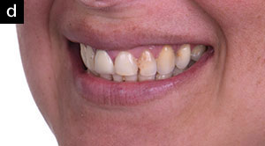
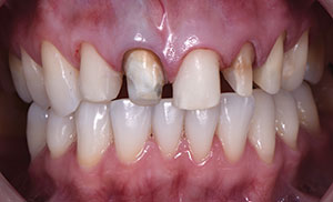
 |
| Figure 1. Initial case. Patient presented with an unadapted crown and a darkened root on tooth No. 8 and failing composite restorations on teeth Nos. 7, 9, 10, 11, and 12. |
The following is a case report that represents these challenges. The aim is to share some of the guidelines used to aid other professionals in the decision-making process during similar cases.
CASE REPORT
A 35-year-old woman in good general health presented with a chief complaint of deteriorated restorations in the upper anterior zone. Clinical and radiographic examination revealed the presence of an unadapted ceramic crown on tooth No. 8 and unsatisfactory composite resin fillings, some of which were associated with secondary caries; discolored teeth due to root canal treatment; and some length discrepancies in the anterior dentition (Figure 1). Tooth No. 7 presented an acceptable endodontic treatment with no symptomatology, while teeth Nos. 10 and 11 presented periapical radiolucencies. The periodontal evaluation found no pathologic probing depths. Occlusal examination revealed normal Class I occlusion.
For a case like this, a number of questions had to be answered during the treatment planning phase:
- Which teeth should be treated?
- Are posts needed?
- What type of restorations are best suited for which teeth?
- What are the clinical and lab considerations for this case?
Number of Teeth Involved
In an aesthetic case, several parameters are considered when deciding which teeth should be included in the treatment plan. Besides the more obvious compromised teeth (for example, due to caries or failing restorations), other factors to be considered are tooth size, shape, proportion, color, and position regarding the overall smile design. A facial analysis completed with intraoral and extraoral photographs is essential. In this case, an insufficient display of the edges of the central incisors, the lack of central dominance, and a flattened smile curve were indicators that an increase of the incisal edges of these 2 teeth would enhance the patient’s smile (Figure 2). A digital smile design was carried out as digital tools can be very helpful for previewing and communication. As shown in Figure 3, aside from the incised length of the 2 centrals, teeth Nos. 11 and 12 were lacking facial volume. Disparities in the gingival margins were dismissed as these were minimal and would allow for “perfect imperfections” that can actually help achieve a more natural-looking result.
 |
 |
| Figure 2a. Lips at rest with insufficient exposure of incised edges. | Figure 2b. Full-smile photograph showing a flattened smile curve, a lack of central dominance, discolored teeth, and an overall unaesthetic appearance. |
 |
 |
| Figures 2c and 2d. Lateral views showing poor aesthetics. |
Although a symmetrical set of restorations allows for a more predictable aesthetic result, the decision was to treat teeth Nos. 7 to 12 with veneers or crowns and treat Nos. 5 and 6 only with teeth whitening. This is because these 2 teeth were not only in perfect health and position within the smile design but also, and very importantly, the patient asked for the minimum number of procedures, where possible.
 |
 |
| Figure 3. Digital smile design of the case. | Figure 4. Preparations before the final impression. Teeth Nos. 7, 9, and 12 were prepped for extended veneers, while teeth Nos. 8, 10, and 11 were prepped for full crowns. Notice the different substrate colors. |
 |
 |
 |
 |
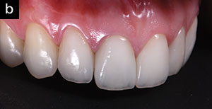
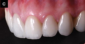
| Figure 5. Intraoral photos of the cemented veneers and crowns. |
Post or No Post
A common dilemma when a clinician is presented with endodontically treated teeth is whether an intraradicular post should be placed or not. It has been demonstrated that posts can cause root fractures during function due to str
ess. The strength of the tooth is directly related to the amount of remaining intraradicular dentin. In other words, a post can actually weaken the tooth by having dentin removed from the canal. Therefore, the use of posts should be limited to endodontically treated teeth where there is insufficient tooth structure remaining. Thus, the purpose of the post is strictly to support a core foundation for the future crown restoration. In anterior teeth, with a limited access opening and strong dentin walls, a post is not needed.
When the decision has been made to place a post, the next step is to select the material and technique. Prefabricated, fiber-reinforced posts and composite resin cores have become very popular because of the simplified procedure, which can be completed chairside. Also, they exhibit a compatible modulus of elasticity with dentin (thus reducing the predisposition to root fracture) and are more aesthetic than their cast counterparts. However, proper selection and a meticulous clinical procedure are critical in achieving long-term success. For instance, a crucial consideration is to have a minimum amount of 1 to 2 mm of coronal sound tissue above the cementoenamel junction to act as a ferrule around the post. This has been proven to prevent root fracture and post fracture or dislodgment.
In the case presented here, teeth Nos. 10 and 11 were selected for fiber-reinforced posts following these criteria.
Veneers or Crowns
The amount and quality of remaining tooth tissue are essential when deciding between all-ceramic crowns and veneers in the anterior dentition. The key requirement for choosing ceramic veneers is that the tooth preparation should be confined within the enamel shell or display at least 50% to 70% of the enamel. The reason for this is that veneers rely only on adhesion (not retention), and it is well known that the highest bond strength occurs in enamel. Also, it has been reported that ceramic veneer debonding can occur when dentin accounts for 80% of the tooth substrate. Therefore, when the tooth’s facial aspect is mostly dentin, a full crown may be a better treatment choice.
A careful examination of the lingual surfaces is also very important. If these are worn through to dentin, or exhibit extended carious lesions or large restorations, a crown may be more suitable than a veneer. In the event of a large diastema or interproximal lesions, extended veneers are also an excellent treatment option.
On another note, endodontically treated teeth do not necessarily need crowns. If the marginal ridges are preserved and the above criteria are met, a veneer could well serve as a final restoration in a tooth with root canal treatment. If a post has been placed, then a crown should definitely follow.
For the above reasons, the decision in this case was to treat teeth Nos. 8, 10, and 11 with full ceramic crowns and teeth Nos. 7, 9, and 12 with ceramic veneers.
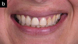
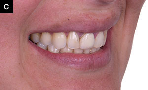
 |
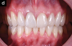
| Figure 6. Extraoral photos of the cemented veneers and crowns. Note the improved smile curve, incisal exposure at rest, and overall aesthetics. |
Ceramic System
Although the descriptions and indications of all the ceramic systems available today are beyond the scope of this paper, it is important to say that the ceramic chosen to restore a case of combined full-ceramic crowns and veneers must fulfill certain requirements. All restorations (crowns and veneers) should be fabricated using the same ceramic system to ensure a homogenous aesthetic outcome. Because ceramic veneers rely entirely on adhesion, it is essential that the ceramic system used permits a chemical bond to the ceramic; in other words, adhesive cementation must be feasible. Another important consideration is the aesthetics. The optical behavior of the ceramic should mimic that of the natural dentition. A range of translucent to more-opaque cores should be available. Also, the ceramic restorations should possess excellent mechanical properties.
For these reasons, the ceramic system chosen for this case was pressed lithium disilicate glass ceramic (IPS e.max [Ivoclar Vivadent]).
Clinical Steps
After approval of the proposed treatment plan, the lower teeth—as well as the upper teeth, which would remain untreated by ceramic restorations—were bleached using 10% carbamide peroxide gel overnight for a period of 2 weeks. The final shade was registered as 1M1 using the VITA 3D Master scale (VITA Zahnfabrik). After root canal treatment, fiber-reinforced posts (Whitepost [FGM, Brazil]) were adapted and cemented on teeth Nos. 10 and 11 with self-adhesive resin cement (RelyX U200 self-adhesive cement [3M]). Old composite fillings and remaining decayed tissues were removed and restored using a universal bonding agent (Single Bond Universal Adhesive [3M]) and composite material (Filtek Z350 XT [3M]).
A set of cylindric, tapered, round-end diamond burs were used to prep teeth Nos. 8, 10, and 11 for crowns (All Ceramic Prep Kit 0955 [Shofu Dental]). The depth of each reduction was controlled using a silicone guide fabricated from the previously obtained diagnostic wax-up, which followed the smile design from the diagnostic phase. The final crown preparations were approximately 1.5 mm deep to create sufficient room for the ceramic material. A slight subgingival chamfer margin was created to aid in masking the teeth’s discoloration at the gingival area.
A smaller cylindric, tapered, round-end diamond bur was used to prep the veneers for teeth Nos. 7, 9, and 11 using the same silicone index for guidance. The final preparations were 0.5 to 0.8 mm deep, and the termination was left at the gingival margin.
At this point, photographs of the prepped teeth were taken for communication with the ceramist (Figure 4). It is of the utmost importance that the ceramist sees the variety of stump shades that he or she will have the difficult task of unifying with the final restorations.
Prior to impression taking, a retraction cord soaked in astringent solution was placed at the bottom of the sulcus (No. 00 Ultrapak [Ultradent]). A 2-step impression technique was used with polyvinyl siloxane silicone material (Panasil Putty Soft and Panasil Initial Contact Light [Kettenbach LP]). The retraction cord was removed before the second impression with the light material. A bite registration material (Occlufast [Zhermack]) was used for this purpose.
Provisional restorations were fabricated using a silicone matrix from the diagnostic wax-up and self-cured temporary resin (ALIKE [GC America]). Intraoral and extraoral photographs were taken to check on the different aesthetic parameters and then sent to the ceramist.
Try-in and cementation of the newly fabricated lithium disilicate (IPS e.max) veneers and crowns occurred 10 days later. The provisional restorations were removed, and the preparations were cleaned. The adaptation of the restorations was checked first (dry try-in) and again after a transparent try-in paste was used so that the patient could assess the aesthetics with the aid of a mirror.
After approval, all ceramic restorations were treated internally with hydrofluoric acid for 20 seconds, washed profusely, dried, and silanated. First, the crowns of teeth Nos. 8, 10, and 11 were cemented using RelyX U200 self-adhesive cement, color A2, and photocured for 40 seconds on the buccal and 40 seconds on the palatal aspect. Because the final color of a veneer can be further adjusted by the color of the cement (which is less likely for a crown), different try-in pastes were used prior to bonding. Teeth Nos. 7, 9, and 12 were then conditioned with phosphoric acid at 37.0%, rinsed, and dried, and universal adhesive was applied (Single Bond Universal [3M]). The vene
ers were luted in place with A1 veneer resin (RelyX veneer [3M]).
DISCUSSION
The color matching of combined veneers, crowns, and natural teeth can certainly be very challenging, especially when darkened substrates are added to the equation. While some dentists may find it easier to place crowns on all the teeth involved, despite this being a more radical approach, the author believes that an effort should be made in today’s dentistry to preserve the most tooth structure possible. However, the patient should be well-informed of the aesthetic difficulties associated with the treatment; presented with different viable treatment options, from most conservative to most aggressive; and made well aware of the advantages and disadvantages of each.
Without a doubt, the success of a case like this lies greatly in the ceramist’s knowledge and craftsmanship. Here, the ceramist used LT BL3 ingots for all the restorations, as this was her preference (other ceramists might choose to work with ingots with different opacities depending on the substrate color). She used a cutback technique and internal characterization with IPS e.max Essence powders (Ivoclar Vivadent) to increase the value, especially in the teeth with darker substrates.
Nonetheless, in the final photos of the case presented above, the highly trained eye will find slight mismatches in value in some of the restorations. An effort will be made to continue to overcome this difficulty. However, a minimally invasive approach was used; patient satisfaction was achieved beyond her expectations; and health, function, and aesthetics were restored back to the patient (Figures 5 and 6).
Acknowledgment:
The author would like to acknowledge ceramist Paola Chinchay (Lima, Peru) for her work in this case.
Dr. Ochoa graduated at the top of her class from Cayetano Heredia University in 2006. She has a degree in Occlusion and Oral Rehabilitation from Universidad Científica del Sur and has received extensive training in Brazil and the United States. She is the co-founder of Infinity Dental Clinic and Infinity Advanced Dental Learning in Lima, Peru. Dr. Ochoa is a Member of the American Society for Dental Aesthetics and a Diplomate in the American Board of Aesthetic Dentistry. In her work, Dr. Ochoa employs an interdisciplinary, facially driven, and minimally invasive philosophy and utilizes digital tools for the planning and communication of all her cases. She can be reached at [email protected] or at the Instagram handle @dra.paolaochoa.
Disclosure: Dr. Ochoa reports no disclosures.
Related Articles
The Modern Aesthetic Mixed Restorative Case
Using Digital Technology for Complex Aesthetic Challenges
Minimizing Patient Visits During a Pandemic: Demonstration of a Combination-Technique Protocol


