In today’s society, an attractive smile is critical to a pleasing appearance. It is an invaluable asset that creates a feeling of self-confidence and self-esteem. According to Gürel, “The pleasant smile is perceived as harmonious only when the various lines, proportions, and structures are in visual balance with each other. The already existing status of the smile can be improved by changing these proportions, creating illusions, and minimizing the negative visual tension produced by improperly aligned teeth, gingiva, and the lips.”1
Excessive gingival display (EGD), often referred to as a “gummy smile,” would be an example of negative visual tension created by normal proportions being out of balance. Ideally, in an attractive smile, 2.0 mm or less of gingival tissue would be displayed. In fact, a study conducted by Kokich Jr et al2 compared the perceptions of dentists and lay people to altered dental aesthetics and found that a gingival display of 3.0 mm or greater was considered to be unattractive and not visually appealing.
EGD is actually a descriptive term and not a diagnosis. Since multiple factors may be responsible for the condition, it is always important to determine the etiology (or etiologies) before developing a treatment plan.3 Etiologies may be both dentoalveolar and non-dentoalveolar in nature.4 Dentoalveolar etiologies include altered passive eruptions, wear and compensatory eruptions, and anterior dentoalveolar extrusions. Non-dentoalveolar etiologies include short upper lips, hypermobile lips, and vertical maxillary excess.
Due to the complexity of cases involving excessive gingival display and the possibility of having more than one etiology, an interdisciplinary treatment approach is often indicated that may involve periodontists, orthodontists, oral surgeons, plastic surgeons, and the restorative dentist.5 The following case report will illustrate the importance of determining the etiology and then the utilization of a coordinated interdisciplinary approach to achieve a successful result for a patient with short teeth and EGD.
CASE REPORT
Diagnosis and Treatment Plan
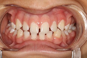
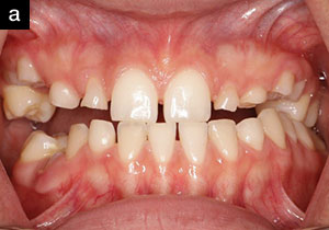
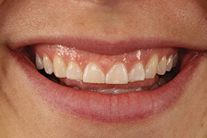
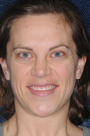
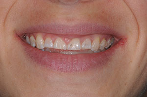
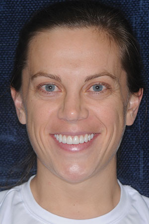
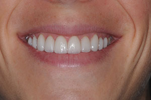
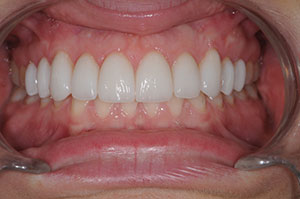
A 31-year-old female presented for a consultation because she was unhappy with the appearance of her smile. Specifically, she felt that she showed a lot of gum tissue when smiling and that her front teeth were short (Figures 1 to 3). Two of the most common etiologies of short teeth and excessive gingival display are altered passive eruption (APE) and wear with compensatory eruption.
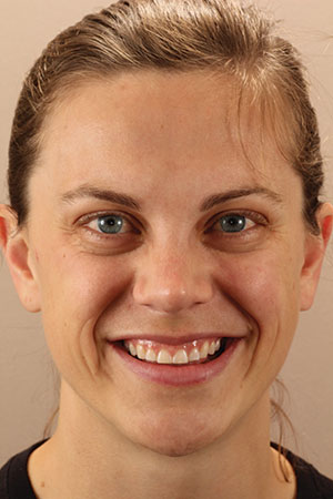 |
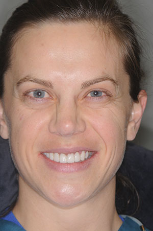
| Figure 1. Preoperative full-face smile. |
 |
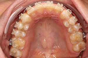 |
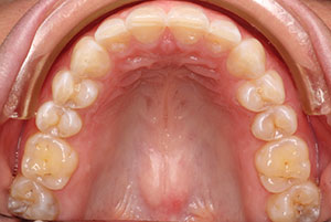
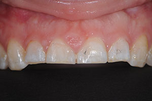
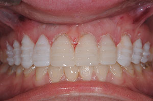
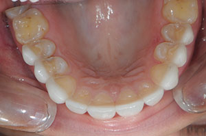
| Figure 2. Pre-op close-up smile. | Figure 3. Pre-op occlusal view. Note wear on the incisal edges. |
In cases of APE, the teeth appear short because gingival tissues do not recede to a normal position and actually cover part of the clinical crown. However, the incisal edges would not exhibit any signs of wear.6 APE can usually be successfully treated with simple gingivectomies or periodontal crown lengthening procedures with osseous resection. Compensatory eruption cases may also be treated with a combination of crown lengthening and restorative dentistry, though this combination treatment can result in a less than ideal crown-to-root ratio and emergence profile due to the necessity of having to place a restoration on the root.7 Therefore, when treating excessive gingival display due to compensatory eruption and incisal wear, it might be more beneficial to consider a treatment plan that combines orthodontic intrusion and all-ceramic restorations.8
The clinical exam revealed short teeth (7.5 mm) with evidence of incisal edge wear and compensatory eruption that contributed to the increased gingival display. In addition, the patient exhibited some hypermobility of the upper lip, further enhancing the excessive gingival display when smiling. A thorough discussion of treatment options included the pros and cons of crown lengthening and orthodontic intrusion, as well as soft-tissue procedures to limit lip elevation. Because of the diagnosis of wear and compensatory eruption, orthodontic intrusion (in lieu of the option of crown lengthening) was recommended. The patient agreed to an orthodontic evaluation to discuss intrusion, but declined a consultation to discuss possible soft-tissue procedures to limit lip elevation.9 In addition to the orthodontic consultation, the treatment plan included all-ceramic veneers on teeth Nos. 4 to 13.
Clinical Protocol: Orthodontics and Restorative Phases
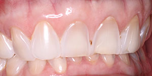
After accepting and completing orthodontic treatment (Figures 4 to 6), the patient returned to begin the restorative phase. The orthodontist had successfully intruded the incisors, decreasing the amount of gingival display and creating the space necessary for ideal length all-ceramic veneers (Figures 7 to 10).
The first step of the restorative phase was to determine incisal edge position. This is a critical step when increasing incisal length, and, although arbitrary to some degree, the case can be evaluated aesthetically, functionally, and phonetically with the provisionals before the definitive restorations are fabricated.10 Initially, composite was added to the central incisors to increase the length to 11.0 mm and to display about 3.0 to 4.0 mm of tooth with the lips at rest. Although this incisal edge position and the length of the centrals were arbitrarily chosen based upon proven smile design principles, they can be evaluated with a fair degree of certainty while the patient is wearing provisional restorations.
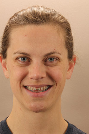 |
| Figure 4. Orthodontic intrusion phase, full-face view. |
 |
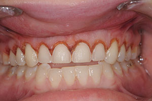
 |
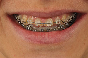
| Figure 5. Orthodontic intrusion phase, close-up smile view. | Figure 6. Orthodontic intrusion phase, occlusal view. |
 |
| Figure 7. Post-orthodontic intrusion, pre-restorative full-face smile. |
 |
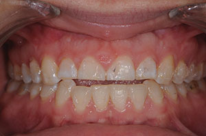 |
| Figure 8. Post-orthodontic intrusion, pre-restorative close-up smile. | Figure 9. Post-orthodontic intrusion, pre-restorative retracted view. |
Normally, the central incisor mockup is sufficient to convey the necessary information to the dental laboratory team to fabricate a diagnostic wax-up and a putty matrix that will be used to make the provisionals. However, in this particular case due to time constraints, a lab-fabricated diagnostic wax-up and putty matrix was not possible. Instead, all 10 teeth (Nos. 4 to 13) were mocked up using composite resin without acid-etching the teeth. No local anesthetic was used for this mockup step so that the smile, speech, and function could be evaluated preoperatively for ideal length, shape, and facial contours. An impression of the completed mockup was taken and set aside to be used later as a matrix for the provisionals.
Next, depth cuts were made in the mockup to allow the most conservative and minimally invasive preparation possible (Figure 11). After removing the mockup composite, a diode laser (Picasso Lite Plus [AMD LASERS]) was used for minor recontouring to create ideal gingival height symmetry. Then, using the depth cuts as a guide, the teeth were conservatively prepared using a single-use round-ended tapered coarse diamond (1116.10 NeoDiamond [Microcopy]) (Figure 12). After completing the preparations, a full-arch polyether impression (Impregum Soft [3M]), an occlusal record, and a stick bite were taken. The shade of the prepared teeth (stump shades) along with a written lab prescription detailing the goals of treatment, material selection, and desired shade (BL2/BL3, taken before preparation of the teeth had begun) were forwarded to the laboratory team. Using the impression of the pre-op mockup, the provisionals were fabricated (Tuff-Temp Plus [Pulpdent]).
After 2 days, the patient returned to evaluate the provisional restorations for aesthetics, phonetics, and function. After minimal adjustments, photos (Figure 13) and an impression were taken of the (patient- and doctor-approved) provisionals. These were also forwarded to the laboratory team to serve as communication tools, providing a blueprint to follow while fabricating the definitive restorations.
The patient returned in 3 weeks to have the lithium disilicate (IPS e.max Press [Ivoclar Vivadent]) all-ceramic veneers, that had been cut back and layered to optimize aesthetics, delivered. The patient had experienced no postoperative problems. The provisionals were removed, and the veneers were tried in with water to evaluate fit, color, and interproximal contacts.
Next, the teeth were isolated using a rubber dam (Non-Latex Dental Dam [COLTENE]) in preparation for cementation using a resin cement. The intaglio surfaces of the restorations were easily cleaned using a universal cleaner (Ivoclean [Ivoclar Vivadent]), then rinsed and dried with oil-free air and treated with silane. The restorations were set aside (organized by tooth number). The isolated teeth were etched with phosphoric acid gel (Select HV Etch, containing the antimicrobial ingredient BAC [BISCO Dental Products]) using the total etch technique for 15 seconds each, rinsed, and lightly air dried.
 |
 |
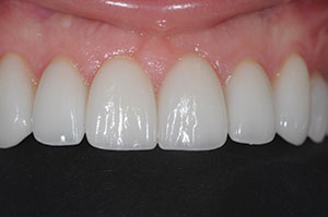
| Figure 10. Post-orthodontic intrusion, pre-restorative 1:1 close-up view. | Figure 11. Depth cuts were made in the composite resin mockup to ensure minimally invasive preparations. |
 |
| Figure 12. Diode laser recontouring was done around the finished all-ceramic preparations. |
A universal adhesive (ALL-BOND UNIVERSAL [BISCO Dental Products]) was applied, the solvent was evaporated, and then the adhesive was light cured. The translucent shade, which was a light-cured resin cement (Choice 2 [BISCO Dental Products]), was placed on all 10 teeth, and the veneers were seated simultaneously. Excess cement was cleaned off with cotton rolls and brushes. The veneers were tack cured at the gingival margin, allowing the cement to be removed interproximally by flossing the contacts. All the restorations were light cured (Bluephase 20i [Ivoclar Vivadent]) from the facial and lingual directions for 40 seconds.
The rubber dam was removed. The occlusion was checked using articulating paper and a T-Scan Novus (Tekscan) and then adjusted where needed. The restorations, where adjusted, were polished with fine and super-fine grit rubber cups and points (CeraMiste [Shofu Dental]). Finally, the patient was appointed for a post-op evaluation and photographs (Figures 14 to 18).
 |
 |
| Figure 13. The patient and doctor approved the provisional restorations, seen here after careful evaluation and any needed adjustments. | Figure 14. Postoperative full-face view. |
 |
 |
| Figure 15. Post-op close-up smile. | Figure 16. Post-op retracted view. |
 |
 |
| Figure 17. Post-op 1:1 close-up view. | Figure 18. Post-op occlusal view. |
IN SUMMARY
Excessive gingival display (gummy smile) is not a diagnosis. It is a descriptive term. When the amount of gingival tissue displayed while smiling is 3.0 mm or greater, it is widely considered to be unattractive and can often be a source of embarrassment or accompanied by a loss of self-esteem. Gummy smiles can be both dentoalveolar and non-dentoalveolar and may have more than one etiology. That is why determining the etiology is important before formulating a treatment plan and deciding on a course of action. More than one treatment option may exist to reach the desired result, and this should always be explained and thoroughly discussed with the patient.
In the case presented in this article, a diagnosis of incisal wear with compensatory eruption and mild hypermobility of the upper lip was determined to be the cause of the patient’s gummy smile and short teeth. Treatment options were explained and discussed, and, although she elected not to pursue any procedures to limit lip elevation, a successful outcome was achieved for this patient using orthodontic intrusion and all-ceramic lithium disilicate veneers.
Acknowledgement
The author would like to recognize the contribution of the orthodontist in the case presented, Dr. David Sarver (Birmingham, Ala), and for the artistry demonstrated in the all-ceramic veneers skillfully created by Mr. Gary Vaughn, CDT (Corr Dental Designs, Roseville, Calif).
References
- Gürel G. The Science and Art of Porcelain Laminate Veneers. Chicago, IL: Quintessence Publishing; 2003.
- Kokich VO Jr, Kiyak HA, Shapiro PA. Comparing the perception of dentists and lay people to altered dental esthetics. J Esthet Dent. 1999;11:311-324.
- Robbins JW. Differential diagnosis and treatment of excess gingival display. Pract Periodontics Aesthet Dent. 1999;11:265-272.
- Gottesman E. Excessive gingival display: addressing multiple etiologies for optimal esthetics outcomes. Perio-Implant Advisory. September 12, 2012. http://www.perioimplantadvisory.com/articles/2012/09/excessive-gingival-display-addressing-multiple-etiologies-for-optimal-esthetic-outcomes.html. Accessed November 5, 2017.
- Spear FM, Kokich VG, Mathews DP. Interdisciplinary management of anterior dental esthetics. J Am Dent Assoc. 2006;137:160-169.
- Dolt AH III, Robbins JW. Altered passive eruption: an etiology of short clinical crowns. Quintessence Int. 1997;28:363-372.
- Kois JC. Altering gingival levels: The restorative connection part 1: biologic variables. J Esthet Dent. 1994;6:3-9.
- Kois JC. Anterior wear: orthodontic and restorative management. In: Cohen M, ed. Interdisciplinary Treatment Planning, Volume II: Comprehensive Case Studies. Chicago, IL: Quintessence Publishing; 2012:189-210.
- Allen EP. Use of mucogingival surgical procedures to enhance esthetics. Dent Clin North Am. 1988;32:307-330.
- Cranham J. The horizontal position of the maxillary incisal edge: the key to optimum esthetics, phonetics, and function. Contemporary Esthetics and Restorative Practice. 2006;10:22-24.
Dr. Dudney is a 1977 graduate of the University of Alabama, Birmingham School of Dentistry. He is an active member of the Alabama Dental Association, the AGD, the ADA, and the Birmingham District Dental Society. He is an accredited member of the American Society for Dental Aesthetics and is a Diplomate of the American Board of Aesthetic Dentistry. He is clinical director for the California Center for Advanced Dental Studies’ live-patient, hands-on continuum. Formerly, he was the clinical director for the Rosenthal Group’s Aesthetic Advantage hands-on continuum and taught at the New York University College of Dentistry and the Eastman Dental Clinic in London, UK. Dr. Dudney has been a speaker at several major dental meetings and has authored several articles on aesthetic and restorative dentistry. He can be reached via the email address tedudneydmd@aol.com or via the website thomasdudney.com.
Disclosure: Dr. Dudney reports no disclosures.
Related Articles
Creating a More Youthful Appearance: Multidisciplinary Treatment Planning
Interdisciplinary Treatment Expanded
Full-Mouth Black Triangle Treatment Protocol