Many times our patients present to the office with particular life or economic circumstances that either outline or even dictate the treatment plan. Most of what the prudent dentist recommends is the very best treatment option to help defray future costs of remakes of failures due to compromised treatment. However, excellent treatment alternatives exist that have the potential to provide the patient high-quality, aesthetic, and long-lasting treatment without incurring considerable expense.
Our elderly patients are examples of these types of case scenarios. Additionally, in many instances, their particular edentulous situation may not lend itself well to providing a balanced occlusion for their restoration. In particular are those who present with bilateral distal extension mandibular removable partial dentures (RPD) with either selected or fully edentulous maxillas. With the loss of mandibular alveolar support in these cases, we often find that the potential for fracture of any remaining teeth is inevitable.
Much of what we do for our elderly patients sacrifices aesthetics for function. Additionally, we frequently find ourselves reacting to the forces of occlusion and parafunction on their remaining dentition via the extraction of fractured teeth and consequently adding teeth to their existing partial prosthesis. Obviously, with this treatment, the patient not only loses the tooth, but also more importantly risks the loss of additional alveolar support after the extraction. Many times, we have found that fractured teeth leave an intact subosseous root structure that can serve as support for the added-on partial denture tooth. The risk of additional root decay is weighed against the complete removal of the root and subsequent loss of the supporting alveolus.
A frequent occurrence is the fracture of the tooth with a pre-existing crown restoration along with the remaining ferrule, which many times leaves the intact root. The remaining root structure does not always leave the dentist with the option of rebonding or recementing the crown. The very best treatment option in this instance, if there is not enough remaining ferrule, is root canal treatment, possible crown elongation, and re-restoration of the tooth with a new crown. However, these types of invasive treatments may not be feasible or indicated in many elderly patients.
This leaves us with the less desirable option of extraction and adding the tooth to the partial denture over the extraction site. If the remaining root structure has no pulp exposure or has had previous endodontic treatment and the area can be sealed with the concurrent removal of caries, why not leave it? Informed consent is provided for the patient in addition to instructions for home care for the remaining root structure. The addition of the new denture tooth to the pre-existing partial denture is the only remaining possible dilemma. If the fractured crown was custom stained to match the adjacent dentition, the option of utilizing the crown as the “denture tooth” is much more viable than a stock denture tooth. With the increased bond strengths to various surfaces available to us with state-of-the-art adhesives, we now have the ability to add the crown to the partial denture using a “cone” of opaque porcelain over a specially designed metal substructure that can be laser-welded to the pre-existing partial framework.
This article describes a case in which this technique was used.
CASE REPORT
 |
| Figure 1. Preoperative condition illustrating fractured core and crown on tooth No. 7. |
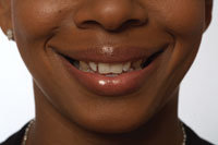
An elderly lady fractured off a beautiful Empress crown to the gingival level (Figure 1). Her removable partial denture was all the posterior occlusal support she had. There was not enough of a ferrule to rebond/recement the fractured tooth/crown, and the patient refused to have endo-dontic treatment, a post and core, and a new crown restor-ation. She additionally needed a full porcelain-to-metal crown under the partial clasp on tooth No. 5.
 |
| Figure 2. Facial view of tooth No. 7 crown illustrating ideal aesthetics and contours suited for adding to patient’s existing removable partial denture. |
Because of her economic situation and her medical condition, she was not a candidate for ideal treatment, which would either be elongation, orthodontic extrusion, endodontic treatment, post and core restoration and a new porcelain crown, or implant replacement. The existing Empress crown for tooth No. 7 was well shaded (Figure 2) and matched her adjacent dentition extremely well. After all the options were discussed, it was decided that there was a better chance of having a more aesthetic and possibly equally functional result with the old crown bonded to the partial denture versus a new denture tooth added to the RPD.
 |
| Figure 3. View of intaglio of crown after removal of all organic debris, sandblasting, and treatment with hydrofluoric acid. It is ready to bond to the “receptor cone.” |
After removing a slight amount of caries on the re-maining root, a small drop of composite (Heliomolar, Ivoclar Vivadent) was placed in the conventional manner over the root stump. It was finished and polished using Brassler ET burs and Astropol Polish-ing System (Ivoclar Vivadent). Subsequently, the fractured tooth structure inside the all-porcelain crown was removed, and all remaining biologic material and adhesive material were removed from inside the existing crown. The intaglio of the crown was then sandblasted to condition and improve the bondability (Figure 3). A bite registration of the patient’s maximum intercuspation was taken using Blu-Mousse (Parkell). An impression with Provil (Her-aeus Kulzer) was obtained of the upper and lower arches with the existing removable partial dentures in place. The lab was then instructed to process an “undercoping” or a “receptor cone” for the cleaned out crown that was to be subsequently added to the partial denture while seated in the mouth.
 |
| Figure 4. Palatal view of prepared site for add-on of minor connector to removable partial denture. |
 |
 |
| Figure 5. Palatal view of wax pattern ready for casting for newly constructed minor connector. | Figure 6. Cast minor connector and receptor cone before laser-welding it to major connector of removable partial denture. |
 |
 |
| Figure 7. Facial view of cast minor connector before laser-welding to partial framework. | Figure 8. Chris Mote, CDT, of Smile Science Lab, in Cleveland, Ga, utilizing Laserstar laser welder. |
After the lab received the model with the partial denture, the bite registration and opposing model, the final impression for the crown for tooth No. 5, and the old Empress crown from tooth No. 7, provisions for the add-on were made on the partial denture to accept the minor connector for the “receptor cone,” which was designed by the laboratory (Bill Thomas, CDT, from Smile Science Dental Lab, Cleve-land, Ga, Figure 4). Initially, the lab prepared a small, antirotational notch in the Empress crown from tooth No. 7 to help with the construction of the receptor cone (Figure 3). The new receptor cone was then fabricated from a resin pattern in the lab inside the crown by Chris Mote, CDT, of Smile Science Lab and cast in nickel-free, nonprecious alloy (Figures 5 to 7). The cone and minor con-nector were then laser-welded to the palatal strap major connector of the partial denture using the Laserstar Compact Welder (Laserstar Technol-ogies). The laser welder (Figure 8) has revolutionized the ability to make repairs and add-ons to all types of metal structures in dentistry. The advantage is that there is very little heat developed during the welding process.
 |
Figure 9. Facial view of opaqued receptor cone after laser-welding to framework. |
 |
Figure 10. Palatal view of opaqued receptor cone on framework off of model. |
 |
Figure 11. Palatal view of laser weld after being added to framework off of model. |
 |
Figure 12. Facial view of opaqued receptor cone on model. |
 |
Figure 13. Palatal view of receptor cone welded to framework on model. |
The receptor cone was later opaqued using opaque porce-lain (IPS d.SIGN, Ivoclar Vivadent) and sprinkled with porcelain margin powder to improve bondability (Figures 9 to 13). The bond between the crown and the opaqued receptor cone on the RPD appears to be as good or better than one to dentin, particularly on a compromised root stump.
 |
 |
| Figure 14. Facial view of framework with receptor cone in the mouth for try-in. | Figure 15. Palatal view of framework in the mouth for try-in. |
After trying in the partial denture in the mouth to confirm the fit of the receptor cone over the root stump (Figures 14 and 15), the intaglio of the porcelain crown and the receptor cone were then prepared chairside as if we were bonding porcelain to porcelain. This includes 37% phosphoric and then hydrofluoric acid etch on both surfaces, silane application (Por-celain Silane, Microdose Series, Premier Dental Products), and Prime and Bond NT (DENTSPLY Caulk) applied to both surfaces. The partial was inserted in the mouth, and after lubricating the root stump, the crown was luted onto the opaqued cone on the RPD with Variolink II cement (Ivoclar Vivadent, Figure 16). Additionally, the crown under the clasp on tooth No. 5 was inserted in the conventional manner using RelyX Unicem resin cement (3M ESPE). The occlusion required minimal adjustment, and the patient was dismissed. The patient was extremely pleased with the final results, particularly be-cause of the treatment’s noninvasive nature and optimum aesthetics (Figures 17 to 20).
 |
Figure 16. Palatal view of crown being bonded to receptor cone in the mouth. |
 |
Figure 17. Facial view of crown added to receptor cone on partial. |
 |
Figure 18. Tissue surface view of crown and receptor cone after bonding. |
 |
Figure 19. Side view of crown added to receptor cone on partial. |
 |
Figure 20. Final facial postoperative view of crown added to receptor cone and partial denture. |
CONCLUSION
As patients present with challenging situations that tax the creative mind of the restorative dentist, common sense mixed with the use of the proper technology preside. If the procedure as described above is properly carried out, the result that can be attained can be as aesthetic, and perhaps as strong, as bonding a porcelain crown onto an existing tooth. Additionally, the ability to match more exactly a porcelain crown versus a stock denture tooth to adjacent dentition is considerably easier with this type of add-on to a pre-existing removable partial denture. Laser welding has provided dentistry with an excellent tool to develop techniques such as the one described. The beam can be adjusted down to approximately 0.2 mm for pinpoint accuracy. Plus, the amount of heat produced during laser welding is negligible, helping to prevent the need either to protect or to replace the nonmetal components of the partial denture.
The procedure described was done with no local anesthetic and was minimally invasive, which is ideal with a patient who cannot, will not, or should not be treated in a more conventional manner. Ideal treatment is not always conventional, as in the case above, however, with proper planning and the correct choice of materials and techniques, one can offer options that were never available to us before now.
Dr. Voller maintains a private practice in Kittanning, Pa, that focuses on aesthetic and reconstructive dentistry and orthodontics. He is a clinical instructor in dental digital photography and on the advisory board of directors of GenR8Tnext.com. Dr. Voller lectures nationally and has published several articles on full-mouth reconstruction and aesthetic dentistry. He can be reached at (724) 543-4948 and (724) 664-5960, or at drvoller.com or vollersmiles.com.