


Twenty-five years after their inception, posterior composites remain unpredictable. In comparison to amalgam restorations, posterior composites show significantly higher failure rates, are more costly, take longer to place, have more postoperative symptoms, leak, stain, chip, and cause food impaction (Figures 1 to 3). My former operative dentistry instructor (a legend in the Pacific Northwest) continues to build his legacy today at the University of Washington School of Dentistry. He recently shared this opinion with me: he “hates” posterior composites and hates to teach the technique.
In this article, I will explore a technique that addresses the most serious shortcomings of posterior composite restorations.
WHAT’S WRONG WITH THIS PICTURE?
 |
|
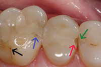
Figure 1. These recently placed posterior composites demonstrate the often woeful state of direct composite restorative dentistry. Black arrow: A carious fissure was missed; lack of magnification was the likely culprit. Blue arrow: “Minimally invasive” class II cavity shape creates impossible C-factor problems. Red arrow: Incremental loading leaves seams and voids that allow subsequent fracture. Green arrow: Proximal tooth was iatrogenically gouged and now has a carious lesion penetrating into dentin. |
 |
 |
|
Figures 2 and 3. Higher magnification views of Figure 1. |
Between 2005 and 2007, I lectured alongside Dr. Gordon Christensen and Derek Hein at the Clinical Research Associates (CRA) Dentistry Update. A good cross-section of restorative dentists from around the United States and Canada attend these lectures. At each city (and also at my private lectures) I have asked attending dentists (now numbering more than 6,000) this question: “How many of you feel that posterior composites are holding up as well as amalgams?” Only a few hands go up. More than 95% have concluded that posterior composites are inferior to amalgams, yet most have stopped doing amalgam restorations and are placing posterior composites. Then I ask the attendees, “So if amalgams are better, then why are we doing posterior composites?” The answers vary, but are less than inspirational.
Endodontists joke that posterior composites are the primary killer of pulps, and that leaking composites are their “number one” referral source. Most studies have shown that class I and class II composites have a significantly higher failure rate than amalgam restorations.1-4 The AMA, ADA, FDA, US Public Health service, CDC, NIH, and WHO have all declared amalgam safe. In light of this evidence and overwhelming opinion, how can we in good conscience continue to place posterior composites? So, let’s ask one more time, what’s wrong with this picture?
Why are we content to provide a posterior composite restoration that essentially cripples the tooth in the name of aesthetics, knowing that there is no proven systemic health benefit compared to amalgam? Many restorative dentists have simply given up on amalgam and composite and spend more than $100,000 for a CAD/CAM unit. They choose porcelain inlays and onlays as successors to amalgam rather than struggle and compromise with posterior composites.
The current state of posterior composite restorations poses empirical arguments for amalgam or porcelain alternatives. However, by the conclusion of this article I will provide 2 very good reasons why posterior composites can bless, rather than curse, the tooth.
THE G.V. BLACK ERA
 |
|
Figure 4. Extracted bicuspid with a conservative G.V. Black class II features classic iatrogenic fracturing pattern. |
To understand how clinicians can be influenced by a cultural and scientific icon, it is helpful to look at medical history. Claudius Galen was a Roman physician who boldly devised a medical model that doctors followed for 14 centuries. Though his medical judgments were remarkably advanced for his day, today, not surprisingly, we know that most of Galen’s theories and treatments were completely wrong, and that the rest were seriously flawed. In a parallel to Galen’s example, G.V. Black was a consummate dentist/scientist, and his exquisite designs for cavity preparation were a huge step forward for dentistry. Unfortunately, we are discovering today that those cavity shapes weaken the posterior dentition and lead to fracturing in even the most conservative applications5,6 (Figure 4).
In an unpublished 2-year study between 2001 to 2003, I utilized 16x magnification to evaluate each posterior tooth that was treated for replacement of an amalgam or posterior composite. I classified and documented incomplete fractures. Here is what I found:
- Sharp internal line angles are only a small part of the problem.
- Joining the occlusal to the interproximal is the worst possible design for crack avoidance and the most common area for crack initiation.
- Most fractures initiate in dentin at the line angles.
- Interrupted cavities were more crack resistant than connected cavity preparations.
In the May 2007 issue of Dentistry Today, I highlighted the widespread problem of amalgam tooth preparations that predispose the tooth to fracture (The Epidemic of Cracked and Fracturing Teeth). One of dentistry’s myths is that amalgam expansion causes tooth fracture. Expansion failures have never been proven. The fracture problem does not originate with amalgam, per se. It originates in iatrogenic G.V. Black cavity preparations. And just as many of us feared, we are seeing the same pattern of fracturing in teeth with posterior composites now that enough time has elapsed to assess their longevity.
THE SIMONSON TRANSITION
 |
 |
|
Figure 5. The gingival margin in this class II composite demonstrates the pervasive problem of microleakage. There was unfavorable C-factor at the margin creating suck-back. Uncured and contaminated flashing results from a metal matrix that blocks light-curing and visualization. |
Figure 6. Case in Figure 5 is retreated with Clark class II filling techniques and instruments, overcoming multiple problems. |
Dr. Richard Simonson is widely recognized as a pioneer in new cavity preparation shapes for minimally invasive, bonded, resin-based posterior composites. In spite of his innovations, the G.V. Black preparations that I was taught in school 20 years ago have been only slightly modified for posterior composites in the typical dental practice and in most dental schools.
As we study and document the many problems with posterior composites we find the following:
(1) Composite is a poor biological space filler. It needs to be sealed 360° and from inside to out (Figures 5 and 6).
(2) Unlike amalgam and gold foil techniques, “packing composite into a hole” is not a predictable method. Excellent clinicians have been dealt an unfair hand when it comes to class II composites. Most of the features of the traditional cavity preparations such as parallel walls and resistance and retention form work against posterior composites. What we have observed at CRA and under the microscope is that polymerization shrinkage cannot be eliminated, only mitigated. The best margin is no margin, and when composite extends slightly past the cavo-surface margin, it is generally well-sealed with no white line. When we polish back to the margin, the white line often appears. “Composite sealing” with thin resins applied after filling the cavity may reduce wear. However, trying to seal an imperfect margin after the fact is futile. As I have explored these white lines, they generally extend completely to the pulpal floor, far beyond the reach of a sealer.
(3) C-factor has been oversimplified and remains a significant problem.
(4) Posterior composites should go “on” not “in” the tooth.
(5) Minimally traumatic dentistry should be considered as an upgrade of minimally invasive dentistry. Well-meaning dentists are promoting minimally invasive dentistry. The best long-term outcomes are more important than the race to minimize the micrograms of tooth structure that are removed. For example, the tunnel preparation preserves the enamel of the marginal ridge but unnecessarily weakens the tooth and impedes clinical visualization. Incomplete caries removal combined with excessive tooth weakening are unacceptable casualties of the noble mission to save marginal ridge enamel.
INTRODUCING THE MODERN POSTERIOR COMPOSITE (THE CLARK CLASS II)
 |
|
Figure 7. Diagrams of the Clark class II (a), the slot preparation created by Simonson and others (b), and the original G.V. Black class II (c). |
 |
 |
|
Figure 8. Preoperative view of an everyday occurrence. A decade after amalgam was placed the tooth is deteriorating as the fracture spreads. |
Figure 9. Composite placed in a traditional, parallel-walled cavity cannot “splint” the coronal tooth structure. In contrast, when the calla lily shape is created, we will engage nearly all of the enamel rods of the occlusal surface. |
 |
 |
|
Figure 10. Clark class II is claustrophobic without magnification and unrestorable with conventional peripherals. With advanced magnification and new tools, there are new possibilities. The Bioclear matrix prototype and Interproxi-mator are pictured. They create an all-translucent system for buccal-lingual curing. |
Figure 11. Low magnification view of postoperative result. |
 |
 |
|
Figures 12 and 13. High magnification postoperative views. |
The Clark Class II is a radical departure from our notions of preparing and restoring posterior teeth (Figure 7). The goal of first-time interproximal caries restoration is to avoid connecting the occlusal to the interproximal, which is a concept that Simonson first advocated. The next evolution of this design is the saucer shape with serpentine/disappearing margins. The final change is discarding and replacing old filling techniques, matrix systems, and curing techniques.
Figures 8 to 13 demonstrate a clinical example of this radical departure from our notions of preparing and restoring posterior teeth. The first bicuspid in Figure 8 is from a 28-year-old male. It shows an early incomplete coronal fracture, based on the magnified view of the mesial marginal ridge and according to the guide that Drs. Sheets, Paquette, and I published in the Journal of Esthetic and Restorative Dentistry and most recently in Dentistry Today. Earlier in my practice, I would have turned this class I (occlusal) amalgam into a class II or MO composite or amalgam, because I would not have seen the fracture undermining the buccal cusp that is not visible at less than 10x. In addition, I suspected but did not understand that turning this class I into a traditional class II with a mesial box form would further weaken this already iatrogenically compromised tooth. There is now a better approach, one that does not necessarily involve an indirect procedure such as a crown or onlay.
As we continue with the bicuspid in Figure 8, the occlusal is treated first, and a calla lily shape that engages the bulk of the occlusal table is prepared and restored (Figure 9). The calla lily class I will be explored in future articles. The interproximal is then addressed separately to simplify the process and to control C-factor. Note the saucer shape on the mesial (Figure 10). This flattened cavity shape requires a completely new filling protocol and peripherals. Instead of metal sectional matrix bands, wedges, and separators, we utilize transparent anatomic sectional matrix bands and translucent Interproximators (both Bioclear Matrix System) and a single-load technique with an injection-molded process where resin, flowable composite, and then paste are loaded in sequence without stopping to light-cure the individual components. The restoration is light-cured with one or multiple curing lights from occlusal, buccal, and lingual with this fully translucent system. The result is a seamless, rounded restoration that delivers breath-taking results (Figures 11 to 13). Better is rarely faster in dentistry, but our test clinicians report both (better/faster) once they are past the learning curve.
CAN THESE THINGS LAST?
 |
|
Figure 14. SEM of 3-year follow-up of one of the author’s cases evaluated in the study. Filtek Supreme (3M ESPE) paste combined with a flowable technique in a molar shows good wear resistance. White arrow shows paste composite/flowable composite interface. Red arrow shows flowable composite/enamel interface. |
Early posterior composites showed unacceptable wear. Microfills like Heliomolar (Ivoclar Vivadent) had excellent wear resistance but mediocre strength. Marginal ridge fracture was common. Many modern composites now exhibit excellent strength and wear resistance. In several studies, composite/enamel bonding has exhibited very lengthy in vitro success that does not deteriorate over time.7 The key is that the initial bond must be exquisite and engage large areas of enamel, such as seen in enamel-based porcelain and composite veneers.
In another unpublished study, CRA scientists assisted me for an in-office recall study. We documented patients with minimally traumatic class I composite restorations that had been in service for 3 to 7 years. In 107 posterior teeth, 100% of the composites were retained. Excess wear was present in some samples that utilized flowable composite alone. The combination paste/flowable cases showed the best wear resistance in SEM evaluation (Figure 14), and slight staining was present in less than 5% of all samples.
SUMMARY
As we introduce the concepts summarized in this article to practicing dentists, they show a broad spectrum of responses from shock and disbelief to sheer exuberance. As these cavity shapes are implemented we will see a dramatic reduction in the rate of tooth fracturing. We also anticipate that these restorations will outlast the class II amalgams that have served so well in the past. This very brief article can only touch on the dramatic differences of this minimally traumatic and incredibly durable direct composite. A full instructional DVD together with a textbook and hands-on courses are available and recommended.
References
- DeRouen TA, Martin MD, Leroux BG, et al. Neurobehavioral effects of dental amalgam in children: a randomized clinical trial. JAMA. 2006;295:1784-1792.
- Van Nieuwenhuysen JP, D’Hoore W, Carvalho J, et al. Long-term evaluation of extensive restorations in permanent teeth. J Dent. 2003;31:395-405.
- Sjogren P, Halling A. Survival time of Class II molar restorations in relation to patient and dental health insurance costs for treatment. Swed Dent J. 2002;26:59-66.
- Mjor IA, Dahl JE, Moorhead JE. Placement and replacement of restorations in primary teeth. Acta Odontol Scand. 2002;60:25-28.
- Clark DJ, Sheets CG, Paquette JM. Definitive diagnosis of early enamel and dentin cracks based on microscopic evaluation. J Esthet Restor Dent. 2003;15:391-401.
- Clark D. The epidemic of cracked and fracturing teeth. Dent Today. May 2007;26:90-95.
- Swift EJ Jr, Friedman MJ. Critical appraisal: porcelain veneer outcomes, part II. J Esthet Restor Dent. 2006;18:110-113.
Dr. Clark founded the Academy of Microscope Enhanced Dentistry, an international association formed to advance the science and practice of microendodontics, microperiodontics, microprosthodontics, and microdentistry. He is a course director at the Newport Coast Oral Facial Institute in Newport Beach, Calif. He is co-director of Precision Aesthetics Northwest in Tacoma, Wash, and an associate member of the American Association of Endodontists. He lectures and gives hands-on seminars internationally on a variety of topics related to microscope-enhanced dentistry. He has developed numerous innovations in the fields of micro dental instrumentation, imaging, and dental operatory design. Dr. Clark is proud to join with Clinical Research Associates in the Update Series lectures and also to participate in the important research at its world class facility in Provo, Utah. He is also developing new techniques and materials to better restore endodontically treated teeth, including the Endo-Restorative Casting. A 1986 graduate of the University of Washington School of Dentistry, he can be reached at drclark@microscopedentistry.com and lifetimedentistry.net.
Disclosure: Dr. Clark has financial interest in several of the products mentioned in this article. The Clark class II and injection-molded technique are intellectual property of the author patent pending and may be used solely with permission.


