


INTRODUCTION
Cervical lesions, which have been found to be present in 85% of the population, represent a major problem for dentists to restore with composite resin materials due to the varying adhesive properties of the tooth structure, the biomechanical aspects of the cervical area, and difficulties in accessing and isolating the area to be restored.1,2 The incidence also may be higher in individuals retaining their permanent teeth, as the aging population is increasing.1 Additionally, at a time when people are maintaining their natural teeth longer, the likelihood of developing caries in Class V areas also increases.3
When Class V cervical lesions are noncarious in nature, they are classified as abfractions, with an appearance characterized by a loss of hard dental structure near the cement-enamel junction. The lesions’ shape may resemble a wedge with an inward-pointing apex.1 The cause of abfractions is thought to be occlusal stress that produces cervical cracks and, subsequently, predisposes the tooth surface to the effects of erosion and abrasion.4 Although critical literature reviews suggest that abfraction is a hypothetical component of cervical wear, it is important to determine causative factors for noncarious lesions, as treatments range from eliminating the aggravating agents to specific restorative procedures.4,5
Gingival Recession
Typically involving at least one tooth surface, gingival recession can lead to root surface exposure at the gingival margin.6 This not only causes aesthetic impairment, but the fear of tooth loss, an increased susceptibility to root caries, and hypersensitivity of the dentin.6 As gingival recession is the displacement of the soft tissue margin, tooth malpositions, high muscle attachment, frenal pull, and occlusal trauma can create the conditions necessary to cause recession and root exposure.7 Another less obvious cause, oral jewelry, also has been linked to gingival recession.8 Studies have shown that piercings in the lip and tongue lead to localized gingival recession as an adverse consequence.8 In one study, individuals with tongue piercings presented an 11-times greater risk of developing gingival recession over the control group.9
Gingival Recession Treatment
Traditionally, gingival recession has been treated with laser therapy, autogenous tissue grafting, flap designs, orthodontics, and guided tissue restoration.10 These types of treatments are not only costly and time consuming, but also may require long, painful recovery for patients.10
Laser treatment has been considered by some as the optimal option for correcting and halting gingival recession.11 When gingival recession is observed in a patient with sensitivity caused by an exposed root, lasers have been used to remove the smear layer from the root surface to expose collagen fibers, which is believed to contribute to improved healing.12 Clinical studies, however, have been unable to find any significant improvements in recession and sensitivity from this type of treatment.12
Tissue grafting is also considered one of the few viable treatment options to correct gingival recession.13 With the advent of tissue grafting techniques, periodontists have been able to correct gingival recession by grafting a patient’s own oral and mucosal tissues.14 This type of procedure, however, requires surgery and can be very costly. Whether using an envelope or tunnel technique, the tissue is grafted around the area of gingival recession.15,16 It is then sutured into place and allowed to heal.17 A protective mouthpiece is often required to allow the graft site to heal properly.17 Grafting does allow for significant increases in gingival and root coverage and has proven to be very effective as a treatment option.
Another technique for correcting gingival recession is a minimally invasive flap design procedure intended for periodontally involved restorations.18 It involves cutting the tissue on 3 sides, leaving the base attached, to open the gingival tissue to allow for cleaning of the roots.18 This procedure often works with guided-tissue regeneration to allow coverage of the root and reduce gingival recession.18 Although this treatment option demonstrates good results, it still involves a surgical procedure and recovery time for the patient.18
Orthodontics may also be used to correct gingival recession, as conditions such as cross-bites and occlusion are seen as causes.19 By using orthodontic appliances to correct the abnormalities in bite and occlusion, studies have shown that gingival recession can be stopped and, many times, reversed.19 These results, however, are typically created through multidisciplinary approaches and not merely with orthodontic treatment and appliances.19 New techniques and materials, which show promising results for root coverage, have proven effective at covering, and in some cases stopping, gingival recession.10
Noncarious Class V Lesions
As the health and appearance of gingival tissue is important to the aesthetics of a smile, many with noncarious Class V lesions and/or exposed tooth structure from gingival recession wish to have their conditions treated without the cost or inconvenience of invasive methods.20 Used to treat noncarious Class V legions, glass ionomer cements, compomers, and composite resins work alone or in combination to correct the aesthetic issues and prevent further damage.1 When unaesthetic Class V lesions display caries, a combination of glass ionomer materials for the internal aspects of the restoration and a resin-based composite material for the surface has been advocated.3 This treatment method is believed to provide aesthetic results while increasing the potential for caries reduction.3
The physical properties of resin-based composites allow a bond to tooth structure, with highly aesthetic results, so many practitioners feel that they are the best materials to use when restoring cervical defects.5 In a recent study, resin composite restorations that were placed to treat noncarious cervical lesions exhibited no secondary caries and far less deficiencies in marginal adaptation than compomer restorations after 3 years.21 There are, however, some challenges in using resin-based composites for Class V lesions.2
When placing resin-based composite restorations in the aesthetic zone, it is good to have an understanding of the composites being utilized, especially with regard to their respective optical and physical characteristics.22,23 When used and placed properly, the polychromatic effects seen in natural teeth can be replicated.22,23 More importantly, producing outstanding composite resin restorations is achieved thorough comprehension of natural tooth morphology and how each component of tooth structure affects aesthetics.24,25
Resin-Based Composites and Gingival Health
In past studies, resin-based composites showed promising results for treating Class V lesions and masking the effects of gingival recession.26 Through observations of composites, it was found that they do not adversely affect gingival health, and that there is typically less inflammatory response to well-finished and contoured composite resins than carious lesions that are left untreated.26 Another study, comparing plaque buildup around newer composite resins and conventional composites, found that there was no significant difference in plaque formation of the 2 materials.27
Unfortunately, when composite resins are applied to teeth presenting with gingival recession, the resulting tooth-colored restorations tend to make the teeth appear very long, leading to an unaesthetic appearance.27 To correct this issue, the need for resins that are gingival-colored has increased.27 Manufacturers have met this demand, creating products that demonstrate the aesthetics of natural gingival tissue.27 Aside from the aesthetic value of these new materials, the composites also allow for minimally invasive procedures to cover the roots and exposed tooth structure caused by the gingival recession.27
These new resin-based composites correct the aesthetic deficits of gingival recession by framing the tooth or teeth with material in a similar pink color to the gingival tissue.20 These gingival-colored composites tend to demonstrate greater color stability and resistance to wear.20 When used in collaboration with the new generation of bonding agents, which enable bonding to metal, porcelain, enamel, and dentin, gingival-colored composites have been proven to enhance the smiles of patients with gingival recession.20 More importantly, this treatment option provides a clinical solution for patients that is aesthetic, economical, and practical.27
Aesthetic Gingival Composite Resins
An aesthetic gingiva-shaded light-cured composite resin (Amaris Gingiva [VOCO America]) was recently introduced, providing practitioners with the option of correcting gingival recession with a minimally invasive and less costly procedure. This pink-colored composite (available in one translucent gingival color and 3 pink flowable opaquers that can be mixed together to better match an individual’s gingival shade) was specifically developed for indications in the cervical area, including composite restorations in gingival colors, V-shaped defects, exposed cervical areas, aesthetic corrections of the gingiva area, primary splinting, and the correction of red/white aesthetics. This restorative material also gives the clinician the ability to mask exposed crown margins to improve aesthetics and patient satisfaction.
The following case presentation will demonstrate the successful use of this aesthetic restorative material.
CASE REPORT
Diagnosis and Treatment Planning
A 67-year-old female patient presented with gingival recession and root sensitivity on teeth Nos. 8, 9, and 11 (Figures 1 and 2). (Tooth No. 10 was congenitally missing and tooth No. 11 had moved in this position.) The patient declined any periodontal surgical grafting procedures to address her situation. A complete examination was performed that included radiographs, photographs, and a review of the patient’s periodontal condition. Occlusal analysis determined that no interferences or contributory factors would require functional and/or occlusal rehabilitation. Also, no pathologies were found that might contraindicate conservative and aesthetic treatment using a gingival-colored composite resin system.
 |
 |
| Figure 1. 1:2 preoperative photo showing teeth Nos. 8, 9, and 11 that presented with gingival recession and root sensitivity. (Tooth No. 10 was congenitally missing and tooth No. 11 had moved into its place.) | Figure 2. Preoperative close-up photo. |
 |
 |
| Figure 3. Shade tabs were used to determine the shade of flowable composite (Amaris Gingiva [VOCO America]). The light shade was used on tooth No. 8, and the darker shade was used on tooth No. 9. | Figure 4. View of the selected shades that were placed. |
 |
 |
| Figure 5. A slight bevel preparation was made. | Figure 6. After the bevel preparation was made, a single-step, self-etching adhesive (Futurabond DC [VOCO America]) was placed. |
 |
 |
| Figure 7. The adhesive was light-cured. | Figure 8. After light-curing the adhesive, light Opaquer was applied and then light-cured. |
 |
 |
| Figure 9. Sulcular anatomy was created using the Nature shade of the gingival composite. | Figure 10. The Nature shade was sculpted and light-cured. |
Clinical Protocol
The patient was anesthetized, her lips and cheeks were properly retracted, and the immediate surrounding dentition was isolated with cotton rolls. Shade tabs were used to determine the appropriate shade of the pink opaque flowable composite and the translucent pink sculptable composite (Figures 3 and 4). A slight bevel preparation was then made on teeth Nos. 8 and 9 (Figure 5). After the bevel preparations were complete, a single-step, self-etching adhesive (Futurabond DC [VOCO America]) was placed (Figure 6). This adhesive was left on the preparation for 20 seconds, lightly air-dried for 5 seconds, and then light-cured for 10 seconds (Figure 7). When used properly for noncarious cervical lesions, it has been demonstrated that single-step self-etching adhesives have acceptable clinical retention-to-dentin rates.28
The Opaquer shade of the gingival composite (light for tooth No. 8, dark for tooth No. 9) was applied and then thinned out with a brush and light-cured (Figure 8). Then, the Nature shade (ie, final pink overlay) of gingival composite was placed and deliberately over-bulked to begin the process of developing the sulcular anatomy (Figure 9). This layer was sculpted and also light-cured (Figure 10).
 |
 |
| Figure 11. The surface texture of the restorations was refined using an ultrafine yellow flame diamond bur No. 863 EF (Brasseler USA). | Figure 12. View of the final surface texture that was refined with the diamond bur. |
 |
 |
| Figures 13 and 14. View of the restorations after finishing and polishing. |
 |
 |
| Figure 15. An unfilled resin surface sealant (Easy Glaze [VOCO America]) was applied to the restoration. | Figure 16. The surface sealant was light-cured. |
 |
| Figure 17. View of the final restorations after application and light-curing of the surface glaze. |
 |
 |
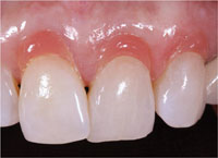
| Figures 18a and 18b. The final restorations. (Note in Figure 18a that the gingival zeniths of teeth Nos. 8, 9, and 11 were now symmetrical and at equal heights.) |
Tertiary anatomy was achieved using an ultrafine yellow flame diamond No. 863 EF (Brasseler USA) (Figures 11 and 12). Using a dry technique, the precise contour of the pink addition (artificial gingiva replacement) material was created. Then, fine cross matching was performed with a very dull bullet nose diamond No. 8850 KR (Brasseler USA), and a No. 649 super green stone (Brasseler USA). This was followed by polishing using a Jiffy (Ultradent Products) polishing brush and point with polishing paste and a goat hair wheel (Figures 13 and 14). To impart the most lifelike appearance to the artificial gingival replacement, a staccato motion was used with a dry goat hair wheel.
The restorations were assessed following placement of the translucent composite layer. A Surface Glaze (Easy Glaze [VOCO America]) was used to simulate the appearance of gingival vitality (Figures 15 and 16). Following assessment, the artificial gingival replacements underwent a 360° polymerization for 60 seconds each (Figure 17).
Upon completion of the procedure, the patient was very happy with the aesthetics. It should also be noted that the gingival zeniths of the adjacent teeth were now symmetrical (Figures 18a and 18b).
CONCLUSION
Cervical lesions and gingival recession present major challenges to dentists and dental manufacturers, as many patients present with some form of this dental issue. With this, gingival surgery is often required with a referral to a periodontist and gum surgery to correct the aesthetic deficiencies caused by the gingival assymetry. Aside from the cost, recovery, and pain to the patient, general dentists also lose potential income. With the development of new gingiva-shaded composite resin materials, general dentists can now directly apply these to the cervical area of the tooth/teeth to correct the appearance of gingival recession. Products like this are not only cost-effective, but also minimally invasive, leading to patient satisfaction, more income for the dentist, and highly aesthetic results.
References
- Ceruti P, Menicucci G, Mariani GD, et al. Non carious cervical lesions. A review. Minerva Stomatol. 2006;55(1-2):43-57.
- Allegri, MA Landi L, Zucchelli G. Non-carious cervical lesions associated with multiple gingival recessions in the maxillary arch. A restorative-periodontal effort for esthetic success. A 12-month case report. Eur J Esthet Dent. 2010;5:10-27.
- Christensen GJ. The ‘new’ operative dentistry. J Am Dent Assoc. 2006;137:531-533.
- Bartlett DW, Shah P. A critical review of non-carious cervical (wear) lesions and the role of abfraction, erosion, and abrasion. J Dent Res. 2006;85:306-312.
- de Melo FV, Belli R, Monteiro S Jr, et al. Esthetic noncarious Class V restorations: a case report. J Esthet Restor Dent. 2005;17:275-284.
- Slutzkey S, Levin L. Gingival recession in young adults: occurrence, severity, and relationship to past orthodontic treatment and oral piercing. Am J Orthod Dentofacial Orthop. 2008;134:652-656.
- Ustun K, Sari Z, Orucoglu H, et al. Severe gingival recession caused by traumatic occlusion and mucogingival stress: a case report. Eur J Dent. 2008;2:127-133.
- Er N, Ozkavaf A, Berberoğlu A, et al. An unusual cause of gingival recession: oral piercing. J Periodontol. 2000;71:1767-1769.
- Pires IL, Cota LO, Oliveira AC, et al. Association between periodontal condition and use of tongue piercing: a case-control study. J Clin Periodontol. 2010;37:712-718.
- Kassab MM, Badawi H, Dentino AR. Treatment of gingival recession. Dent Clin North Am. 2010;54:129-140.
- Dilsiz A, Canakci V, Ozdemir A, et al. Clinical evaluation of Nd:YAG and 685-nm diode laser therapy for desensitization of teeth with gingival recession. Photomed Laser Surg. 2009;27:843-848.
- Dilsiz A, Aydin T, Canakci V, et al. Root surface biomodification with Nd:YAG laser for the treatment of gingival recession with subepithelial connective tissue grafts. Photomed Laser Surg. 2010;28:337-343.
- Saha S, Bateman GJ. Mucogingival grafting procedures—an update. Dent Update. 2008;35:561-562, 565-568.
- Khuller N. Coverage of gingival recession using tunnel connective tissue graft technique. J Indian Soc Periodontol. 2009;13:101-105.
- Park JB. A two-stage approach using an autogenous masticatory mucosal graft and an autogenous connective tissue graft to treat gingival recession: a case report. J Int Acad Periodontol. 2010;12:45-48.
- Abundo R, Corrente G, des Ambrois AB, et al. A connective tissue graft envelope technique for the treatment of single gingival recessions: a 1-year study. Int J Periodontics Restorative Dent. 2009;29:593-597.
- McLeod DE, Reyes E, Branch-Mays G. Treatment of multiple areas of gingival recession using a simple harvesting technique for autogenous connective tissue graft. J Periodontol. 2009;80:1680-1687.
- Trombelli L, Simonelli A, Pramstraller M, et al. Single flap approach with and without guided tissue regeneration and a hydroxyapatite biomaterial in the management of intraosseous periodontal defects. J Periodontol. 2010;81:1256-1263.
- Seehra J, Fleming PS, DiBiase AT. Orthodontic treatment of localised gingival recession associated with traumatic anterior crossbite. Aust Orthod J. 2009;25:76-81.
- Zalkind M, Hochman N. Alternative method of conservative esthetic treatment for gingival recession. J Prosthet Dent. 1997;77:561-563.
- Pollington S, van Noort R. A clinical evaluation of a resin composite and a compomer in non-carious Class V lesions. A 3-year follow-up. Am J Dent. 2008;21:49-52.
- Terry DA, Geller W, Tric O, et al. Anatomical form defines color: function, form, and aesthetics. Pract Proced Aesthet Dent. 2002;14:59-67.
- Terry DA. Dimensions of color: creating high-diffusion layers with composite resin. Compend Contin Educ Dent. 2003;24(2 suppl):3-13.
- LeSage B, Milnar F, Wohlberg J. Achieving the epitome of composite art—advanced concepts in shade selection and layering composite. J Cosmetic Dentistry. Accepted July 2008.
- Milnar FJ. Altering tooth inclination and overall esthetics with direct composite veneers, gingival contouring, and enamelplasty. Inside Dentistry. 2006;2:46-49.
- Blank LW, Caffesse RG, Charbeneau GT. The gingival response to well-finished composite resin restorations. J Prosthet Dent. 1979;42:626-632.
- van Dijken JW, Sjöström S, Wing K. The effect of different types of composite resin fillings on marginal gingiva. J Clin Periodontol. 1987;14:185-189.
- van Dijken JW, Sunnegårdh-Grönberg K, Sörensson E. Clinical bonding of a single-step self-etching adhesive in noncarious cervical lesions. J Adhes Dent. 2007;9 suppl 2:241-243.
Dr. Milnar, a graduate of the University of Minnesota School of Dentistry, maintains a full-time practice in St. Paul, Minn, emphasizing appearance related dentistry. He is an accredited member of the American Academy of Cosmetic Dentistry and a Board Examiner for accreditation. Dr. Milnar has published numerous articles about the direct placement of composites, shade selection, and porcelain materials. Dr. Milnar is also co-founder of the Minnesota Academy of Cosmetic Dentistry. He lectures extensively on the subject of direct composite restorations, shade selection, and porcelain materials. He can be reached at (651) 645-6111 or frank@milnardds.com.
Disclosure: Dr. Milnar has received financial and materials/product support from VOCO America, Inc.


