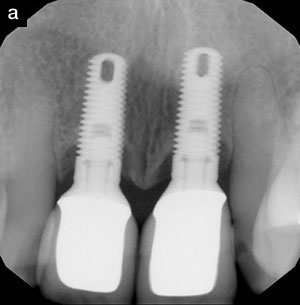
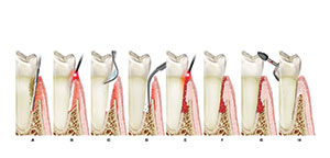
Advanced periodontitis in the anterior maxilla may be associated with an unaesthetic smile. When severe attachment loss is present, comprehensive cosmetic reconstruction cannot take place until the periodontal disease has been treated. As opposed to treating this situation by tooth extraction and prosthetic replacement, an alternative approach can be periodontal regeneration. The surgical treatment of periodontal disease has evolved from a focus on resection to a strong emphasis on regeneration. Considerable histologic and clinical evidence has been gathered over the last 2 decades indicating that the regeneration of periodontal tissues lost as a result of periodontitis can be achieved in humans.1,2
A variety of approaches aimed at periodontal regeneration have been developed. Bone grafts and guided tissue regeneration have all been used with varying degrees of success to regenerate lost attachment in deep, intrabony defects.1,2 Histologic observations and controlled clinical trials have demonstrated that some of the available regenerative procedures may result in healing that can be termed periodontal regeneration.3-6 Clinically, several factors may affect the extent of clinical attachment gain and bone regrowth as a clinical outcome.7-10 Nevertheless, it is this technical sensitivity and variability in regenerative success that fostered an interest in the possible modulation of the periodontal wound healing process using biochemical mediators.
As the understanding of the embryologic and biochemical development of the periodontal attachment apparatus progressed, the hypothesis was suggested that cellular mediators expressed by Hertwig’s epithelial root sheath play a role in the genesis of the periodontal ligament.11 Subsequently, a series of animal studies led to the identification of enamel matrix proteins in the development of the root and the adjacent periodontal ligament.12,13 In light of earlier observations, it was suggested in the late 1980s that growth factors could be used to achieve periodontal regeneration.14
In the “aesthetic zone” of the anterior maxilla, several challenges must be overcome to have a successful, functional, and aesthetic regenerative outcome. Amongst the different clinical obstacles in regenerative therapy, primary closure over the treated area is one of the most important. A technique to preserve the papillae during flap elevation, and thereby allow for primary closure of the flap in interdental areas, was introduced by Takei and co-workers.15 This technique was used initially with osseous grafting. Later, Cortellini and co-workers16 reported a modified papilla preservation technique in association with a nonresorbable (ePTFE) barrier membrane to minimize the incidence of membrane exposure and as a result increase the degree of regeneration. Recently, a further modification of the papillary preservation technique was reported.17 This latest modification was described as a means to obtain and maintain primary closure over a barrier membrane in narrow or posterior interdental spaces.
All of the previously mentioned regenerative procedures led to substantial improvement in the treatment outcome. An approach to periodontal regeneration utilizing an enamel matrix-derived amelogenin growth factor,18,19 combined with ideal flap design involving papillary preservation, has been developed. The following case demonstrates the successful management of periodontitis in the anterior maxilla with papillary preservation and an amelogenin growth factor.
CASE REPORT
The patient, a 60-year-old white male, presented with chief complaints of “My gum is very tender” and “I would like to improve my smile.” His medical history was noncontributory. He denied use of prescription medications. His dental history was notable for prior periodontal therapy (nonsurgical debridement in 1993; localized surgical debridement in 1996) and inconsistent periodontal supportive care since that time.
Clinical Presentation
The patient’s clinical examination revealed a periodontium that was generally healthy, with the exception of an erythematous and edematous papilla between teeth Nos. 6 and 7. Tooth No. 6 was not mobile. A diastema was present between teeth Nos. 6 and 7. Tooth No. 6 also had a 9-mm probing depth, 1 mm of recession relative to the facial restorative margin, and the presence of a purulent exudate upon probing. Subgingival calculus was not detected upon probing. The radiograph of tooth No. 6 suggested a mesial 1-wall intraosseous defect (Figures 1 to 5).
 |
 |
| Figure 1. Facial view of dentition. Note healthy gingival color and texture, discrepency in gingival line, exposed prosthetic margin, disharmony in prosthetic tooth form, and porcelain color. | Figure 2. Close-up view of teeth Nos. 6 and 7. Note gingival redness and swelling extending to the mucogingival junction. |
 |
 |
| Figure 3. Palatal view of teeth Nos. 6 and 7. | Figure 4. Mesial 9-mm probing depth on tooth No. 6. |
 |
|
| Figure 5. Periapical radiograph displaying bone loss pattern indicative of a mesial 1-wall intraosseous defect. | |
Treatment Options
The following treatment alternatives were presented to the patient: (1) isolated, nonsurgical debridement and application of a locally delivered antibiotic; (2) open flap debridement; and (3) papillary preservation open flap debridement and growth factor-modulated tissue regeneration. After discussing the treatment alternatives, the regenerative treatment was selected.
Surgical Procedure
After administration of local anesthesia, sulcular incisions were made with a 6400 scalpel blade (Sable). The papilla was incised from the palatal aspect and reflected with the facial flap (Figure 6). Following elevation of the mucoperiosteal flap, burnished subgingival calculus and a 6-mm 1-wall intraosseous defect were found on the mesial root surface of tooth No. 6. Following ultrasonic and hand instrumentation of the root surface, the root surface was chemically biomodified with a neutral pH buffered ethylenediainetetraacetic acid (EDTA) gel (PrefGel, Biora, Figures 7 and 8). After 2 minutes, the root surface was washed with sterile saline, and an amelogenin growth factor (Emdogain, Biora) was applied to the root surface and allowed to remain undisturbed for 2 minutes (Figure 9). The proteins present in the gel competitively bind to the root surface. It is important that the area be isolated from saliva and blood during the 2-minute application interval. The flaps were then replaced, and a tension-free wound closure was completed using 5-0 and 6-0 resorbable vertical-mattress sutures (Figures 10 and 11).
 |
 |
| Figure 6. Palatal view of sulcular incision extending across interdental papilla between teeth Nos. 6 and 7. This papilla will be reflected to the facial with the facial flap. | Figure 7. Facial view demonstrating root surface preparation. |
 |
 |
| Figure 8. Neutral pH buffered ethylenediaminetetraacetic acid (EDTA) gel applied to root surface of tooth No. 6. | Figure 9. Porcine-derived amelogenin growth factor applied to root surface of tooth No. 6. |
 |
 |
| Figure 10. Facial view of immediate postsurgical result. The surgical wound has been closed using vertical-mattress sutures. Note the absence of papillary soft-tissue loss. | Figure 11. Palatal view of immediate postsurgical result. Note primary closure of interdental osseous defect site. |
The patient returned for routine 2-week and 2-month postsurgical follow-up (Figures 12 through 15). At the conclusion of the 60-day surgical follow-up appointment, the patient was placed into the periodontal recall system and returned to the office for supportive periodontal care on a 3-month schedule. The area was not probed until 6 months had elapsed following treatment. The patient’s periodontal evaluation at that time revealed a gingiva of normal color and texture, no gingival swelling, a 3-mm probing depth with no bleeding on probing, and most importantly, no additional gingival recession beyond that already present (Figures 16 and 17).
 |
 |
 |
 |
| Figures 12 through 15. Facial and palatal views of treated area at 2-week and 2-month surgical follow-up appointments. Note resolution of gingival hyperemia and a return of normal gingival color, contour, and texture. | |
 |
 |
| Figures 16 and 17. Two views of the treated area at 6-month supportive periodontal therapy appointment. Figure 17 demonstrates a mesial 3-mm probing depth on tooth No. 6 and no additional gingival recession. | |
CONCLUSION
Severe periodontitis diagnosed in the anterior maxilla often is a difficult clinical problem for clinicians. In the past, a surgical periodontal approach to treatment may have resulted in a poor aesthetic result. Due to the perceived potential for a negative aesthetic outcome, severe periodontitis in this area often is not treated, or limited treatment is suggested. This case has demonstrated the use in the anterior maxilla of a papillary-preserving surgical approach with regeneration. In a complex multidisciplinary case, the treatment described could be followed by orthodontic therapy and restorative treatment to restore anterior aesthetics fully.
References
1. Caton JG, Greenstein G. Factors related to periodontal regeneration. Periodontol 2000. 1993;1:9-15.
2. Karring T, Nyman S, Gottlow J, et al. Development of the biological concept of guided tissue regeneration: animal and human studies. Periodontol 2000. 1993;1:26-35.
3. Bowers GM, Chadroff B, Carnevale R, et al. Histologic evaluation of new attachment apparatus formation in humans. Part 1. J Periodontol. 1989;60:664-674.
4. Nyman S, Lindhe J, Karring T, et al. New attachment following surgical treatment of human periodontal disease. J Clin Periodontol. 1982;
9:290-296.
5. Gottlow J, Nyman S, Lindhe J, et al. New attachment formation in the human periodontium by guided tissue regeneration. Case reports. J Clin Periodontol. 1986;13:604-616.
6. Cortellini P, Tonetti MS. Focus on intrabony defects: guided tissue regeneration. Periodontol 2000. 2000;22:104-132.
7. Tonetti MS, Pini-Prato G, Cortellini P, et al. Effect of cigarette smoking on periodontal healing following GTR in infrabony defects. A preliminary retrospective study. J Clin Periodontol. 1995;22:229-234.
8. Rosen PS, Marks MH, Reynolds MA. Influence of smoking on long-term clinical results of intrabony defects treated with regenerative therapy. J Periodontol. 1996;67:1159-1163.
9. Trombelli L, Kim CK, Zimmerman GJ, et al. Retrospective analysis of factors related to clinical outcome of guided tissue regeneration procedures in intrabony defects. J Clin Periodontol. 1997;24:366-371.
10. Kornman KS, Robertson PB. Fundamental principles affecting the outcomes of therapy for osseous lesions. Periodontol 2000. 2000;22:22-43.
11. Slavkin HC. Towards a cellular and molecular understanding of periodontics. Cementogenesis revisited. J Periodontol. 1976;47:249-255.
12. Hammarstrom L. Enamel matrix, cementum development and regeneration. J Clin Periodontol. 1997;24:658-668.
13. Schonfeld SE, Slavkin HC. Demonstration of enamel matrix proteins on root-analogue surfaces of rabbit permanent incisor teeth. Calcif Tissue Res. 1977;24:223-229.
14. Lynch SE, Colvin RB, Antoniades HN. Growth factors in wound healing. Single and synergistic effects on partial thickness porcine skin wounds. J Clin Invest. 1989;84:640-646.
15. Takei HH, Han TJ, Carranza FA Jr, et al. Flap technique for periodontal bone implants. Papilla preservation technique. J Periodontol. 1985;56:204-210.
16. Cortellini P, Prato GP, Tonetti MS. The modified papilla preservation technique. A new surgical approach for interproximal regenerative procedures. J Periodontol. 1995;66:261-266.
17. Cortellini P, Prato GP, Tonetti MS. The simplified papilla preservation flap. A novel surgical approach for the management of soft tissues in regenerative procedures. Int J Periodontics Restorative Dent. 1999;19:589-599.
18. Tonetti MS, Lang NP, Cortellini P, et al. Enamel matrix proteins in the regenerative therapy of deep intrabony defects. J Clin Periodontol. 2002;29:317-325.
19. Trombelli L, Bottega S, Zucchelli G. Supracrestal soft tissue preservation with enamel matrix proteins in treatment of deep intrabony defects. J Clin Periodontol. 2002;29:433-439.
20. Rees TD. Drugs and oral disorders. Periodontol 2000. 1998;18:21-36.
Dr. Young graduated from the Indiana University School of Dentistry. After completing an advanced education in general dentistry at the Irwin Army Hospital in Fort Riley, Kan, Dr. Young practiced general dentistry for 5 years. He completed his residency in periodontology at the University of Michigan. Dr. Young maintains a private practice limited to periodontics in Des Moines, Iowa. His practice has an emphasis in aesthetic periodontal regeneration, cosmetic periodontal reconstruction, and dental implants. He can be reached at (515) 224-1771 or ryoungdsm@aol.com.


