Each year in the United States, thousands of patients receive radiation therapy for cancer. All dental practices have patients who have undergone, are undergoing, or will undergo radiation therapy as part of comprehensive cancer treatment. While the theoretical basis for radiation treatment of cancer was developed decades ago, new delivery systems have been introduced, and protocols are constantly being revised. This article discusses current concepts in radiation therapy, with an emphasis on those treatments that relate to oral health.
Dental care is an important consideration for radiation therapy patients for several reasons: (1) head and neck cancer patients who have excellent oral health are less likely to have severe complications from their cancer radiation therapy than those patients in poor oral health1; (2) dental treatment before, during, and after radiation therapy requires special considerations; and (3) radiation therapy to the head and neck region is associated with specific oral pathology.2
Among the current issues regarding dental care of radiation therapy patients are the question of bacterial endocarditis (BE) prophylaxis for patients who have previously received radiation therapy to the left breast or chest wall, the utilization of hyperbaric oxygen (HBO), amifostine (a sulfhydryl scavenger) therapy, and precautions with regard to the daily use of chlorhexidine. Also, dental professionals should be alert to 2 newer technologies—gamma knife technology and intensity modulated radiation therapy (IMRT)—that have implications for oral healthcare.
Ionizing radiation is commonly employed in the treatment of malignancies. Experience and research have identified those cancers that respond and those that do not respond to irradiation. For many tumors, radiation therapy alone can be curative. For other tumors, ionizing radiation is most efficacious when used in conjunction with surgery and/or chemotherapy.
Radiation may be delivered interstitially via seeds of iodine125, cesium, or similar isotopes. More commonly, radiation is delivered by an external beam of x-ray, cobalt, high-energy electrons, or high-energy photons. Standard therapy passes a radiation beam through the tissues, with each treatment (“fraction”) delivered at a dose of 120 to 220 centigray (cGy). These fractions are given once (QD) or twice (BID) daily, 5 days per week until the total dose is completed.
Several recent advances in radiation therapy have implications for patients’ oral health. First, IMRT is a new technique, allowing delivery of external beam radiation treatments at multiple angles with varying degrees of intensity within each beam.3 This method allows dose contours to be “carved” very precisely around structures. IMRT is designed to deliver higher doses of radiation to the targeted tumor and lower doses to the surrounding tissues, thus moderating the radiation damage to normal structures. In the case of tumors of the head and neck, IMRT often enables the radiation oncologist to preserve the function of at least one parotid gland during treatment, thereby preserving significantly more salivary gland function than with previous techniques.
Use of gamma knife stereotactic radiosurgery has also received recent attention.4 This technology uses pencil-thin beams of cobalt-60 irradiation directed through tiny collimators to focus energy with pinpoint precision to target lesions in the brain or other tissues and organs. Fields as small as 4 mm can be treated with accuracy to 0.1 mm. The geometry of these beams is such that little energy is delivered to peripheral structures, so for treatment of a brain lesion, the oral effects are negligible. The gamma knife is also used to treat trigeminal neuralgia by focusing the beams on the nerve as it exits the brainstem.
Another advance in the field of radiation oncology is the use of amifostine, a radioprotective medication. Amifostine is a thiophosphate that is activated to produce an active thiol metabolite. The metabolite circulates through the bloodstream and scavenges the damaging reactive oxygen species generated by radiation or cisplatin chemotherapy. Studies in patients receiving radiation therapy to the head and neck region have shown that those receiving amifostine had much better postradiation salivary flow and decreased neck fibrosis.5 Amifostine is administered subcutaneously 20 minutes prior to each radiation treatment.
SIDE EFFECTS OF RADIATION THERAPY
Common side effects of head and neck radiation therapy are dermatitis, mucosal changes, candidiasis, loss of taste, salivary gland dysfunction, radiation caries, soft-tissue necrosis, scar tissue formation, and osteoradionecrosis. These side effects have important implications for care of patients in the dental office.
Dermatitis. Redness of the skin and local loss of facial hair are common side effects of external beam radiation. Depending on skin texture, the dermatitis may form an outline of the radiation field. The practical significance is that the skin may be sensitive to pressure and neck positioning during dental treatment. In severe cases, the skin overlying the zygoma or the mandible may experience necrosis.
Mucosal changes. The death of the basal cells of the oral mucosa occurs when daily radiation is delivered in fractions of 180 to 220 cGy. The severity of mucositis is dose related. Since the maturation sequence requires about 2 weeks, one can anticipate up to a 2-week delay between commencing radiation therapy and the onset of mucositis. With mucositis, the oral mucosa first becomes swollen, red, ulcerated, and potentially susceptible to infection. With-in 2 to 6 weeks post-irradiation, the mucosa recovers, and most of the signs and symptoms resolve. Erythema and dryness are the most common persistent changes.
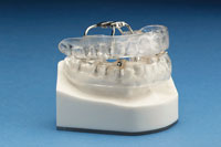
Back-scatter of the radiation beam due to a metal dental restoration may create a localized mucositis, especially on the buccal mucosa or tongue. Many radiation oncologists recommend the fabrication of custom acrylic bite blocks covering teeth that have metallic restorations, using flanges to displace the tongue and buccal tissues.
On December 15, 2004, the FDA approved palifermin (Kepivance), which is said to decrease the incidence and duration of mouth and throat mucositis. Palifermin is a recombinant human keratinocyte growth factor (rHuKGF) that stimulates the growth of epithelial cells, leading to more rapid replacement of epithelial cells damaged by chemotherapy and radiation. Palifermin has been approved for patients receiving high-dose chemotherapy, with or without radiation therapy, for hematologic malignancies.6
Since most cases of mucositis are reversed with the cessation of irradiation, treatment is generally palliative. Topical rinses of liquid or viscous local anesthetics, rinses of baking soda and salt in warm water, or compounds of Maalox/Benadryl/Xylocaine viscous may provide comfort. Severe mucositis, with or without infection, is a primary cause for interruption of the prescribed regimen of irradiation.
 |
| Figure 1. Candidiasis and mucositis secondary to radiation therapy. |
Candidiasis (moniliasis, thrush). An uncomfortable overgrowth of Candida albicans may cover the oropharynx and esophagus (Figure 1). The infection may present as diffuse red patches (erythematous candidiasis); removable white plaques (pseudomembranous candidiasis); raw, red and white patches in the corners of the mouth (angular cheilitis); or as denture stomatitis (chronic atrophic candidiasis).7 Mouthrinses may be helpful in reducing the discomfort. Alkaline mouth rinses of salt and baking soda, 0.5% povidone iodine rinses, or Maalox/Benadryl/Xylocaine viscous may also be effective. These rinses do not, however, control the infection. Nystatin (Mycostatin) has been recommended for years; however, resistance is now making nystatin less effective than in the past. Amphotericin B (Fungi-zone) has also been employed for years, but is not available in the United States. Clorimazole (Mycelex), ketoconzole (Niz-oral), and fluconazole (Diflucan) are now used for the treatment of candida infection, with varying levels of success.
Loss of taste (dysgeusia). Many patients experience a change in taste or a complete loss of taste during radiation therapy. The loss of “salt” perception is particularly problematic, since patients tend to add significant quantities of salt to bland-tasting food, with potential effects on blood pressure. Taste perception often improves approximately 4 months after the completion of radiation therapy. Zinc sulfate supplements may increase taste perception and improve salivation.8
 |
 |
| Figure 2. Radiation caries and xerostomia secondary to external beam radiation therapy. | Figure 3. Radiation caries and candidiasis secondary to radiation therapy. |
Xerostomia. Dry mouth frequently occurs during and after radiation therapy, especially when the parotid glands and the submandibular glands are directly in the field of irradiation. Associated with xerostomia is a shift in the pH in the mouth toward a more acidic environment (pH 5.5 or less) and a highly cariogenic oral flora. Under these conditions, the teeth rapidly demineralize, especially in the cervical and incisal/occlusal surfaces. This demineralization is termed “radiation caries” (Figures 2 and 3). Radiation caries may involve all of the remaining teeth, not just those in the direct line of radiation.
In some patients, the risk of radiation caries may be reduced by meticulous oral hygiene, daily topical fluoride applications at home, and frequent professional dental care. A home-care regimen recommended to patients is the following:
(1) floss each tooth and brush teeth with toothpaste
(2) place 0.4% stannous fluoride or 1.1% sodium fluoride in disposable or freshly cleansed flexible custom fluoride trays
(3) place trays with fluoride over the teeth for 5 minutes
(4) do not rinse mouth, do not eat, and do not drink for 30 minutes after application
(5) repeat daily for as long as needed.
Following head and neck irradiation, dental treatment should be approached cautiously. Remineralizing gel (Revive) may be useful. Professionally applied topical fluoride and dental prophylaxis should be routine. Dental restorations using amalgam and glass ionomers are desirable, and composites are not recommended. Haveman9 compared amalgam and glass ionomers in xerostomic patients who used topical fluoride gel daily and those who did not. In fluoride users, amalgam and glass ionomers showed no significant difference in caries incidence. In fluoride nonusers, glass ionomers (fluoride-releasing materials) reduced the incidence of caries. Wood10 showed that in patients who used daily topical sodium fluoride, glass ionomer cements failed, while amalgam restorations did not. He further showed that in patients who did not use topical fluoride, glass ionomer cements did not fail, while amalgam restorations did fail. McComb11 studied glass ionomers and resin composite restorations in xero-stomic head and neck radiation patients. Composite restorations failed in patients who used fluoride as well as in patients who did not use fluoride. Based on these studies, resin composite is not recommended—while amalgam is recommended—for patients who may be compliant with the use of topical fluorides; glass ionomer is recommended for patients who may be noncompliant.
Endodontic therapy should be considered to preserve teeth or tooth roots even if the roots are not restorable. Teeth may be extracted from areas receiving less than 5,000 cGy. If extractions are unavoidable from regions receiving more than 5,000 cGy, one long-established protocol recommends that HBO be considered prior to and after extractions.12 Other clinicians rarely prescribe HBO therapy.13
Soft-tissue necrosis. Ischemia of the oral tissues following radiation therapy may increase the incidence of sloughing of the soft tissues of the oral cavity. This problem usually localizes to the site of pre-existing periodontal lesions or as the result of denture irritation. Such soft-tissue necroses may precede osteoradionecrosis. Appropriate denture care and/or periodontal care may prevent this complication.
Scar tissue formation. Fibrosis of the muscles of mastication may produce trismus or deviation of the mandible during opening. Further, post-irradiation neck dissection surgery may be more difficult when the tissues of the neck are scarred from irradiation. Active jaw opening exercises during and after radiation therapy may reduce trismus in some patients.
Salivary gland dysfunction. Radiation exposure may produce fibrosis, fatty degeneration, acinar atrophy, and/or cellular necrosis of the salivary glands. The critical dose of radiation for such changes is not known; however, the serous acini are more sensitive than the mucinous acini, so that the saliva decreases in quantity and thickens.14 If both parotid glands are exposed to radiation, xerostomia is often severe. Xerostomia produces marked dryness of the mucosa, reduces the protective moisturizing of the teeth, and creates an environment for rapid demineralization of the teeth.
Currently, amifostine (Ethyol) is prescribed by many radiation oncologists to be given just prior to each radiation therapy treatment. In many cases, amifostine markedly improves salivary gland function during and after irradiation. If xerostomia does occur, a patient’s comfort may improve with the following actions:
•avoiding alcohol-based mouthrinses
•avoiding alcohol intake
•taking frequent sips of water or lemon water
•pilocarpine 5 mg QID
•zinc sulfate 220 mg BID
•glycerine swabs before meals
•special food preparation, such as pureed solids, and avoiding abrasive foods
•avoiding high-sugar-content foods
•artificial saliva solutions (Moi-Stir, Salivart, Xerolube).
 |
Figure 4. Osteoradionecrosis and sequestration of the mandible. |
 |
Figure 5. Maxillary osteoradionecrosis (asymptomatic) secondary to radiation therapy. |
 |
Figure 6. Cutaneous fistula secondary to osteoradionecrosis of the mandible. |
Osteoradionecrosis (ORN). Ischemia and fibrosis secondary to irradiation in the region of the head and neck may lead to avascular necrosis of the mandible or maxilla and the soft tissues near the jaws. Sequestration, secondary infection, pathologic fracture, fistulization, and drainage may result (Figures 4 to 6). The following are risk factors for ORN: radiation portals that include teeth or jaw bones; total irradiation above 5,000 cGy; local trauma from dental extractions or periodontal treatment; and smoking. When assessing risk, changes in the radiation oncology treatment plan should be kept in mind. On occasion, the total dosage is greater than initially scheduled due to a change in the treatment plan, the need to re-treat an area, or the use of brachytherapy, such as the implantation of radioactive “seeds.”
The most common co-factor for ORN is the extraction of a tooth from an irradiated jaw.2 It is a common misconception that teeth may be safely extracted from irradiated bone several years after radiation therapy. In fact, after radiation therapy, the bone remains vulnerable to ORN.15
ORN is not totally avoidable; however, some precautions may reduce an individual’s risk of ORN. Prior to radiation therapy, all grossly carious teeth and teeth with advanced periodontal disease should be removed. Mandibular teeth in an area scheduled to receive 6,000 cGy or more direct irradiation should be removed. Tension-free closure, closure without raising a local flap, antibiotic prophylaxis, and a 21-day healing period prior to commencing radiation therapy should improve the outcome.
In the past, some dentists and radiation oncologists advocated the removal of most teeth, if not all, prior to irradiation. Currently, the recommendation is the removal of nonrestorable teeth, those with advanced periodontal disease, mobile teeth with furcation involvement, residual roots, necrotic teeth, or mandibular teeth receiving high levels of direct radiation.16 Patients who demonstrate adequate oral hygiene, can be expected to receive regular dental care, and express a desire to maintain their teeth should not undergo removal of sound teeth that are not in the radiation field.
In the case of patients who present with rapidly expanding tumors or massive tumors that require immediate radiation therapy, dental extractions should be deferred and performed in the first 4 months following the completion of radiation therapy. Experience seems to show that extractions, periodontal procedures, and even dental implants may be safely completed during this immediate post-irradiation period without the use of HBO.17
From the fifth month following irradiation, dental extractions should be avoided if possible. It is sometimes preferable to treat teeth or residual roots endodontically that would normally be treatment planned for extraction. If post-irradiation extractions are absolutely necessary, this surgery is relatively safe during the first 4 months after radiation therapy. After this point, the risk of ORN increases and is always above pre-irradiation levels. If extractions are unavoidable and the bone housing the teeth received greater than 5,000 cGy of radiation, the use of HBO should be considered. The following is a common regimen:18
(1) presurgical treatment is administered in a “dive” chamber producing 2.4 to 2.6 atmospheres for 90 minutes daily for 20 dives (days)
(2) antibiotic prophylaxis (phenoxymethyl penicillin V is preferred)
(3) chlorhexidine or List-erine rinses
(4) minimally traumatic extractions without reflection of a tissue flap
(5) postsurgical HBO at 2.4 to 2.6 atmospheres for 90 minutes daily for 10 dives (days).
For future extractions or routine oral surgical procedures, there is no need to repeat HBO, since HBO confers “permanent” improvement in the vascularity.
Some investigators have proposed the use of ultrasound therapy in place of HBO.19 Ultrasound appears to induce angiogenesis, thereby revascularizing ischemic bone. It may be utilized as a prophylactic measure prior to oral surgery as long-wave ultrasound at intensities of 15 to 30 mW/cm2 for 45 kHz for 20 presurgical and 20 postsurgical 10-minute sessions. Ultrasound may also be used as part of the treatment regimen for osteoradionecrosis using 40 to 50 10-minute sessions until healing is complete. Note that some investigators consider HBO to be adjunctive and not the standard of care with respect to dental extractions in irradiated jaws.16
Post-irradiation, removable prosthetic appliances can be considered. Some patients may be unable to tolerate any prosthesis. Others will tolerate appliances with soft lining materials, while some will tolerate conventional prostheses.
Topical fluoride should be applied to the remaining teeth every day, and professionally applied fluoride should be accomplished 2 to 4 times annually. This regimen may be required for the life of the patient.
 |
| Figure 7. Pathologic fracture of mandible secondary to osteoradionecrosis. |
If ORN occurs, the extent of the necrosis and the response to therapy determine the aggressiveness of the treatment. Many cases will respond well to 30 HBO dives, followed by gentle removal of sequestered bone, followed by 10 further HBO dives. Other cases will respond poorly or degenerate further and will require wide surgical debridement, antibiotics, and oral antiseptics. Still other cases will produce jaw fractures, cutaneous fistulae, or lytic jaw lesions requiring jaw resection (Figure 7).
ORAL EVALUATION PRIOR TO RADIATION THERAPY
Oral evaluation and care should be routine for all patients who will receive radiation therapy to the head and neck, including edentulous patients. This includes the following:
(1) dentate patients—full series of dental radiographs and a panoramic radiograph
(2) edentulous patients—a panoramic radiograph and problem-focused intraoral radiographs
(3) periodontal evaluation and control of periodontal disease for dentate patients
(4) if oral hygiene is excellent, caries control and restorative dentistry
(5) endodontic care for teeth with a necrotic pulp that are considered restorable but are outside of the radiation field
(6) extraction of all hopeless or questionable teeth in the field when more than 5,000 cGy of irradiation is planned; extraction of mandi-bular teeth directly in the field scheduled to receive 6,000 cGy or greater; extractions should be completed 21 days (if possible) prior to initiation of radiation therapy
(7) dental prophylaxis, professional fluoride application, flexible fluoride tray preparation, and detailed home-care instructions
(8) evaluation of prostheses for undercuts, irregularities, and abrasive projections
(9) removal of orthodontic appliances in the field of radiation
(10) when needed, fabrication of custom acrylic bite blocks to cover metal restorations and displace the tongue and buccal tissues.
ORAL CARE DURING RADIATION THERAPY
Care of the mouth during irradiation consists of palliative treatment of mucositis, xerostomia, moniliasis, dysgeusia, and herpetic lesions. The patient’s mouth may become sore, dry, inflamed, and ulcerated so that dental treatment is difficult. Neverthe-less, patients should be encouraged to apply fluoride to the teeth daily and follow the prescribed oral hygiene regimen.
Chlorhexidine should be used with caution. While some studies have promoted chlorhexidine for the prevention and treatment of radiation-related mucositis,20 other studies have noted potential problems. First, in randomized studies, chlorhexidine was no more successful in preventing mucositis than sterile water or a mouthrinse of one-half teaspoon of salt and one-half teaspoon of baking soda in 8 ounces of water.21 Second, chlor-hexidine may precipitate the emergence of Gram-negative bacilli infections in the oropharynx.22 Third, the commercial preparations of chlorhexidine contain alcohol, which induces marked discomfort in patients who have mucositis. Nonalcohol chlorhexidine mouthrinses may be compounded by a pharmacist. Fourth, chlorhexidine may interfere with nystatin preparations during antifungal treatment.23 Therefore, the routine use of chlorhexidine during radiation therapy should be avoided.
ORAL CARE FOLLOWING RADIATION THERAPY
Following irradiation, patients should be encouraged to have regularly scheduled dental examinations, dental prophylaxes, professional application of fluorides, and daily self-application of topical fluorides. If surgical treatment such as extractions, endodontic therapy, periodontal treatment, and implant placement are required, the first 4 months following irradiation is a relatively safe time frame for such care. Thereafter, soft-tissue radionecrosis and osteoradionecrosis are more likely to occur following surgical trauma.
Many experienced clinicians are comfortable performing invasive oral procedures following radiation therapy; however, clinical protocols vary. For example, Larsen24 advocates the use of HBO adjunctively to the placement of dental implants, while Keller25 supports the placement of dental implants in irradiated mandibles without HBO.
BACTERIAL ENDOCARDITIS PROPHYLAXIS FOR RADIATION THERAPY PATIENTS
Patients who have had irradiation to the left chest wall or who have received cardiotoxic cancer chemotherapy may develop valvular heart disease as well as coronary vessel disease.26 Fortunately, modern radiation techniques can often avoid these structures in the majority of patients so that a history of left chest-wall radiation therapy without evidence of valvular dysfunction is not an indication for bacterial endocarditis prophylaxis. In those patients who are diagnosed with valvular heart disease, the American Heart Association bacterial endocarditis guidelines27 are appropriate. Therefore, when planning invasive dental procedures, the dentist should consult with the patient’s oncologist regarding the presence of valvular disease and the need for bacterial endocarditis prophylaxis.
CONCLUSION
This article reviews current concepts in radiation therapy with an emphasis on oral healthcare of cancer patients. The goals of this article are to improve understanding of the oral healthcare needs of patients who are scheduled to receive, are receiving, or have received radiation therapy for cancers of the head and neck.
References
1. Jansma J, Vissink A, Spijkervet FK, et al. Protocol for the prevention and treatment of oral sequelae resulting from head and neck radiation therapy. Cancer. 1992;70:2171-2180.
2. Reuther T, Schuster T, Mende U, et al. Osteoradionecrosis of the jaws as a side effect of radiotherapy of head and neck tumour patients: a report of a thirty year retrospective review. Int J Oral Maxillofac Surg. 2003;32:289-295.
3. Intensity modulated radiation therapy. Cancer Treatment Centers of America web site. Available at: http://www.brachytherapy.com/IMRT.html. Accessed November 12, 2004.
4. Gamma knife surgery. International RadioSurgery Association web site. Available at: http://www.irsa.org/gamma_knife.html. Accessed November 10, 2004.
5. Antonadou D, Pepelassi M, Synodinou M, et al. Prophylactic use of amifostine to prevent radiochemotherapy-induced mucositis and xerostomia in head-and-neck cancer. Int J Radiat Oncol Biol Phys. 2002;52:739-747.
6. Questions and answers on palifermin (keratinocyte growth factor). FDA Center for Drug Evaluation and Research web site. Available at: http://www.fda.gov/cder/drug/infopage/palifermin/paliferminQA.htm. Accessed December 26, 2004.
7. Sapp JP, Eversole LP, Wysocki GP. Contemporary Oral and Maxillofacial Pathology. St Louis, Mo: Mosby; 1997:228-231.
8. Siegel MA, Silverman S Jr, Sollecito TP, eds. Clinician’s Guide to Treatment of Common Oral Conditions. 5th ed. Seattle, Wash: American Academy of Oral Medicine; 2001:7-8.
9. Haveman CW, Summitt JB, Burgess JO, et al. Three restorative materials and topical fluoride gel used in xerostomic patients: a clinical comparison. J Am Dent Assoc. 2003;134:177-184.
10. Wood RE, Maxymiw WG, McComb D. A clinical comparison of glass ionomer (polyalkenoate) and silver amalgam restorations in the treatment of class 5 caries in xerostomic head and neck cancer patients. Oper Dent. 1993;18:94-102.
11. McComb D, Erickson RL, Maxymiw WG, et al. A clinical comparison of glass ionomer, resin-modified glass ionomer and resin composite restorations in the treatment of cervical caries in xerostomic head and neck radiation patients. Oper Dent. 2002;27:430-437.
12. Johnson RP, Marx RE, Buckley SB. Hyperbaric oxygen in oral and maxillofacial surgery. In: Worthington P, Evans JR, eds. Controversies in Oral and Maxillofacial Surgery. Philadelphia, Pa: Saunders; 1994:107-125.
13. Maxymiw WG, Wood RE, Liu FF. Postradiation dental extractions without hyperbaric oxygen. Oral Surg Oral Med Oral Pathol. 1991;72:270-274.
14. Rothwell BR. Prevention and treatment of the orofacial complications of radiotherapy. J Am Dent Assoc. 1987;114:316-322.
15. Oh HK, Chambers MS, Garden AS, et al. Risk of osteoradionecrosis after extraction of impacted third molars in irradiated head and neck cancer patients. J Oral Maxillofac Surg. 2004;62:139-144.
16. Sulaiman F, Huryn JM, Zlotolow IM. Dental extractions in the irradiated head and neck patient: a retrospective analysis of Memorial Sloan-Kettering Cancer Center protocols, criteria, and end results. J Oral Maxillofac Surg. 2003;61:1123-1131.
17. Marx RE. Is hyperbaric oxygen still needed in managing radiated patients? Presentation at: The Florida Society of Oral and Maxillofacial Surgeons; November 14, 2004; Tampa, Fla.
18. Marx RE, Johnson RP, Kline SN. Prevention of osteoradionecrosis: a randomized prospective clinical trial of hyperbaric oxygen versus penicillin. J Am Dent Assoc. 1985;111:49-54.
19. Reher P. Ultrasound for the treatment of osteoradionecrosis (letter). J Oral Maxillofac Surg. 1997;55:1193-1194.
20. Ferretti G


