


Cone-beam computed tomography (CBCT) has motivated a new standard of care when it comes to diagnoses and treatments of dental diseases by way of 3-D imaging of the maxillofacial skeleton. For nearly 2 decades, CBCT technology has been available, but only recently has it been commercialized and popularized in the field of digital dentistry.1,2 Two-dimensional diagnostic imaging, such as traditional radiographs, photographs, and video imaging, are essential to dentistry, no doubt, but limited. Magnification, geometric distortion, and projective displacements may all be misconstrued with 2-D imaging.3 In contrast, 3-D volumetric data allows for evaluation of more realistic anatomy without the aforementioned limitations of 2-D imaging.
A Brief Look at How the Technology Works
The complexity of creating 3-D data with CBCT technology lies in the detector and data processing software. CBCT acquires volumetric data by exposing a section of the patient over one detector to generate individual slice images with a single rotation, while traditional CT uses a row of detectors. This data can then be reconstructed into a 3-D volume and processed on commonly available personal computers.4 CBCT machines are generally less cumbersome than traditional CT scanners, yet CBCT results in comparable or better images of the skeletal structures, making it well suited for use in dentistry.
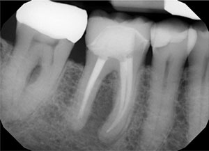 |
| Figure 1. Periapical radiograph of a symptomatic and previously endodontically treated lower right molar. |
Advantages of CBCT
Using CBCT over conventional CT or routine radiography in specific cases offers many advantages in the clinical setting. Not only does CBCT boast image accuracy, it also has a rapid scan time, the ability to interact with data for chairside image display or real-time analysis, and a reduced dose of radiation compared to conventional CT.1 As far as diagnoses and treatments, the 3-D volumes created by CBCT can be exported to third-party software for convenient implant, maxillofacial, or orthodontic treatment planning.
More specifically, in endodontics, CBCT can be used to diagnose periapical lesions, visualize root fractures, and detect root resorption. With periapical lesions, CBCT offers information on their juxtaposition to the maxillary sinus, sinus membrane involvement, lesion location relative to the mandibular canal, classifying the source of the lesion as endodontic or non-endodontic, and more. CBCT can also be used to determine the morphology of roots and associated canals. The data can establish working length and angulation and provide an accurate detection of vertical and horizontal root fractures. In terms of root resorption, CBCT imaging can allow for early detection and identify the extent of a lesion.5 CBCT offers an array of valuable information that can greatly impact treatment planning.
As dentistry sees more and more patients of younger generations, convenience and time-effective planning and diagnostics are imperative. By incorporating 3-D CBCT data into diagnostics, referrals to specialists for imaging are made redundant. The patient can get an almost-immediate diagnosis to determine whether or not the tooth is restorable in the office without wait times for outside specialists. This allows the patient and provider to have a better understanding of timeline, treatment, and finances. Overall, CBCT usage leads to a better clinical outcome and satisfaction for the patient.
CASE REPORTS
In the case examples that follow, CBCT data was used in the diagnosis of various circumstances.
 |
 |
| Figure 2. A CBCT scan was taken to assist in the diagnosis of symptoms on the patient’s mandibular right side. See lucency at the lower right first and second molar not evident on the 2-D radiograph. | Figure 3. Axial view of the root apices of the mandibular right first molar on the CBCT scan. |
 |
| Figure 4. Failing and symptomatic left central incisor. |
Case 1
This case demonstrates the value of a CBCT machine in an emergency situation. The patient came into the office with pain and swelling on a previously root canaled tooth—potentially indicative of root fracture. This is rather common: Approximately 11% to 20% of root-filled teeth need to be extracted due to root fractures.6,7 Traditionally, a general dentist will take a PA radiograph (Figure 1) and then refer this patient to an endodontist for evaluation, but there’s no telling if the patient will go to the referral or wait until the problem is exacerbated. By having a CBCT machine in-office, a full diagnosis is available in a matter of minutes, giving insight as to whether the problem can be resolved with a re-treat root canal or if the tooth is non-restorable (Figures 2 and 3). With the help of CBCT, the general dentist is able make a definitive treatment plan to ensure the patient receives the treatment he or she needs in a timely manner.
 |
| Figure 5. Diagnostic CBCT scan of the symptomatic left central incisor. |
 |
 |
| Figure 6. Patient with Class III occlusion and missing upper right bicuspids and lateral incisor. | Figure 7. Integration of the CBCT and intraoral digital scan for implant planning. |
Case 2
In this case, the patient presented with an unsightly restored tooth in the aesthetic zone with retention issues and accompanied by achy pain and swelling (Figure 4). A CBCT scan was taken to decide whether or not the tooth was savable (Figure 5). In the event that a traditional radiograph was taken, it would not have given a definitive answer to the aforementioned question and would have required a referral for more comprehensible imaging. The CBCT scan accurately depicted a root fracture. In a previous study of 1,821 anterior restorations, the total failure rate was 24.1%, with more than half of those failures due to root fracture.8 The second most common reason for failure was due to aesthetic qualities, both of which attributed to the failure in this specific case. Ultimately, this patient was diagnosed with a root fracture, and the tooth was treatment planned to be extracted, along with an immediate implant placement.
Case 3
CBCT is not only invaluable in diagnostics, but also in treatment planning. The final case presented shows the capabilities of CBCT in terms of implant planning. This case involved a young patient who was missing several permanent teeth. He had a Class III occlusion, which complicated his restorative plan (Figure 6). More often than not, clinicians will use their knowledge and experience to estimate the proper location for implantation. Usage of a CBCT scan to identify critical anatomic boundaries of individual patients and to prevent neurovascular trauma can reduce the risks associated with estimating location.9 The precise volumetric data of CBCT can be integrated into software that can design and retrofit exactly where the implant will be placed (Figures 7 and 8). From this data, a stent can be formed for guided implant placement, making the procedure achievable by a general dentist (Figures 9 and 10). This case illustrates how restoratively driven, fully guided implant placement results in optimal aesthetic and functional outcomes.
 |
| Figure 8. Digital restorative planning for the replacement of the upper right bicuspids and lateral incisor. |
 |
 |
| Figure 9. The surgical guide for implant placement. | Figure 10. The surgical guide in place for fully guided, restoratively driven implant placement. |
CLOSING COMMENTS
In the present age of Zoom and instant consultations, care providers must be able to evaluate current technologies and determine what creates the best clinical outcomes for their patients. The single most important commodity on which patients base treatment acceptance may be convenience. With access to CBCT, diagnosis is almost instantaneous, allowing the general dentist to be at the forefront of patient service and clinical care.
References
- Scarfe WC, Farman AG, Sukovic P. Clinical applications of cone-beam computed tomography in dental practice. J Can Dent Assoc. 2006;72:75-80.
- American Dental Association Council on Scientific Affairs. The use of cone-beam computed tomography in dentistry: an advisory statement from the American Dental Association Council on Scientific Affairs. J Am Dent Assoc. 2012;143:899-902.
- Kapila S, Conley RS, Harrell WE Jr. The current status of cone beam computed tomography imaging in orthodontics. Dentomaxillofac Radiol. 2011;40:24-34.
- Dawood A, Patel S, Brown J. Cone beam CT in dental practice. Br Dent J. 2009;207:23-28.
- Venkatesh E, Venkatesh Elluru S. Cone beam computed tomography: basics and applications in dentistry. J Istanb Univ Fac Dent. 2017;51(3 suppl 1):S102-S121.
- Demarco FF, Collares K, Coelho-de-Souza F, et al. Anterior composite restorations: a systematic review on long-term survival and reasons for failure. Dent Mater. 2015;31:1214-1224.
- Popescu SM, Diaconu OA, Scrieciu M, et al. Root fractures: epidemiological, clinical and radiographic aspects. Rom J Morphol Embryol. 2017;58:501-506.
- Tamse A. Vertical root fractures in endodontically treated teeth: diagnostic signs and clinical management. Endod Topics. 2006;13:84-94.
- Jacobs R, Salmon B, Codari M, et al. Cone beam computed tomography in implant dentistry: recommendations for clinical use. BMC Oral Health. 2018;18:88.
Dr. McMahon is a graduate of the University of Pittsburgh School of Dental Medicine. She maintains a private practice focused on cosmetic dentistry in Pittsburgh. Dr. McMahon is accredited by the American Academy of Cosmetic Dentistry (AACD) and is an invited Fellow of the prestigious American Society for Dental Aesthetics. She is a past clinical instructor in prosthodontics and operative dentistry at the University of Pittsburgh and is currently the director of new product evaluation and a lecturer for Catapult Education. Dr. McMahon frequently lectures across the United States on minimally invasive dentistry and conservative cosmetic dentistry for teenagers and young adults and is a 7-time award winner in the AACD’s Annual Smile Gallery. She can be reached via email at drsusan@wowinsmile.com.
Ms. Kuzemchak is currently a third-year undergraduate at the University of Pittsburgh. She is majoring in natural sciences on the pre-dental track and plans to attend dental school. She can be reached at ark138@pitt.edu.
Disclosure: The authors report no disclosures.
Related Articles
Cosmetic Dentistry in Today’s Selfie Culture: Conservative Treatment for an Adolescent
CBCT Guided Surgery: Accurate, Predictable, and Economical


