|
|
|
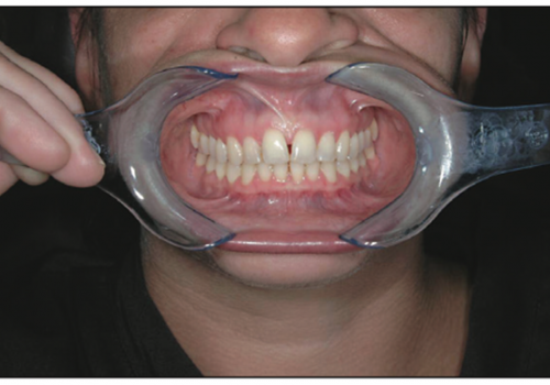
| Figures 1 to 3. Pretreatment and immediate post surgical digital photographs taken with the Kodak DX4900 Camera that are both printed and sent with specimen and also e-mailed digitally to pathology lab to assist in proper diagnosis. |
|
| Figure 4. The digitized microscopic image that is e-mailed from the lab back to the clinician to be incorporated into the patient’s electronic clinical patientrecord ensuring a complete and comprehensive record of the case and treatment. |
Screening for soft tissue pathology, particularly oral cancer, is arguably one of the most important services dentists provide. Dentists trained in oral pathology obtain expertise that other primary care providers, such as general medical practitioners, often lack. Routine dental check-ups provide an opportunity to identify cancerous lesions in their early stages. For this reason, New York State is mandating continuing education in oral pathology to improve dentists’ ability to identify cancers and assist in the development of oral cancer treatment plans.
To effectively screen dental patients for soft tissue pathology, dentists must address several issues. The first is the diagnosis itself. When an oral lesion is found, it is often possible to make a preliminary diagnosis based on the lesion’s visual characteristics, its duration, and other factors. However, in many cases it may be necessary to consult with a specialist in oral pathology, especially when malignancy is suspected.
Another issue is patient acceptance of a diagnosis or possible diagnosis of cancer by a dentist. The public at large often associates dental care with cosmetic procedures and the treatment of cavities and periodontal disease—not with cancer screening. Therefore, when a dentist or staff person discovers a soft tissue lesion, the patient may fail to appreciate the importance of the discovery, and may resist the dentist’s recommendation for biopsy or referral to a specialist.
A final issue, often interrelated to the first two, is procedural. Failing to establish and follow sound clinical procedures can lead to both substandard care, including the inability to properly diagnose suspicious lesions, as well as lowered case acceptance and even increased risk of legal liability. Too often, for example, dentists fail to properly document a patient’s refusal to accept treatment for a suspicious lesion. Should a patient or a patient’s family bring a lawsuit later suggesting that the dentist had not made the importance of a biopsy clear to
the patient, the dentist has nothing but his or her word for defense—and juries tend to feel sympathetic toward cancer patients.
DIGITAL PHOTOGRAPHY’S PLACE WITHIN ORAL PATHOLOGY EXAMINATIONS
Photography promises to help dentists address each of the previously mentioned issues as follows (Figures 1 through 3):
- Photodocumentation can aid diagnosis by enabling dentists to share critical information
with oral pathology specialists.
- Photodocumenting oral lesions can increase treatment acceptance by enabling more effective patient communication.
- Photodocumentation helps ensure that oral lesions are properly recorded.
The value of photography for documenting oral pathology has been raised even further by the availability of high-quality, moderately priced digital cameras for dentistry. Digital cameras enable dental staff members to quickly and easily capture images of oral lesions. In so doing, they help to keep both fixed and variable costs of photography low, while contributing substantially to the practice’s standard of care.
CLINICAL PROCEDURE FOR SOFT TISSUE PATHOLOGY SCREENING
Screening for soft tissue pathology should be integrated into every patient examination, whether performed by the dentist or the hygienist. Soft tissue lesions can be classified as follows:
- Inflammatory. Lesions of this type may be caused by localized irritants, which may be physical in nature (eg, lacerations), chemical (eg, aspirin burns), or infectious agents (bacterial, viral, or fungal).
- Neoplastic. Benign or malignant.
- Developmental. For example, because of growth abnormalities of soft tissue, jaws, bone, or teeth.
- Other. This category includes lesions caused by degenerative or immune deficiency diseases.
To perform soft tissue screening, the patient’s entire mouth should be visually inspected for any abnormality, including the lips; labial and buccal mucosa; tongue (dorsum, lateral borders, and ventral surfaces); floor of the mouth; hard and soft palate; and finally, gingival tissues.
If a lesion is found, it should be documented. To photograph it, the ideal tool is a digital camera that is both high-quality and easy-to-use. Select a camera with high resolution that is able to capture color accurately. Color fidelity will be a function of both the quality of the camera sensor and the camera’s internal software. The camera operation must be straightforward enough so that staff can use it regardless of their photographic skill level. Routine photodocumentation should not be performed by the dentist, but by adjunct staff; otherwise the time spent capturing images will add too much to the practice’s overhead costs.
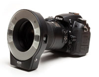
One camera that meets both these criteria is the new Kodak DX4900 Dental Digital Camera Kit. This is a 4.0 megapixel camera with excellent color quality. The camera is equipped with tools, such as a distance guide and dental positioning grid, that help ensure the user will frame views properly, so that any staff member will be able to use it without extensive training. The kit also comes with the Kodak EasyShare Dock II, an interface device that automates the uploading of the digital images to a computer workstation, helping to streamline the imaging process.
To photograph the lesion, place a periodontal probe with millimeter demarcations next to the lesion to record its size. If the lesion is on the tongue or is otherwise easily visible, lip retractors are not necessary. In some cases, a tongue depressor can be used to help make the lesion visible. Spring-loaded lip retractors are also a useful tool because their “hands-free” operation enables the individual doing the exam to take the photograph without requiring another staff member to hold a retractor. It’s best to avoid intraoral mirrors and other metal devices for retraction, since they can cause reflections that may distort the image. Photographic mirrors are another useful tool for imaging some areas of the mouth, such as the palate.
As soon as the image is captured, it should be uploaded to a computer for viewing. It should be checked for clarity, focus, and color. If the image does not accurately and completely document the lesion, it can and should be redone. When it is verified that the image is of sufficient quality, an electronic copy should be saved to the computer workstation or practice network. Copies can also be printed; for highest quality print results, use a photographic-quality, glossy paper such as Kodak DMI Paper for Dental Imaging.
In addition to photographing the lesion, its location should be documented schematically and its characteristics should be recorded textually. The textual description of the lesion should include size, shape, color, consistency, and how long it has been present. Any symptoms associated with the lesion, such as pain, should be noted. An initial classification of the lesion, based on its visual characteristics and location, should be made.
If the lesion appears to be noncancerous, a follow-up exam should be scheduled to take place in about 2 weeks. The scheduling of this follow-up appointment should be noted as part of the patient record. In many cases, the lesion will resolve during that interval. Should the patient cancel the follow-up exam because the lesion has healed, that should also be recorded.
If cancer is suspected, the dentist should recommend that the lesion be biopsied immediately. This recommendation should be noted, along with the patient’s response. Note whether or not the patient appeared to understand the doctor’s recommendation for biopsy. If the patient refuses the doctor’s recommendation, it is important that this refusal be noted to protect the practice from potential liability. If the patient is to be referred to an oral surgeon or other specialist for the biopsy, that should be noted, along with the patient’s acquiescence to the suggested referral.
If any lesion is still evident 2 weeks after initial discovery, it should be re-photographed for comparison purposes and then biopsied.
Biopsies can be performed by the dentist. Traditionally, biopsies have been made using scalpels, either by the dentist or an oral surgeon. Today, options also include laser biopsies, which may often be done without a local anesthetic. Offering this option may help increase patient acceptance because it can be relatively painless. When performing a laser biopsy, care must be taken to remove a border of clinically normal-looking tissue around the lesion. This border must be large enough so that the edges of the lesion are not damaged, and the extent of any cellular abnormality determined.
For certain oral lesions, a brush biopsy can also be used. With this technique, a bristle brush is rotated on the lesion to collect a sample of cells. Using this technique, the entire lesion is not removed, so if the collected cells are found to contain dysplastic or cancerous cells, additional tissue will have to be removed to enable a definitive diagnosis to be made. As such, a brush biopsy is, in cancer cases, but an intermediary step. However, because it is relatively simple to perform and does not require anesthesia, some dentists are more comfortable with it. Patients may also perceive a brush biopsy in more positive terms than they do a surgical biopsy that may raise a treatment acceptance problem, and the brush biopsy can be performed immediately, rather than requiring another appointment with a specialist. The benefit of increased patient acceptance may outweigh the fact that an additional “traditional biopsy” will be required if the brush biopsy is found to show dysplastic cells.
Documenting patient acceptance is as important during the biopsy stage of the screening process as during initial exams. For example, if the biopsy is to be performed on a referral basis, it is important to follow up with the specialist to make sure the patient kept the biopsy appointment. If the patient fails to follow through on the referral, the practice should do two things: telephone the patient to reiterate the treatment recommendation (record the date and time of the call) and if necessary, send a certified letter to the patient, restating the treatment recommendation.
This same procedure should be implemented in cases where a patient does not return for a recommended 2-week follow-up exam of a “suspicious” lesion.
DIAGNOSIS
Once a biopsy is performed, the tissue should be sent to an oral pathology laboratory along with the clinical photographs of the lesion. Having both the tissue and a photograph of the lesion is a great advantage to the oral pathologist. The photograph enables the pathologist to correlate the clinical features of the lesion with its histological characteristics, significantly improving the pathologist’s diagnostic accuracy.
With digital photography, copies of the images can be e-mailed immediately to the pathology laboratory. Some laboratories will find this useful, because they can familiarize themselves with the clinical aspect of a case before the microscopic evaluation of the biopsied tissue is conducted.
For dental practices that have computerized clinical patient records, the oral pathology laboratory can submit the written biopsy reports in digital form as well as the digitized microscopic images (Figure 4). This enables the practice to combine and organize all associated patient records of the pathology in the patient’s electronic clinical record, which can be easily viewed and shared with any other healthcare providers as needed.
PROGNOSIS
Approximately 30,000 US citizens are diagnosed with some form of oral cancer annually. As the baby boomer generation ages, this number may well increase. Sadly, about half of the oral cancers discovered today have already spread, and convey an overall 5-year survival rate of less than 50%. The prognosis for oral cancer has not changed significantly over the years. However, one thing is indisputable: the sooner a cancerous lesion is found, the greater the chances that the patient will survive.
Dentists are in a position, therefore, to effect significant improvements in oral cancer survival rates. Part of the profession’s role is communication, educating patients about oral cancer itself as well as the need for regular screening. Photography may play a role in this; in our experience when dental staff photograph lesions, patients tend to take them more seriously. This, in turn, leads to a significantly higher rate of treatment acceptance. Patients are more willing to accept the doctor’s recommendation for follow-up exams and biopsy.
If photography in general contributes to raising the standard of patient care, digital photography brings additional value to the practice. Digital cameras have become increasingly easy to use, minimizing their impact on practice overhead. At the same time, their image quality has improved significantly, to the point where any dental practice can afford a digital camera capable of producing very high-quality images. As a result, dentists are well-advised to consider integrating digital photography into their standard oral pathology screening procedures.
Dr. Benjamin is the advanced technology editor for Practical Procedures and Aesthetic Dentistry, and the working group chairman for digital camera, and co-chairman of the electronic patient record working group of the ADA Standards Committee on Dental Informatics. His private practice is based in Sidney, NY.
Dr. Aguirre is a professor in the Department of Oral Diagnostic Sciences, School of Dental Medicine at SUNY in Buffalo, NY. He is director of the Advanced Training Program in Oral and Maxillofacial Pathology, and directs the OMA Oral Pathology Laboratory.
Dr. Drinnan is a SUNY Distinguished Service Professor Emeritus in the Department of Oral Diagnostic Sciences, School of Dental Medicine at SUNY in Buffalo, NY.