
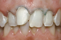
Porcelain laminate veneers were first introduced to the dental profession in 1983 by Faunce,1 Horn,2 and Calamia.3 Since that time, they have become a popular mode of aesthetic dental treatment. They are generally more durable than composite resin veneers, and, when properly formed and placed, are indistinguishable from natural teeth. Light passes through and reflects from a tooth restored with a porcelain veneer in almost the same way as with an unrestored natural tooth (Figure 1). Problems occur, however, when the veneer is inappropriately selected as the mode of treatment, or when some aspects of fabrication and/or placement are less than ideal. The problems most often encountered with porcelain veneers are: failure to meet the patient’s aesthetic expectations, fracture or loss of the restoration, or adverse periodontal consequences following placement. This article reviews the major causes of these problems and offers suggestions for overcoming them.
 |
| Figure 1. Porcelain veneers mimic the aesthetics of natural teeth. |
MEETING THE PATIENT’S AESTHETIC EXPECTATIONS
Patients often select porcelain veneers to improve the appearance of their teeth because they feel aesthetically disfigured. They see their teeth as being the wrong color, the wrong shape, or in the wrong position. They are looking for a conservative method to correct their problems at a reasonable cost and in a reasonably short time. The first step toward satisfying a patient is to obtain a clear definition and understanding of that patient’s personal goals and expectations. The dentist should ask simple, open-ended questions at the initial interview, such as “How can I help you?” or “What would you like me to do for you to improve the appearance of your teeth?” Patients should be encouraged to express their feelings about their teeth and how they would like to change them. Having a patient hold a mirror to view his teeth during this discussion is helpful. The patient and the dentist must understand the limitations of veneers (in comparison to crowns) and assure themselves that the patient’s expectations can be met with this treatment modality.
CONTRAINDICATIONS FOR VENEERS
Porcelain veneers are not appropriate for aesthetic restoration of all anterior teeth. Careful selection of the teeth to receive the veneer is necessary to ensure satisfaction. Long-term success is measured by continued aesthetic satisfaction, durability without becoming dislodged or fractured, the absence of visible surface or peripheral staining, and functional harmony with the other teeth. Some of the contraindications for veneers include the following situations.
Little Enamel Available for Bonding
 |
| Figure 2. After 3 years, the veneer has fractured from the dentin of the prepared tooth but remains bonded to enamel. |
Retention can be assured by selecting teeth that, after proper preparation, provide a substrate of enamel with minimal (if any) dentin exposure. Although materials and methods for bonding resins to dentin are currently popular, neither the strength nor the durability of the resin-dentin bond compares with the bond of resin to etched enamel, particularly when stress is applied to the restored teeth. Resin-enamel bonds are strong and enduring for years, whereas the preponderance of published dental research shows conclusively that over time, under stress, resin-dentin bonds weaken.4-13 In fact, it is difficult to find any research reports that indicate that dentin-resin bond strength of any dentin adhesive agent improves or even remains the same over a period of years. If porcelain veneers are bonded primarily to dentin, even with the best materials and techniques currently available, they become vulnerable to fracture or loss over time (Figure 2). The risk is increased significantly when tensile and torquing stresses are applied to the veneer during normal chewing, or when patients exhibit parafunctional habits such as bruxism.
 |
| Figure 3. Placing veneers on the teeth of bulimic patients results in an incisal margin bonded to dentin. The risk of incisal fracture of the veneer is high over time as stresses are applied to bonded interface. |
Contrary to recommendations made frequently by others writing or lecturing on this subject, I choose not to place porcelain veneers on any tooth that has more than 30% exposed dentin after preparation. Recognizing the instability of resin-dentin bonding under stress, I also do not use veneers in cases where the incisal margin terminates in dentin (such as the typical bulimic patient) (Figure 3). In these cases, ceramo-metal or all-porcelain crowns, rather than veneers, are more predictably durable.
Substantial Shade Changes Are Desired
 |
| Figure 4. Veneers often appear unnatural when opaque porcelain is used to mask very dark teeth. |
In order to preserve a substrate of enamel, the thickness of the veneer often must be less than 1 mm. When the patient’s teeth are very dark colored (eg, severe tetracycline staining), it is impossible to use such a very thin veneer of porcelain to mimic the translucency and general appearance of an extremely light-colored natural tooth (Figure 4). Opaque porcelains and/or relatively opaque resin cements must be used to block out the underlying dark color of the tooth. Opaque veneers lack translucency and appear artificial. It is generally true that the greater the degree of lightening of the teeth desired by the patient, the less desirable veneers are to accomplish that result. All-porcelain crowns are generally thicker than 1 mm, and are more suitable than veneers when major changes in shade are desired.
Mandibular Teeth
Restoring mandibular anterior teeth with porcelain veneers is much more technically difficult than placing them on maxillary teeth. This is due to the small size and thin natural enamel of lower teeth, coupled with inconvenient access and isolation. I do not endorse the use of porcelain veneers to restore mandibular teeth. I prefer crowns instead. The additional circumferential reduction of the lower anterior teeth required by crowns offers more opportunity for developing proper embrasure form, anatomical contours, more predictable retention, and less risk of fracture over time.
Severely Rotated Teeth
Patients with severely rotated anterior teeth often feel that a veneer will provide them with “instant orthodontics.” When a labially rotated anterior tooth is prepared to receive a veneer, the result is almost always removal of all the enamel from the labioverted segment of the tooth. When the preparation is complete, the veneer must fill a large triangular proximoincisal space with a cantilevered segment of porcelain bonded to an adjacent island of dentin. This is a very unfavorable situation that increases the probability of fracture. I feel that “instant orthodontics” should be done using crowns rather than porcelain veneers, except in cases of very minimal rotation where preparation exposes very little dentin or none at all (Figure 5).
Large Class III Restorations
Some patients requesting veneers present with multiple large, defective class III composite or cement restorations that will not be covered on the lingual surface by the veneer. The dentist and the patient must assess the relative expenditure of time, money, and effort involved in replacing these prior to veneering, in comparison with placing a crown that removes or covers them, eliminating these restorations from any further consideration.
Bruxism
 |
| Figure 6. Placing veneers on this bruxing patient’s teeth without concurrent restoration of the lost posterior support makes fracture of the veneers highly likely over time. |
Destructive habits such as bruxism place high stresses on restored teeth. Veneers that lengthen the anterior teeth are at a very high risk for fracture, unless the posterior teeth are similarly elongated to restore the patient’s lost vertical dimension. When veneers are placed on the teeth of bruxing patients, protective guards are often indicated to prevent future fracture (Figure 6).
Based on nearly 17 years of clinical experience with porcelain veneers, which includes many successes and failures, I limit my use of porcelain veneers (in nonbruxing patients) mainly to maxillary teeth of nearly normal color, minimal rotation or malalignment, and minimal exposure of dentin and/or old composite restorations after preparation.
PREPARATION DESIGN
A very important determinant of long-term aesthetic and structural success of the porcelain veneer is the configuration of the prepared tooth, particularly the location of the proximal margins. When preparing a tooth for a laminate veneer, the dentist has three options to position the proximal margins: labial to the proximal contact, halfway through the contact, or lingual to the contact.
When the proximal margin of a porcelain veneer is placed only slightly labial to or halfway through the proximal contact of the adjacent tooth, access for proper finishing is restricted. There is a high probability of visible stain forming along this margin sometime in the future. Such stain is difficult, if not impossible, to remove.
 |
 |
| Figure 7. Preparations that do not break the proximal contacts complicate fabrication and cementation of the veneers, and often result in visible interproximal stain over time. | Figure 8. Stain is evident at the midline because the proximal margin is located slightly labial to the contact. |
Placing the proximal margin halfway through the contact makes it impossible to section the stone model without damaging the margins of the dies. Marginal inaccuracy is almost ensured with this preparation design (Figures 7 and 8).
However, breaking the contact and placing the proximal margins on the lingual surface of the tooth provides many benefits:
•Any proximal marginal stain is not visible.
•The margins are more accessible for finishing.
•There is a greater surface area for bonding the veneer.
•The veneer exhibits a greater resistance to labiolingual flexion because of curvature.
•It is easier for the laboratory to separate the dies without damaging the margins.
•There is reduced likelihood of tearing the impression during retrieval.
•There is reduced likelihood of bonding the veneer to the adjacent tooth during cementation.
•It facilitates a more positive positioning of the veneer before and during cementation.
•The veneer can have improved proximal contours and aesthetics (Figure 9).
 |
| Figure 9. Placing the proximal margins lingual to the contact provides many advantages over preparations that maintain the contacts. |
It is not always possible to break both proximal contacts when preparing teeth for veneers. When veneers are placed on bicuspids and molars with broad contacts, at least one of the proximal margins usually must be placed labial to the contact to allow seating of the veneer from a buccal or facial direction. Any stain that might form at the interface over time will be minimally visible.
PROVISIONAL VENEERS
Provisional (temporary) veneers can be the key to ultimate success with porcelain veneers. They maintain the physical and social comfort of the patient during treatment while permitting both the dentist and the patient to assess the aesthetic, phonetic, and functional consequences of this type of dental treatment.
The major benefits of provisional veneers are reviewed.
Preview of the Future
High-quality provisional veneers afford the dentist and the patient an excellent opportunity to preview the future teeth, helping ensure that there will be no unpleasant surprises at the end of treatment. Provisional restorations that resemble the original disfigured teeth (or do not resemble the final intended product) have no value in predicting the outcome of treatment. Provisional veneers should be used to predict the future; they should not be a reflection of the past.
Assessment of Occlusal Function
Aesthetic dental treatment often includes changes in length and/or shape of the teeth. These changes have both functional and phonetic consequences. A patient may want the incisal length of all the maxillary anterior teeth to be the same. Restoring the teeth in this manner may create occlusal interferences, particularly in latero-protrusion. This can lead to discomfort, incisal fracture of the veneers, or destructive wear of the opposin
g teeth. The patient often cannot foresee or understand this conflict between aesthetics and function.
Provisional veneers constructed according to the patient’s wishes and expectations will reveal this problem as the occlusal interferences created by this configuration of the teeth are seen and felt by the patient. This can help convince the patient that their aesthetic preference is not realistic, or that adjustments of opposing teeth or even extensive restoration of the posterior teeth to increase the vertical dimension may be necessary if treatment with veneers is to be successful.
Identification of Consequences
Veneers that close large diastemata and/or enlarge or lengthen the teeth may cause disturbances in speech that may or may not be accommodated by the patient. This, too, should be assessed with provisional veneers prior to commitment of time, energy, and money to permanent restorations that otherwise would need to be recontoured or replaced.
Accomodation of Patients’ Requests for Major Changes in Shade
Occasionally, patients request treatment that deviates from the usual standards of aesthetics. Perhaps the most familiar example of this is the desire for teeth that are unnaturally white. To some patients this is beauty, although the majority of the population may perceive the teeth as fake or artificial. Provisional veneers made as white as the patient desires give the patient an opportunity to think about this and get reactions from other people before making a commitment to the final outcome. This process will usually clarify the aesthetic acceptability of such a request. Often, sending patients home with “white as snow” provisional veneers is the only way to convince them to modify their expectations and accept more natural-appearing restorations.
FABRICATING PROVISIONAL VENEERS
A variety of techniques for producing provisional veneers are described in the dental literature. Many of these articles give greater emphasis to speed, efficiency, utility, and protection from sensitivity than to their predictive value or their role in preserving and maintaining periodontal health. Splinted provisional veneers frustrate dental hygiene, making the patient vulnerable to periodontal disease. In my view, provisional veneers should resemble the final porcelain veneers as closely as possible. They should be individually bonded to the prepared teeth, permitting the patient to floss and clean the teeth conveniently. The seal of the provisional veneers to the prepared teeth should be sufficient to prevent ingress of bacteria or stain, and should protect the patient from sensitivity. The surface finish should be smooth, conveniently hygienic, and kind to the periodontal tissues.
 |
 |
| Figures 10 and 11. Individual provisional veneers can be made by pressing composite resin onto the tooth, curing it, and shaping it with rotary discs. | |
 |
 |
| Figure 12. A clear template can be made using a wax-up or a cast of the patient’s teeth. | Figure 13. The template and a silicone model of the prepared teeth should fit together precisely. |
 |
 |
| Figure 14. Composite resin can be applied directly to the silicone model without separating agents. | Figure 15. The clear template can be used to form a provisional veneer resembling the preoperative model. |
 |
| Figure 16. Temporary veneers. |
High-quality individual provisional veneers (as well as permanent resin veneers) can be made semidirectly by pressing microfilled composite resin onto the prepared tooth, curing and removing the prototype veneer, then shaping it with rotary discs (Figures 10 and 11). I often find it convenient to make provisional veneers indirectly at chairside using a silicone model of the prepared teeth (Figures 12 through 16). This is done in less than 5 minutes by injecting die silicone (Mach-2, Parkell; Die-Flex, Danville Materials) into an alginate impression of the prepared teeth. Composite resin can be applied directly to the silicone model, light cured, and removed without the need for any separating medium. The prototype veneer is then lifted off, contoured, and polished with rotary discs. This technique (which can be used to make “permanent” composite resin veneers as well) is fast, accurate, and involves very little interaction with or cooperation from the patient during the fabrication process.
If a diagnostic wax-up is available, or if the veneers will closely resemble the shape of the original teeth, a template can be made preoperatively using clear polyvinylsiloxane (PVS) bite-registration material (Clear-Bite, Danville Materials; Memosil, Bayer) in a nonretentive tray. After the composite resin is built up on the silicone model, the clear template can be placed over it, forming a mold that confines and shapes the composite resin to mimic the diagnostic cast or preoperative contours of the teeth. Excess composite resin can be vented through holes drilled through the template prior to light curing through the clear template. The mold is then disassembled and the provisional resin veneers are readily removed and individually finished.
The provisional veneers are attached to the teeth by etching a large central spot of facial enamel followed by rinsing and drying to reveal the characteristic “frosty” surface. They are bonded to the teeth with any unfilled resin or flowable composite. When the provisional veneers are removed, all remnant hardened resin must be removed from the tooth prior to try-in of the porcelain veneers.
In order to avoid unpleasant surprises, unhappy patients, and costly remakes, I recommend delaying the submission of veneer cases to the laboratory until after the patient has had an opportunity to wear the provisional veneers for several days and give comments, pro and con, to the dentist. These comments are relayed to the laboratory in the prescription that is accompanied by a silicone or stone cast of the provisional veneers bonded to the patient’s teeth.
I have been asked on many occasions to serve as a consultant in legal cases related to dental treatment with porce
lain veneers. This experience indicates clearly that a dentist’s failure to provide quality provisional veneers often leads to a violation of the patient’s expectations, expensive remakes, and ultimately lawsuits. Short-cutting the provisionalization stage often validates the lament, “We always seem to have time to do it over, but never have time to do it right.”
TRY-IN, CEMENTATION, AND FINISHING
 |
 |
| Figure 17. This photo of identical veneers illustrates the difference in appearance of wet (No. 8) and dry (No. 9) veneers at try-in. | Figure 18. The same veneers after cementation. |
Previewing the final restoration at the delivery appointment is essential to gaining the patient’s acceptance. If patients first view their veneers outside the mouth prior to try-in, they often feel that “what they see is what they’ll get.” However, the appearance of any veneer that is viewed in one’s hand, on a model, or on a dry tooth will change once it is wet and seated on the prepared tooth. No useful purpose is served if a patient views or passes judgment on a veneer before it is placed on his own wet tooth. A veneer that is much too light when viewed dry may prove to be a perfect shade match when cemented with a clear resin cement. Veneers appear yellow when viewed on yellow stone models; gray when seen on gray, green, or blue stone models; and too light when viewed on any tooth-colored dry surface. A veneer that looks to be a good match when viewed dry will be unacceptably dark after cementation. In all cases, patients will be disappointed at some point in the procedure if they are allowed to inspect the veneers prior to intraoral try-in. If the dentist wants to avoid disappointment, he/she should “just say no” to eager patients who want to see their veneers too soon (Figures 17 and 18).
 |
 |
| Figure 19. Residual salt formed by etching the porcelain must be removed by scrubbing with wet cotton before bonding the veneer. | Figure 20. Gingival inflammation due to ill-fitting veneer. |
Optimal adhesion to the tooth is ensured through proper treatment of both the veneer and the prepared tooth. The bond of resin to etched porcelain is equivalent to that of resin to etched enamel. It is strong and enduring over time, under stress. That bond is compromised if, after etching the porcelain, adherent salts remain on the etched surface. Before cementation, the dentist should dry the veneers and carefully inspect the etched surface for areas of white, frosty powder. If any remains, it should be removed by scrubbing each veneer with a wet cotton pellet, followed by rinsing and drying (Figure 19).
Veneers should not irritate and compromise the health of periodontal tissues. Adverse periodontal responses result from:
•placing gingival margins too deeply into the gingival sulcus.
•laceration of gingival tissues during preparation.
•ill-fitting provisional veneers.
•overcontoured or ill-fitting veneers (Figure 20).
•abraded or underglazed porcelain surfaces that contact the gingival tissues.
•incomplete removal of excess resin cement.
•splinted restorations that interfere with good hygiene.
The gingival tissues can be protected from laceration by retracting them with a relatively large, nonimpregnated cord prior to final preparation of the gingival margin. Placing the margin 0.5 mm incisal to the cord will ensure an equi- or subgingival margin that is accessible for finishing and does not violate the biologic width when the veneer is bonded.
Porcelain veneers do not always meet the dentist’s expectations for precise fit. This can be the result of poor- quality impressions, inaccurate dies, or the method of fabrication. Not all methods of fabricating porcelain veneers are equally accurate. Fusing porcelain onto burnished platinum foil has the least potential for precision fit because there is loss of detail that cannot be reproduced by the platinum foil. It is challenging to make extremely thin veneers (ie, less than 0.5 mm) with pressed porcelain methods. Fusing porcelain powder directly onto refractory dies produces the most precise fit, and most easily permits the technician to make very thin veneers with minimal risk of fracture.
Dentists who place porcelain restorations should become comfortable recontouring and polishing them with rotary wheels at chairside. Using abrasive wheels that are specifically designed for recontouring and polishing porcelain, the dentist can customize and improve the veneers for an optimal aesthetic and periodontal outcome.
Summary
Porcelain veneer restorations require close attention to detail from beginning to end. It is often prudent to go slowly when working with these cases. Patients receiving them have high expectations that go beyond considerations of function alone. Many problems are encountered when porcelain veneers are used to improve the appearance of the teeth. Avoiding and overcoming these problems first requires identification of the causes of the problems, followed by changes in clinical techniques. Success is the result of careful selection of teeth to receive veneers; preparing teeth in a manner that optimizes the aesthetic potential of the veneer; employing techniques that maximize the strength of both the veneer and its adhesive bond to the tooth; utilizing high-quality provisional veneers; insisting on a precision fit; and paying attention to the details of adhesive bonding protocols.
References
1. Faunce FR. Structured ceramics for laminate veneers. CDS Rev. 1987;80:36-38.
2. Horn HR. Porcelain laminate veneers bonded to etched enamel. Dent Clin North Am. 1983;27:671-684.
3. Calamia JR. Etched porcelain facial veneers: a new treatment modality based on scientific and clinical evidence. NY J Dent. 1983;53:255-259.
4. Okuda K, Pereira PN, Nakajima K, et al. Relationship between nanoleakage and long-term durability of dentin bonds. Oper Dent. 2001;26:482-490.
5. Kitasako Y, Burrow MF, Nikaido T, et al. Effect of resin-coating technique on dentin tensile bond strengths over 3 years. J Esthet Restor Dent. 2002;14:115-122.
6. Kitasako Y, Burrow MF, Nikaido T, et al. Long-term tensile bond durability of two different 4-META containing resin cements to dentin. Dent Mater. 2002;18:276-280.
7. Shono Y, Terashita M, Shimada J, et al. Durability of resin-dentin bonds. J Adhes Dent. 1999;1:211-218.
8. Kitasako Y, Burrow MF, Katahira N, et al. Shear bond strengths of three resin cements to dentine over 3 years in vitro. J Dent. 2001;29:139-144.
9. Hashimoto M, Ohno H, Kaga M, et al. In vivo degradation of resin-dentin bonds in humans over 1 to 3 years. J Dent Res. 2000;79:1385-1391.
10. Kato G, Nakabayashi N. The durability of adhesion to phosphoric acid etched, wet dentin substrates. Dent Mater. 1998;14:347-352.
11. Burrow MF, Satoh K, Tagami J. Dentin bond durability after 3 years using a dentin bonding agent with and without priming. Dent Mater. 1996;12:302-307.
12. Burrow MF, Tagami J, Hosoda. H. The long-term durability of bond strengths to dentin. Bull Tokyo Med Dent Univ. 1993;40:173-191.
13. Erickson RL, Glasspoole EA. Bonding to tooth structure: a comparison of glass-ionomer and composite-resin eyestems. J Esthet Dent. 1994;6:227-244.
Dr. Lacy is an associate professor in the Department of Preventive and Restorative Dental Sciences at the University of California School of Dentistry, San Francisco. He can be contacted at (415) 476-5431 or [email protected].

