


Direct posterior composites offer dentists the most simple and cost-effective manner in which to effect an aesthetic change in the posterior of the mouth. However, the placement techniques employed by dentists create myriad potential pitfalls to a successful and long-lasting restoration. One of the most challenging aspects of placing such restorations has been achieving proper interproximal contact in a predictable and reproducible manner. This article presents a clinical protocol designed to facilitate the predictable placement of direct posterior composite restorations, and outlines several material and armamentarium considerations.
BACKGROUND
Over the past 2 decades, patients’ awareness of the aesthetic attributes of dentistry has increased exponentially for the anterior and posterior regions of the mouth. Patients may desire to rid themselves of the many “dark” fillings they have in an attempt to improve their overall dental health or appearance. They may wish to eliminate metal from their mouths when replacing defective or worn restorations or when faced with the need to treat new lesions.
To this end, clinicians have available two categories of restorative alternatives for the posterior region—direct and indirect—each of which presents benefits and contraindications. Such considerations include material composition, physical properties, wear characteristics, shrinkage, and compressive strength, among others. Additionally, the skill of the clinician when executing the preparation design to ensure conservation of tooth structure, applying dentinal adhesives, and bonding the restoration, in addition to the technique sensitivity of placing these materials compared with other, more conventional restoratives (eg, amalgam and cast restorations) may also contribute to a clinician’s hesitation toward selecting such restorative modalities.
Direct posterior composites offer dentists the most simple and cost-effective manner in which to effect an aesthetic change in the posterior of the mouth. They have reached widespread use within the dental community, but not without a pronounced learning curve. Issues related to material properties and handling characteristics, as well as the clinician’s skill, are key to the success or failure of a posterior composite restoration. Dental product manufacturers have developed improvements to the material properties of posterior composites,1-6 in addition to available bonding agents, which have elevated the probability of success when placing direct composites. However, the placement techniques employed by dentists create myriad potential pitfalls to a successful and long-lasting restoration. One of the most challenging aspects of placing posterior composite restorations has been achieving proper interproximal contact in a predictable and reproducible manner.
TECHNIQUE AND MATERIAL CONSIDERATIONS
Clinicians have addressed this challenge in a variety of ways during the past 2 decades in order to provide the best possible direct resin dental treatment for patients. The number of methods and techniques sited in journals and continuing education courses, and discussed in study clubs, is too voluminous to mention without failing to credit all who have continued to modify the technique. Also, the use of acid etching dentin, glass ionomers, flowable composites, and most recently, packable composite resins, has played a role in advancing the exactness of placing direct posterior composite restorations.
However, it must be emphasized that direct posterior composites are not at all like amalgam. Therefore, clinicians must alter their thinking process and adjust their placement techniques in order to maximize the materials to their fullest. Composites are, for the most part, fluid, and assume the shape dictated by dentists based on the manner in which the material is manipulated. Composites also have the unique ability to bond to tooth structure, where as amalgam does not.
The principles of cavity preparation design are much different for composites than amalgams. G. V. Black could have only imagined the benefits that composite resins would offer clinicians, presenting the profession with an ability to be ultraconservative with composite restorations, removing only that tooth structure necessary for eliminating the caries present.7,8 Clinicians can now offer patients tooth-colored restorations that, although not an ideal replacement for enamel or dentin, can yield an aesthetic and functional solution to the problem of finding a modern direct alternative.
However, the manner in which these materials have been placed requires careful consideration, as differences in placement techniques and other armamentarium used affect the longevity and function of the final directly placed posterior composite restoration. Options include following an incremental technique compared with a bulk technique, placing a cold cure composite in the proximal box (in an attempt to achieve a more predictable seal and minimize shrinkage stresses), and selection of effective bonding agents, all of which may collectively determine the success or failure of a direct posterior composite restoration. Furthermore, the selection of curing lights, different matrices, and hand instruments leaves much to be considered. While there is no one “right” way to accomplish a direct posterior composite restoration, techniques will continue to be modified to ensure the creation of the most predictable and reproducible result.
MATERIALS
In the case report presented, a recently introduced direct restorative composite (Heliomolar HB [Heavy Body], Ivoclar Vivadent) was used. This material represents further development in a family of products with a nearly 20-year history of clinical success. The packable consistency enables the material’s placement for posterior restorations, and provides the clinician with time efficiency when placing these traditionally technique-sensitive and time-consuming restorations. The material is available in a range of shades and convenient delivery options, affording clinicians flexible and aesthetically appropriate treatment options. Heliomolar HB is available in nine shades, including seven Vita shades (A1, A2, A3, B1, B2, C2, D2) and two enamel shades (110T and 420T [white and gray]). A screw-type syringe and a new heavy-body cavifil delivery system are also available.
Heliomolar HB is based on microfiller technology, and exhibits the properties dentists and patients demand. Using identical base components found in the original material, Heliomolar HB contains a rheological modifier and similar particle-in-particle technology for a less-sticky, packable, nonslumping consistency ideal for posterior restorations. Specifically, the material demonstrates low wear, exceptional aesthetics, and high polishability, in addition to excellent shade harmony and stability and continuous fluoride release. When placed in combination with Heliomolar Flow as an initial base layer or by itself, the heavy-body formulation enables clinicians to achieve exceptional contacts. Similar to other Heliomolar products, the heavy-body consistency demonstrates low sensitivity to ambient light, providing the dentist with adequate working time.
CASE PRESENTATION
 |
| Figure 1. A class II amalgam with defective margins requiring a new restoration. |
A 44-year-old male patient presented for a periodic oral examination, at which time margination was detected around the existing class II amalgam restoration on tooth No. 4 (Figure 1). Options were presented to the patient to replace the defective filling with another amalgam, a cast gold inlay, a ceramic or composite indirect inlay, or a direct composite restoration. The patient chose to proceed with a direct composite resin based on the physical characteristics, aesthetics, and costs discussed, and a desire to have a non-metal restoration.
PREPARATION PROTOCOL
 |
| Figure 2. Removal of amalgam with a 330 pear-shaped bur and enamel cavosurface bevels applied with a 40-µm diamond. |
The tooth was anesthetized with 1.8 cc of 2% Carbocaine with 1:20,000 levonordefrin (Eastman Kodak). A rubber dam was placed to aid in the isolation of the site. This is certainly an option for the clinician, and many times can assist in keeping the field dry and free of potential contamination. However, studies and surveys conducted with practicing dentists continue to show that the vast majority do not use a rubber dam for direct restorations on a routine basis.9-11 The MO amalgam was removed using a 330 pear-shaped bur (Brasseler). The existing base material and caries were excavated and verified using a caries detecting dye to leave a sound preparation. Rounded internal line angles were preferred for this direct composite restoration because of the fluid nature of the composite material and its ability to dissipate stresses upon function. Cavosurface bevels were placed in the enamel on the occlusal surfaces, and the proximal enamel line angles were rounded using a flame-shaped 40-µm grit diamond (Axis Dental) to eliminate loose and fractured enamel rods that might have compromised the etch pattern and marginal integrity of the resin12,13 (Figure 2).
MATRIX SELECTION AND PLACEMENT
The appropriate matrix was then attached to not only contain the restorative material, but also to function in several ancillary capacities as well. It is important to note that a matrix should act as a mold because, as previously mentioned, composite resins are more fluid or free flowing than amalgam.
Most manufacturers today package flowable composite resins in small syringe delivery systems. Unidose compules are used in the case of a traditional or heavy-bodied composite that is injected into the cavity preparation. Therefore, a matrix with a preformed shape that mimics the contours of a natural tooth offers the advantage of increasing chairside efficiency.
 |
 ; ; |
| Figure 3. Scanning electron microscope showing the smooth surface transferred by the Mylar strip to the composite. | Figure 4. Scanning electron microscope showing the striated surface of the composite when cured against a hand-contoured stainless steel band. |
Another useful attribute of a matrix is a smooth surface that allows transference to the proximal area of the composite. Although there are many matrices on the market for class II composites, those made of clear plastic (Mylar) allow this phenomenon to occur to its fullest. A metal matrix band that is hand contoured with a ball burnisher or other similar instrument in an attempt to create a fuller interproximal contact will demonstrate a rough surface, and the composite will “impression” this irregular surface because of the consistency of the resin material (Figures 3 and 4).
A third desirable attribute of a matrix is its ability to increase the access of light energy into the tooth preparation by enabling curing of a restoration from the buccal and lingual aspects through the tooth itself, rather than only from the occusal aspect.14-16 A metal matrix, especially a full circumferential one, does not allow this to happen. If nothing else, a matrix that increases the access of light into the preparation adds to chairside efficiency by enabling the use of two lights simultaneously to harden the composite.
When selecting a matrix, another factor for consideration is the size and number of surfaces to be restored. The matrix selected for a conservative or average size MO restoration may not be as user-friendly when restoring a MOD with a wide proximal box buccal-lingually. In the latter case, a band that meets the above criteria and can adapt in many ways to the specific indication is a valuable asset.
 |
| Figure 5. Sectioned Contact Molar band matrix held in place with a segmented wooden wedge and the Palodent ring. |
One matrix that meets these criteria is the Contact Molar band (Ivoclar/Vivadent). As packaged, it is a full circumferential Mylar band with a preformed and contoured interproximal shape designed to be placed in a Toffelmire retainer, lending itself to all sizes of tooth preparations. In this particular case, a sectional piece was cut off the full band, inserted interproximally, and secured with a contoured wooden wedge (Premier Dental) that was cut in half so only the small diameter end was used. This allowed better seating of the Palodent matrix retainer (DENTSPLY Caulk), because the length of the traditional sized wedge many times interferes with the jaws of the Palodent retainer fully engaging the prepared and adjacent tooth. The Palodent retainer, combined with the sectioned piece of the Contact Molar band and the downsized wooden wedge, created a system that contained the composite materials, promoted photo-initiation from the buccal/lingual and occlusal aspects, formed an anatomically correct proximal shape, and facilitated achieving tight contact between the restoration and the adjacent tooth (Figure 5).
ADHESIVE PROTOCOL
 |
 |
| Figure 6. Scrubbing the preparation with 5% NaOCl (sodium hypochlorite) on a disposable brush to remove any surface debris. | Figure 7. Total etch time of 15 seconds, then rinse thoroughly and lightly air dry. |
 |
| Figure 8. Application of the dentin bonding agent (Prime & Bond NT) according to the manufacturer’s directions. |
Any remaining debris was cleaned from the tooth surface using sodium hypochlorite on a stiff bristle brush in a scrubbing motion for 15 seconds, rinsed with a stream of water, and lightly air dried (Figure 6). A total etch was achieved by using a 37% phosphoric acid (Ivoclar/Vivadent) placed on the enamel for the first 5 seconds, then on the dentin for the next 10 seconds, for a total etch time of 15 seconds (Figure 7). The preparation was thoroughly rinsed with a water/air mix for 10 seconds, and air dried to a gloss finish to the dentin. A fifth-generation, single-bottle, dentin-bonding agent (Prime & Bond NT, DENTSPLY Caulk) was applied to the dentin according to the manufacturer’s specifications using a disposable applicator (Figure 8). Because of the volatile nature of the solvents used as the primers in these agents, it was best to not place these materials in a dispensing well prior to use. As an alternative, many manufacturers offer single-dose packaging of their bonding agents (eg, Excite, Ivoclar/Vivadent, and Optibond Solo, Kerr Corp), which enables convenient and reliable delivery of the product to the tooth.
PLACEMENT PROTOCOL
 |
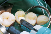 |
| Figure 9. Syringe a small amount of the flowable composite (Heliomolar Flow) into the preparation, but do not cure. | Figure 10. Syringe the heavy-bodied composite (Heliomolar HB) into the proximal box to create a hydraulic action, forcing the flowable composite into all margins and fine recesses to help achieve superior sealed cavosurface margins. |
The preparation was filled by first syringing a small amount of a flowable composite (Heliomolar Flow, Ivoclar/Vivadent) and spreading it around with a fine needle tip (Figure 9). Without curing the flowable composite, a heavy-bodied “packable” microfilled composite (Heliomolar HB) was syringed on top of the flowable, creating a hydraulic action within the cavity system created by the matrix (Figure 10). This forced the flowable resin into all margins and fine recesses of the cavity preparation in order to help achieve superior sealed margins at the cavosurface interface.
This new generation of “packable” composites demonstrates less shrinkage than traditional composites as a result of the higher percentage of filler, which increases viscosity and condensability, thus potentially contributing to the creation of a tighter contact. The increase in filler content should ultimately yield improvements in physical properties, such as occlusal wear.
 |
| Figure 11. To ensure tight proximal contact, plunge the Composite Contact Former (CCF) into the uncured packable composite. The instrument is used as a fulcrum off the axial wall against the proximal wall of the adjacent tooth, and exposed to a visible light source from the buccal and lingual aspect for 20 seconds. |
To ensure a tight interproximal contact, the Composite Contact Former (CCF) (American Eagle) was placed into the uncured composite, and the instrument was used as a fulcrum off the axial wall of the preparation, applying pressure to the proximal wall of the adjacent tooth (Figure 11). This action created a tight interproximal contact, positioned in the correct location. With the instrument held securely in place, the composite was light cured for 20 seconds through the buccal and lingual walls of the tooth using a visible light source (Astralis 7, Ivoclar/Vivadent) of at least 1,000 mw/cm2.
 |
 |
| Figure 12. Removal of the CCF shows a hollow was created when the proximal contact was formed. | Figure 13. Add a small increment of flowable resin and additional packable composite to fill the hollow. Cure for an additional 20 seconds from the buccal, lingual, and occlusal. |
Following polymerization, the CCF was removed to reveal a “hollow” space (Figure 12). This space was backfilled with a small increment of flowable resin, followed by another layer of the packable composite, and then contoured and condensed with a composite condensing and shaping instrument (P-1 Plugger, Ivoclar/Vivadent). The restoration was then cured for an additional 20 seconds from the buccal, lingual, and occlusal surfaces (Figure 13).
FINISHING AND POLISHING
 |
 |
 |
| Figures 14, 15, and 16. After initial shaping and contouring with carbide burs, final finishing and polishing are completed using a series of cups and points (Astropol) to create a long-lasting luster. |
 |
| Figure 17. The finished Heliomolar HB restoration. |
After removal of the retainer, wedge, and matrix, initial shaping and contouring of the composite were accomplished using 12-bladed spiral-shaped carbide burs in a variety of shapes (Axis Dental). Final finishing and polishing were completed using a series of cups and points, progressing from gray to green to rose (Astropol, Ivoclar/Vivadent) (Figures 14, 15, and 16). The highly polishable nature of the microfilled Heliomolar HB contributed to a final finish that will endure for many years in the oral cavity (Figure 17).
CONCLUSION
The heavy-body consistency of Heliomolar HB enables the dentist to maintain total control when manipulating the material, contributing to full and tight contacts. Its improved handling, polishability, and wear resistance represent significant advantages to clinicians that enable them to provide their patients with durable, easily placed aesthetic posterior restorations.
The “Recipe for Success” presented, which is based on a fundamental technique used in practice for several years, enables predictable placement of a direct composite specifically formulated for posterior restorations. To summarize, successful placement of this heavy-body posterior composite requires the clinician to:
•Prepare marginal enamel surfaces with a rotary diamond instrument.
•Secure an appropriate matrix.
•Clean and scrub surfaces with an antimicrobial agent.
•Total etch the enamel and dentin.
•Apply a fifth-generation dentin bonding agent.
•Syringe the composite materials into the preparation.
•Polymerize with sufficient light energy.
•Finish and polish in a time-efficient manner.
The “Recipe for Success” is the culmination of an evolutionary process of years working with direct resins to develop a technique that has resulted in negligible postoperative sensitivity and predictable, reproducible results.
Acknowledgment
The author would like to acknowledge Paul C. Belvedere, DDS (Edina, Minn) for his pioneering efforts in the field of composite dentistry and the numerous innovations he has brought to the profession, including the invention of the Contact Molar band and the Composite Contact Former.
References
1. Lambrechts P, Braem M, Vanherle G. Buonocore memorial lecture. Evaluation of clinical performance for posterior composite resins and dentin adhesives. Oper Dent. 1987;12:53-78.
2. Dietschi D. Composite resins: the transition from traditional to modern dentistry. Pract Periodontics Aesthet Dent. 1996;8:600-601.
3. Hoang A, Koh S, Bebermeyer R, et al. A review of condensable composite. J Gt Houst Dent Soc. 1999;71:15-17.
4. Talib R. Dental composites: a review. Journal of Nihon University School of Dentistry. (Matsudo, Japan.) 1993;35:161-170.
5. Freedman G. Condensable composites: the new paradigm in amalgam alternatives. Dent Today. 1998;17:72-74.
6. Behle C. Flowable composites: properties and applications. Pract Periodontics Aesthet Dent. 1998;10:347, 350-351.
7. Summitt JB, Della BA, Burgess JO. The strength of class II composite restorations as affected by the preparation design. Quintessence Int. 1994; 25; 251-257.
8. Simonsen RJ. Conservation of tooth structure in restorative dentistry. Quintessence Int. 1985;16:15-24.
9. Joynt RB, Davis EL, Schreier PH. Rubber dam usage among practicing dentists. Oper Dent. 1989;14:176-181.
10. Marshall K, Page J. The use of rubber dam in the United Kingdom: a survey. Br Dent J. 1990;169:286-291.
11. Gergely EJ. Rubber dam acceptance. Br Dent J. 1989;167:249-252.
12. Opdam NJ, Roeters JJ, Kuijis R, et al. Necessity of bevels for box only class II composite restorations. J Prosthet Dent. 1998;80:274-279.
13. Nebot D, Goldberg M, Fortier JP, et al. Bonding and enamel prisms. Importance of cavity margin prep for posterior composites. Actual Odontostomatol. 1989;43:609-618.
14. Belvedere PC, Lambert DL. Resin-bonded margins in direct posterior composites. Dent Today. 1997;16:64, 68-71.
15. Cvitko E, Denehy G, Boyer DB. Effect of matrix systems and polymerization techniques on microleakage of class II resin composite restorations. Am J Dent. 1992;5:321-323.
16. Belvedere PC. Controlling shrinkage using TEP: trans-enamel polymerization. Dent Today. 1995;14:92, 94-97.
Dr. Lambert maintains a private practice in Edina, Minn, emphasizing aesthetic and sports dentistry. Besides being involved with numerous dental organizations, he is a fellow of the American College of Dentists and the Pierre Fauchard Academy, a board member for the Academy for Sports Dentistry, and an accredited member of the American Society for Dental Aesthetics. An independent consultant, evaluator, and researcher for various dental product manufacturers, Dr. Lambert has presented numerous lectures and hands-on seminars in the United States, Europe, and Latin America for universities, dental societies, and study clubs. He can be reached at (952) 922-9119 or ddssmile@aol.com.
Disclosure: The author has presented lectures that were cosponsored by Ivoclar and DENTSPLY Caulk.


