This article describes treatment for 2 periodontally and endodontically involved mandibular central incisors, using the eventually extracted incisors as a pontic section in a bonded, lingually retained prosthesis. The materials, technique, and rationale for providing this treatment are discussed.
CASE REPORT
 |
| Figure 1. Pre-op radiograph. |
The patient, a 28-year-old teacher, first appeared on an emergency basis due to swelling in his chin region and mobility of his lower central incisors. A periapical radiograph (Figure 1) revealed considerable bone loss around the 2 incisors. Antibiotics were prescribed at that visit, which provided temporary relief. Endodontic and periodontic referrals were made, and both specialists felt that any treatment they rendered would not be successful. With the considerable amount of bony destruction evident in the area, it was felt that post-extraction healing of the lesions would result in a probable deformed anatomical ridge structure, which would not be conducive to titanium implants. Thus, implants and ridge augmentation were considered an “iffy” option. Cost would also be a factor, and consequently, that option was eliminated.
 |
 |
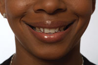
| Figure 2. Pre-op facial view. | Figure 3. Periodontal probe showing pocket depth. |
There was a greater than 12-mm pocket on the lower right central incisor (Figures 2 and 3). Both arches were intact, and there was no mobility evident on either the mandibular lateral incisors or mandibular cuspids. Pocket depths around those teeth were considered within accepted “normal” limits.
 |
| Figure 4. Preparation of teeth. |
After discussing the treatment options, it was decided to pursue a fixed bonded resin-retained bridge (Maryland bridge-type design) utilizing the existing teeth as the pontic section for the bridge. Due to the poor anatomical root structure that lateral incisors possess, the abutment preparations were extended to include the more preferable anatomical root structure and length provided by the cuspids. To increase the surface area for retention and “cradle” the teeth involved, vertical lingual grooves and horizontal cingulum notches were paralleled for all teeth, including the hopeless central incisors, in preparation for the bonded lingual retainer that would extend between both cuspids (Figure 4).
 |
| Figure 5. Casting. |
Rexillium III (Pentron), in spite of its nickel and beryllium content, was selected as the metal of choice for the prosthesis due to its rigidity in thin casting situations, its history of success in the literature (with articles dating back to Thompson and Leviditis), as well as previous positive results by the operating dentist (Figure 5). A hydrocolloid impression was taken, a model poured, and parallelism of the vertical lingual grooves was confirmed with a surveyor prior to being sent to the dental laboratory (Professional Dental Arts). The outline of the prosthesis was marked on the models, incorporating as much of the anatomical coronal lingual surface as possible but ending approximately 1 mm below the lingual/incisal line angle both to enhance light transmission in that area and to hide the prosthesis from a normal line of sight.
Upon return of the prosthesis from the laboratory, a tight fit of the prosthesis was confirmed. The prosthesis snapped into place, and even though incisal hooks were requested to assist in correct placement of the fixed bridgework, it was evident that dislodgment of the prosthesis would require some effort even without cement. The lower central incisors were extracted as atraumatically as possible, and the sockets were gently curetted.
The root surfaces of the extracted central incisors were curetted to remove any remnants of the periodontal ligament and scaled to eliminate calculus accretions on both teeth. The roots were then horizontally sectioned at an estimated subgingival level to compensate for the eventual post-extraction bony shrinkage of the alveolar ridge in this region. Both the extracted teeth and the splint were sandblasted (Micro-etcher, Danville Manufacturing) on their lingual surface, and the teeth were etched in 2 applications for 2 minutes total time with 37% phosphoric acid according to the manufacturer’s instructions.
 |
 |
| Figure 6. Teeth sectioned and bonded in place. | Figure 7. Restoration in place, sealing the ends of the teeth. |
The teeth were bonded to the metal using Panavia 21’s opaque resin cement (Kuraray), and the resultant air-inhibited layer was “sealed” with Oxyguard (Kuraray, Figure 6). This now became the pontic section of the bridge/splint. Next, the pulp chamber on tooth No. 25 was broached and debrided from the apical end. Both teeth received apical preparations for bonded composite restorations to seal off the apical end of the roots using Optibond FL (Kerr) and Tetric Flow (Ivoclar Vivadent), even though the root canal on the left central incisor had calcified (Figure 7).
 |
 |
| Figure 8. Facial view with rubber dam in place. | Figure 9. Lingual view with rubber dam in place. |
The incisal hooks used to help position these 2 teeth as pontics on the prosthesis were then ground off, and a rubber dam was placed and inverted into the gingival sulci to isolate the abutment teeth, thus providing predictability of the bonding procedure (Figures 8 and 9). Once isolated, the lateral incisors, cuspids, and splint were sandblasted, and the teeth were etched and bonded according to the manufacturer’s instructions, similar to what had been done previously with the pontic section. The incisal hooks used to help position the bridge were severed from the cuspids. Excess cement was removed from the margins of the prosthesis with special emphasis directed to the gingival interproximal areas to avoid any gingival irritation from residual cement. Finally, the rubber dam was sectioned and removed; the bite was checked for any excessive contacts that might have occurred from any tooth movement during the procedure.
 |
 |
| Figure 10. Postoperative labial view at 24 hours. | Figure 11. Postoperative lingual view at 24 hours. |
Several points concerning this case need to be highlighted. The choice of restoration was far less expensive than constructing a 6-unit splint with porcelain-fused-to-metal pontics. As good a technician as the CDT is, even he or she would have had a difficult time fabricating pontics with the anatomical shape and internal shading nuances that the patient’s own natural teeth possessed. In addition, the patient avoided the occasional embarrassment of “missing” anterior teeth that a removable partial denture dictates. Lastly, psychologically, the patient must realize that he has kept at least part of his own teeth by doing this procedure. Unless the patient and his teeth are observed from a decidedly superior position, the lingual metal retainers are not visible at normal conversational distances and angles (Figures 10 and 11). The author has done several restorations of this nature, all without debonding to date.
Dr. Heher served 3 years active duty in the US Navy and has been in private practice emphasizing aesthetic and restorative dentistry for more than 32 years in Salisbury, Md. He can be reached at (410) 749-0320.
To comment on this article, visit the discussion board at dentistrytoday.com.