Rx for the Durable Partial Denture: Proper Maintenance Plan

Dentistry Today

0 Shares

After a new partial denture is delivered, educating the patient regarding what is required to maintain the partial and preserve the health of the remaining oral structures is very important. Prescribing a maintenance plan and explaining to the patient the effect that time has on the partial and the surrounding periodontium will commit the patient to come in for scheduled checkups. After the newly constructed partial is delivered to the patient, it will undergo a process of adaptation for a few weeks. It is quite normal for the partial to appear slightly tight during the delivery. In a short time, the clasps will alter the position of the abutments as the saddles fully adapt to the ridge. As a consequence, with bilateral free-end saddles, the lingual bar may drift closer to the tissue and the clasps will loosen. In the patient’s terms, this is called “getting used to.”
With properly constructed partials, the remedy for “getting used to” is quite simple. Tightening the clasps will restore the retention, and positioning the lingual bar slightly away from the tissue will prevent its contact with the tissue. The effects of not maintaining the partial are much different. Therefore, it is important to understand how changes take place so a proper maintenance plan can be prescribed.

MAINTENANCE PROBLEMS

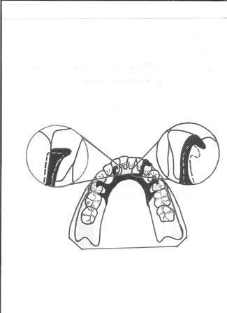
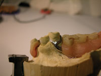
Figure 1. Cingulum or incisal rests drift away from the teeth, resulting in gaps.

A properly constructed partial derives support from the ridge or saddle area and the abutment teeth or clasps and attachments. As the ridge resorbs, the saddle area support decreases, resulting in increasing stress on the abutment teeth, or clasps and attachments. If the balance is not restored, damaging effects will result. As the ridge resorbs, the clasps begin to move the abutment teeth, causing portions of the saddle base to readapt to different areas of the ridge. Likewise, this movement directs the major connector toward the tissue. If the major connector is a lingual bar, then it will begin to press against the tissue. If the major connector is a double bar or lingual plate, then the metal will drift away from the incisors while the lower portion of the connector will impinge upon the tissue. As seen in Figure 1, the cingulum or incisal rests placed for indirect retention will also drift away from the teeth, resulting in constantly increasing gaps.
When a bridge supports a free-end partial, the sturdiness of the bridge will not allow the saddle base to readapt to different areas of the ridge. This places severe stress on the clasps or attachments, causing them to break. After a clasp or attachment that supported a free-end saddle breaks, the patient cannot continue wearing the broken partial. As soon as a clasp or an attachment that supports a free-end saddle breaks, the rotational force on the other quadrant’s abutments will cause the clasp or attachment to break as well.
When individual abutments provide the retention, the rotational force on the other quadrant will orthodontically move the clasped abutments, and the existing partial will develop a permanent poor fit. For patients who decide to have the partial replaced, but for aesthetic reasons cannot give up the broken partial, before the impression is taken for the new partial a properly sized wire arm must be added to the partial to prevent further distalizing of the unsupported saddle.
I have personally experienced a situation where, after remaking the framework 3 times, the doctor (for whom we never had a remake) still could not get the new frame to fit the mouth. To look for answers, I began to mix and match the failed frameworks to the first, second, and third model. I discovered that the abutment teeth on one side were positioned slightly differently on every model. After consulting the doctor, I found out that the patient was wearing a partial with a missing clasp on the side of a free-end extension. Between the time the impression was taken and the framework constructed, the orthodontic movement on the other quadrant’s clasped abutments was significant enough for the new framework not to fit in the patient’s mouth.

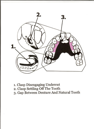
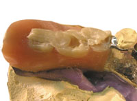
Figure 2. If a partial denture is not maintained, it may become unsalvageable.

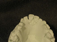
Figure 3. A partial denture case that experienced settling due to loss of ridge support.

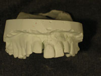
Figure 4. This neglected partial denture is beyond repair.

As can be seen in Figure 2, if the partial is not maintained, after a certain point it becomes unsalvageable. After the ridge support lessens and the partial begins to settle, as illustrated by No. 1 in Figure 2, the clasps on the most distal abutments begin to disengage the undercuts, and spaces between the rests and the teeth develop. Illustrated by No. 2 in Figure 2, the clasp on the central incisor begins to settle downward and becomes non-retentive. If the doctor attempts to restore the retention by adjusting the clasp, the severity of the undercut will prevent the partial from seating, or after a painful snap, from being removed from the mouth. Illustrated by No. 3 in Figure 2, the cingulum rests will settle off both centrals, and a diastema and a drop between the right central incisor and the denture tooth will develop. Figure 3 shows a model of an actual case that has experienced such settling. Since the I bar clasp on the right central has settled off the tooth and was impinging on the tissue, the doctor removed it. As seen in Figure 4, this patient has neglected the partial for too long, and repairing it was no longer an option.

Figure 5. This lower partial denture (model) was neglected and needed relining for years. The patient could no longer wear it due to impingement on the tissue.

Figure 6. Occlusal view of partial denture (model) shown in Figure 5.

Seen in Figure 5 is a lower partial that has experienced similar neglect. By looking at the space underneath the anterior saddle area, one can see that this partial needed a reline for years. As a result, the bar clasp and the lower portion of the major connector started impinging on the tissue until it became unbearable for the patient to continue wearing the partial. Figure 6 shows the occlusal view of the same partial. The settling caused a portion of the rest to break, and the denture teeth lost contact with the opposing teeth. Someone previously placed self-curing acrylic on top of the denture teeth to restore contact. To save money, this patient asked the doctor to replace the denture teeth and acrylic and use the same framework. Using a frame that impinges on the tissue is throwing away good money after bad. After such a procedure is completed and after numerous, time-consuming adjustments, the patient may tell the doctor that there was nothing wrong with the old partial that a simple adjustment could not fix. Now a partial that served him well for the past 20 years is ruined. Therefore, it is helpful for the doctor to have these types of photos on hand, which can be used to convince the patient that a new partial is in order.
Having photos available to show not only your clinical accomplishments, but also problematic cases that relate to the patient, can persuade the patient to make the right decision.

MAINTENANCE PLAN

After the finished partial is delivered to the patient and the final adjustment is made, a personalized maintenance plan for the patient needs to be prescribed. As part of a 6-month office visit, changes in the ridge should be monitored. This is best accomplished by examining the following:

Contacts Between the Denture and Opposing Teeth

Contacts between the denture teeth and the opposing teeth are best determined by placing articulating paper no thicker than 0.0008 of an inch, or 20 µm, between the opposing quadrants and pulling the paper with the teeth in centric. Every tooth should catch the paper. If it is determined that the contacts have loosened, it is a sign that the edentulous ridge is beginning to change.

Space Underneath the Saddle Base and Ridge

Every 6 months after the contacts between the opposing teeth are determined, without using adhesives impression material should be placed inside the saddles and the patient instructed to bite fairly hard in centric relation. The amount of impression material inside the saddles will show the amount of change that has taken place. If the material is very thin or nonexisting inside the saddles, the partial is fine until the next office visit. If the impression material shows up thicker in some areas and thinner in others, the saddles need to be relined.
During this procedure it is important that the patient bites into centric relation correctly and that the teeth in the partial have good contacts with the opposing teeth. No contact with the opposing teeth or the wrong centric relation will prevent the partial from fully seating and result in false thickness of the impression material.
I have seen instances where patients, by brushing their teeth with a hard toothbrush while wearing the partial, have worn off the buccal, labial, lingual, and occlusal surfaces of the denture teeth. The wear on the occlusal surfaces of the denture teeth eliminated all contacts with the opposing teeth. The lack of opposing contact will prevent the partial from seating fully, causing the impression material to be falsely thicker. If this is not recognized and the saddles are relined, then many other problems will develop.

Retention of the Clasps

Retention of the clasps can also be linked to ridge changes. After the partial is relined, tightening the clasps should restore the retention. Upon delivery of an immediate partial, the patient should be instructed to avoid applying pressure to the area of the extraction. In addition, the patient should be instructed to come in for a reline as soon as the extraction site heals. Chairside relines can be done quickly and efficiently until the time that a final and more permanent reline is possible.
A prescribed maintenance plan for partial dentures places a major responsibility for properly maintaining the prosthesis with the patient. If the patient fails to follow the prescribed maintenance plan and years later comes in with the partial showing gaps between the abutment and denture teeth, or broken parts on the partial, the dentist can honestly say that the responsibility for these problems rests with the patient. And if a patient insists on having an unrepairable partial repaired, it is best to refuse to do such inferior work and insist on providing quality dentistry.


Mr. Saunaitis is a former dental laboratory owner, educator, publisher, and researcher. With more than 25 years of experience, he has developed techniques that better integrate crowns, bridges, and implants with the removable sector of dental technology. He has been published in various dental journals and newspapers and has appeared on radio and television. He has combined much of his research into a full-day slide presentation entitled “Forum for Prosthetic Dentistry.” To be notified about upcoming events or to reach the author, e-mail tsaunaitis@msn.com