


INTRODUCTION
Intraoral viral lesions are a common occurrence in the dental practice and may be of herpes simplex virus (HSV) or human papillomavirus (HPV) origin. Treatment has traditionally been palliative in nature, using chemotherapeutic agents (antiviral) either topically applied or systemically administered. The literature has reported use of the diode laser as an alterative or adjunctive treatment modality in the treatment of viral lesions intraorally to provide both pain management and speed lesion resolution.
Oral viral lesions may be found intraorally on freely movable tissue (buccal mucosa, tongue, floor of the mouth) or on attached gingiva and also extraorally on the lips. Lasers have been documented as an alternative or adjunctive treatment to both decrease lesion duration and speed healing as well as provide improved patient comfort during lesion outbreak. Diode lasers have been used 2 different ways with regard to viral lesion treatment, used in both contact and non-contact modes, with success being reported in the literature.
Studies have supported the benefit of low-level laser therapy (LLLT) in decreasing pain related to the lesions and improving lesion healing in comparison to other treatment modalities being utilized. The mechanism of action that has been theorized is that laser irradiation “acts in the final stage of HSV-1 replication by limiting viral spread from cell to cell and that laser therapy acts also on the host immune response unblocking the suppression of pro-inflammatory mediators induced by accumulation of progeny virus in infected epithelial cells.”1 LLLT has been reported clinically as longer-lasting viral suppression therapy. Additionally, LLLT study results utilizing the diode laser reported a delay in reoccurrence of herpetic lesion outbreaks, with the median recurrence-free interval of 37.5 weeks in the laser-treated group and 3 weeks in the placebo group. LLLT has been reported with an immediate outcome, providing improved patient comfort.
Recurrent aphthous stomatitis (RAS) is a multifactor immunologic inflammatory condition of the oral cavity characterized by painful, recurrent single or multiple lesions that are shallow, round, or ovoid ulcerations of the mucosal tissues. The assessed literature demonstrates the benefits of laser therapy mainly due to immediate analgesia and ability to speed up an RAS healing process. These lesions when biopsied frequently, demonstrate a virus present in the lesion. The virus may be the primary etiological factor or secondary to bacterial initiation of the lesions. But, as with HSV, these lesions are painful and have demonstrated improvement when treated with LLLT.
LLLT is utilized in a non-contact mode with an uninitiated tip. Low power of less then 1 W, is used and treatment is geared to energy absorption in the tissue to exert its effects. The diode laser tip is defocused 5.0 to 8.0 mm from the lesion and advanced slowly toward the area ending 2.0 to 3.0 mm away from the lesion. Continual movement is done over the lesion from the periphery to the center moving away from the lesion if the patient feels warmth. The setting is initially put at 0.6 W continuous or 1.2 W pulsed for 30 to 45 seconds and utilized with a refractory period of 15 to 20 seconds between reapplication of the laser on the same lesion allowing the tissue to cool down. Treatment is performed without the need for local anesthetic. A second and third pass with the laser is recommended to decrease the pain associated with the lesion. After each subsequent pass, the power is increased, with the second pass performed at 0.7 W continuous (or 1.4 W pulsed) and a third pass at 0.8 W continuous (or 1.6 W pulsed).
Diode lasers have also been utilized for lesion eradication via direct contact with the diode laser tip. Therapeutic efficacy of laser therapy in treating oral HPV lesions has been reported in the literature. This appears to be applicable to other viral oral lesions such as HSV. Lesions that were detected were excised by a diode laser using an average power of 2.1 W in a continuous wave mode with an initiated tip. Complete lesion healing was observed in most cases (95.4%). No adverse effects were reported. The diode laser is a valuable tool for viral lesion eradication, but it also reduces relapses.
Today, the diode laser is widely used in oral pathology to excise lesions. Excision of a lesion to allow histological diagnosis of the specimen is possible as an alternative to excision with a scalpel blade. Specimens larger than 3.0 mm excised with the diode laser were not significant in terms of stromal changes or vascular stasis that could affect histology of the specimen. While epithelial and stromal changes were significantly more frequent in specimens with a size below 3.0 mm, the histological diagnosis was not achievable in 46.15% of them. Data confirms that the diode laser is a valid therapeutic instrument for excising oral lesions larger than 3.0 mm in diameter, but induces serious thermal effects to the specimen in small lesions (below 3.0 mm). From a clinical standpoint, it is suggested that specimens taken have a diameter of at least 5.0 mm in order to have a reliable reading of the histological sample.
The use of diode lasers for viral vaporization of the lesion in urological applications for genital warts caused by HPV has been reported. This can also be applied to treatment of viral lesions intraorally. No incidences have been reported of postoperative infection or complications from the laser diode vaporization of viral lesions.
Laser diode vaporization can be considered as an alternative method for treating lesions, with satisfactory results in terms of pain and aesthetics and minimal recurrence of the lesions. When studied 20 days following laser excision of the lesion, the virus load of the area where the lesion was located was greatly reduced.2 This appears to indicate that use of the diode laser in excision of viral lesions may prevent recurrence of the lesion by eradication of the residual virus that would not occur with other excision methods. Several advantages the diode laser excision of these viral lesions include improved hemostasis, lack of the need for sutures, wound sterilization, and minimal post-op pain and edema.
CASE REPORT
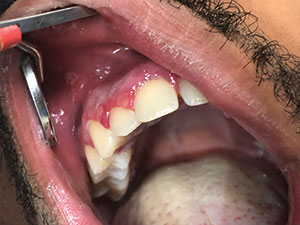
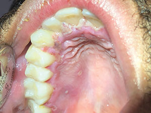
A 32-year-old male presented with the chief complaint of generalized oral pain of 2 days duration on the maxillary arch. A review of the medical history found no current or past medical conditions reported. The patient described the sensation on the gingival tissue on the buccal and palatal aspects of the arch as a burning sensation that was constant and hampering daily activities such as sleeping, eating, and speaking. Examination noted an ulcerated multiple area on the buccal mucosa confined to the attached gingiva and papilla (Figure 1); it did not cross the midline on the facial (Figure 2). Further examination of the palatal aspect of the maxilla noted lesions predominately on the right side of the arch (Figure 3) with ulceration of the papilla and viral-like lesions on the hard palate extending to the tuberosity. The diagnosis, based on clinical appearance and patient-reported symptoms, was HSV outbreak. The patient was informed of the clinical findings, diagnosis, and recommendation to use a diode laser to aid in lesion healing as well as manage the pain associated with the viral outbreak.
 |
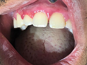 |
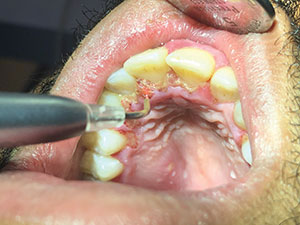
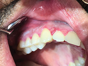
| Figure 1. Initial presentation of inflammation of the buccal-attached gingiva and papilla with multiple noted viral lesions. | Figure 2. The initial viral lesions and associated inflammation appeared to not cross the facial midline. |
 |
 |
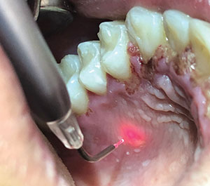
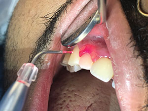
| Figure 3. The palatal viral lesions on the right side of the papilla and severe ulceration of the palatal aspect of the papillae with generalized inflammation over the soft tissue. | Figure 4. The diode laser tip is held several millimeters away from the affected soft tissue (non-contact) at 0.7 W to precondition the tissue. |
 |
 |
| Figure 5. Preconditioning continues on the affected tissue on the palatal aspect of the arch. | Figure 6. The diode was used in non-contact with a non-initiated tip on the more severely ulcerated lesions requiring greater wattage. |
 |
 |
| Figure 7. The diode was used in non-contact at increasing power (higher wattage) on the affected areas of soft tissue. | Figure 8. At 24 hours post-laser treatment, the patient presented for evaluation, and less inflammation was noted, along with the absence of previously felt pain and some resolution of the ulcers. |
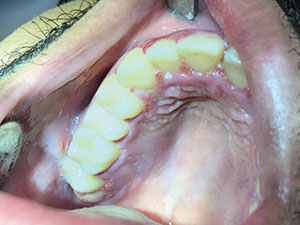 |
| Figure 9. At 24 hours post-laser treatment, the palatal tissue demonstrated significant improvement, and ulcerations on the papilla greatly improved. |
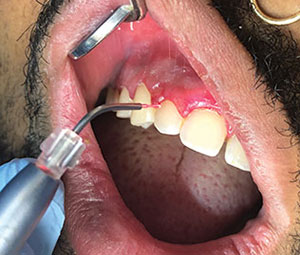
A diode laser (Epic X [BIOLASE]) was set at 0.7 W in continuous wave (aphthous ulcer mode), and, in a non-contact manner, the tip was moved over the affected areas, beginning on the buccal aspect of the arch to condition the tissue (Figure 4). The tip was kept in motion and held several millimeters away from contact with the tissue. In addition, high-speed suction was held near it to help cool the tissue being treated. Preconditioning was continued on the palatal affected soft tissue (Figure 5). Severely ulcerated areas, as found on the palatal papilla of this patient, may be treated by non-contact of the tissue with a non-initiated diode tip at greater power (wattage) while keeping the tip in motion so that tissue is not further damaged (Figure 6). The power was increased to 1.3 W, and the process was repeated on all affected areas (Figure 7). This was again repeated at 1.5 W and, finally, 1.9 W to complete treatment of the viral lesions and of the adjacent soft tissue.
It is important that the tip be kept in motion so that energy will not be concentrated and generating heat that may burn the tissue being treated. Depending on the tissue (the amount of keratinization) and tissue location, higher power can be tolerated. Attached gingival and palatal tissue will tolerate higher wattage than the more delicate tissue of the lip. Since this treatment protocol is performed without anesthesia, as power is increased with each pass, feedback from the patient aids the practitioner in use of maximal wattage and distance between the tip and tissue. The patient should feel some warmth in the area as the diode tip is used in a non-contact manner; however, he or she should not feel any burning sensation. Should that sensation start to be felt, the tip can be moved 2.0 to 3.0 mm further away to defocus the beam so the increase of power on that tissue can be avoided.
The patient was instructed to return the next day for evaluation and possibly a repeat of the diode laser treatment for his viral lesions. When the patient presented the following day, he stated that all his pain had resolved and the burning sensation that had been present for several days and up until laser treatment was also gone. Normal activities, including eating, had resumed without discomfort. Upon examination, it was noted that there was some resolution of the viral lesions and that they did not appear as inflamed or ulcerated (Figure 8). The ulcerated palatal papilla had demonstrated significant healing (Figure 9). The patient returned once again at 1 week post-treatment, and he stated that the previous pain and burning sensation had not returned and his mouth continued to feel “normal.” Upon visual examination, it was noted that the affected areas were reduced in size, and this corresponded with the patient’s indications that the resolution of any pain and burning sensation from the viral outbreak was proceeding.
CLOSING COMMENTS
Viral oral outbreaks are painful experiences for the patient. Historically, before the advent of diode laser technology and the clinical techniques described herein, treatment of such outbreaks has been palliative in nature with patients having to deal with the discomfort while the virus ran its course over the subsequent 7 to 10 days. Diode lasers have demonstrated a useful treatment in pain management and accelerating healing of the viral lesions. Treatment often is accomplished in a single visit, but may be supplemented, as needed, 24 hours after the initial visit with repetition of the diode laser treatment if pain remains.
References:
- Donnarumma G, De Gregorio V, Fusco A, et al. Inhibition of HSV-1 replication of laser diode-irradiation: possible mechanism of action. Int J Immunopathol Pharmacol. 2010;23:1167-1176.
- Baeder FM, Santos MT, Pelino JE, et al. High-power diode laser versus electrocautery surgery on human papillomavirus lesion treatment. J Craniofac Surg. 2012;23:702-705.
Editor’s Note: This is an abridged version of this article. The full-length, fully referenced article will be available for a 2-hour FAGD/MAGD-credit-hour course at dentalcetoday.com in September 2019.
Dr. Javid graduated from the University of Southern California (USC) and received his DDS from USC School of Dentistry. He served on the faculty of USC in the advanced education in general dentistry program before opening his private practice, Doctor Smile Dental Group. Dr. Javid is a Diplomate of the International Congress of Oral Implantologists, a Fellow of the California Implant Institute, an Associate Fellow of the World Congress Laser Institute, and a clinical phlobotomy technician. He can be reached at onecure@aol.com.
Dr. Kurtzman is in private general practice in Silver Spring, Md. A former assistant clinical professor at the University of Maryland, he has earned Fellowships in the AGD, the American Academy of Implant Prosthodontics, the American College of Dentists, the International Congress of Oral Implantologists, the Pierre Fauchard Academy, and the Association of Dental Implantology; Masterships in the AGD and ICOI; and Diplomate status in the ICOI and the American Dental Implant Association. He has lectured internationally, and his articles have been published worldwide. He has been listed as one of Dentistry Today’s Leaders in Continuing Education since 2006. He can be reached at dr_kurtzman@maryland-implants.com.
Disclosure: The authors report no disclosures.
Related Articles
Know the Risks Before Adding Phlebotomy to Your Practice
6-Part Series Explores the Evolution of Comprehensive Care
A Guidance System for the Partially Edentulous Arch


