
INTRODUCTION
A 2010 review concluded that the failure rate of PFM fixed partial dentures (FPDs) due to veneering porcelain fracture was 3% after 3 years of service.1 Although the reported incidence of veneering porcelain fracture is low, when it occurs in a long-span FPD, the consequences can be tolling for both the dentist and the patient. If the dentist chooses to replace the FPD, the patient must undergo the financial burden of paying for the replacement, the time required for multiple appointments, and the possible disappointment if the replacement is less satisfactory than the original FPD. The dentist will face the challenge of removing the previous FPD without damaging the underlying tooth structure or causing a pulpal response. Therefore, many clinicians would choose to repair the fractured veneering porcelain rather than replace the FPD.
One method of repairing a fractured PFM restoration is with composite resin. The method of composite resin bonding will be dependent on the location of the porcelain fracture.2 If the fracture occurs without exposure of the metal core, the porcelain should be etched with hydrofluoric acid and treated with a coat of silane prior to applying a bonding agent and a composite resin. If the fracture exposes the metal core and the veneering porcelain, the dentist will have 2 surfaces to bond. In this case, the metal surface should be roughened with silica-coated Al2O3 particle abrasion (CoJet Sand [3M] in a handheld microblaster CoJet Prep [3M]) prior to application of silane. These particles can roughen the metal surface to increase the bonding area while depositing silica in the metal to allow chemical bonding with silane.
When replacing veneering porcelain with a direct composite, the success of the repair is predominantly dependent on preventing the composite from chipping or wearing. A recent study reported higher fatigue resistance of resin composites than a veneering porcelain subjected to repeated masticatory simulating forces.3 These results indicate that it is less likely that the composite material will fracture than the original veneering porcelain. However, the weakest link of a composite repair will be the bond between the composite resin and the surface of the FPD. Therefore, a composite resin repair will often fail due to adhesive fracture along the bonded surface.
Aside from the mechanical properties of the composite repair, the optical properties of the composite resin should be considered, as these may limit the aesthetic outcome of the repair. A recent study reported higher gloss and stain resistance of a polished feldspathic porcelain material than a polished nanofilled composite resin.4 These results indicate that it may be difficult or impossible for the repair composite resin to match the polish of the surrounding porcelain. Throughout time, the composite resin repair will also be more likely to stain than remaining porcelain. Another aesthetic challenge with composite repair is choosing a material with enough translucency to match the surrounding porcelain but sufficient opacity to cover any exposed metal.
With the advent of high-strength ceramics such as zirconia, dentists have another option for the repair of PFM FPDs. Zirconia has 4 times the reported fracture toughness of a composite resin and more than 6 times the fracture toughness of leucite-reinforced porcelain material.5 Wear of zirconia is minimal, and it is more wear-friendly to opposing enamel than veneering porcelain.6 In addition, recent studies have reported methods for achieving a chemical bond to zirconia.7
This patient case study describes the clinical steps for repairing a fractured FPD with a bonded zirconia onlay.
CASE REPORT
Diagnosis and Treatment Planning
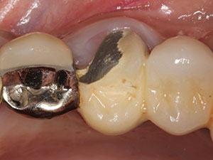
A 45-year-old female patient presented to the comprehensive care clinic at from University of Alabama at Birmingham School of Dentistry with the chief complaint of, “my bridge chipped.” Full-mouth radiographs and a panoramic radiograph were taken and intra- and extraoral examinations were done. The intraoral examination showed a long-span PFM FPD from teeth Nos. 10 to 15 with abutment teeth Nos. 10, 11, 13, and 15 and a fracture of the veneering porcelain on the buccal and occlusal surface of crown No. 13 (Figure 1). The fit of the margin over the abutments was considered clinically acceptable. Treatment plan options were provided to patient, including replacement of the FPD, composite resin repair, and repair with a bonded zirconia onlay. For financial reasons, the patient was only interested in fixing the chipped porcelain on abutment No. 13. After discussing the advantages of each treatment option with the patient, she opted to fix the chipped porcelain over the PFM abutment No. 13 by using a monolithic zirconia onlay.
 |
 |
| Figure 1. Initial presentation of a fractured crown on tooth No. 13. | Figure 2. Buccal preparation of tooth No. 13. |
 |
 |
| Figure 3. Lingual preparation of tooth No. 13. | Figure 4. Application of the universal adhesive (Scotchbond Universal Adhesive [3M]) to the metal coping. |
Clinical Protocol
At the first appointment, the existing porcelain was removed carefully from the tooth No. 13 abutment with a medium diamond (856.31.014 [Brasseler USA]) until the metal framework was exposed. A light 0.5-mm chamfer margin was prepared on the metal framework at the facial and lingual margins and at the junction between the neighboring teeth (Figures 2 and 3). A minimal 1.0-mm occlusal clearance was ensured between tooth No. 13 and the opposing tooth (No. 20). All line angles were smoothed, and retraction cord was used to retract the gingival tissue from the margin. A final impression was taking using a vinyl polysiloxane impression material (Aquasil Ultra Monophase and XLV [Dentsply Sirona Restorative]) and the impression was then poured with Type V dental stone (Jade Stone [Whip Mix]). The maxillary master cast and the mandibular cast were mounted on a semi-adjustable articulator (model 2340 [Whip Mix]), and a laboratory prescription for a polished (unglazed) monolithic zirconia onlay was sent to the laboratory team.
When the restoration returned from the lab, marginal fit of the onlay was ensured intraorally using a fit checker (FIT CHECKER ADVANCED [GC America]). Adjustments were performed on the metal coping until the fit of the onlay was acceptable. The exposed metal coping was cleaned with a mixture of plain flour pumice and distilled water, rinsed, and then air-dried.
The zirconia onlay was bonded to the metal coping using a dual-cure resin cement (RelyX Ultimate [3M]). In order to create a bond between the metal coping and the resin cement, a universal adhesive (Scotchbond Universal Adhesive [3M]) containing the monomer 10-methacryloyloxydecyl dihydrogen phosphate (10-MDP) was applied to the surface of the metal coping (Figure 4). The 10-MDP is claimed to bond to metals by the manufacturer with some evidence in the literature.8,9 Scotchbond Universal is an 8% by weight filled adhesive; therefore, it was thoroughly air dispersed in order to prevent film thickness at the margins. The universal adhesive was then light-cured for 10 seconds with an LED curing light (Bluephase G2 LED Curing Light [Ivoclar Vivadent]) (irradiance = 1,100 mW/cm2).
 |
 |
| Figure 5. Alumina (Al2O3) particle abrasion of the zirconia onlay. | Figure 6. Application of the universal adhesive to the zirconia onlay. |
 |
 |
| Figure 7. Bonded zirconia onlay from buccal. | Figure 8. Bonded zirconia onlay from lingual. |
The zirconia onlay was particle abraded with plain Al2O3 since this treatment has been shown to remove salivary contamination and increase surface area for bonding (Figure 5).10,11 The universal adhesive was then applied to the internal aspect of the onlay (Figure 6) and air-evaporated. The resin cement (RelyX Ultimate [3M]) was applied to the internal aspect of the onlay and it was seated on the metal coping. Gross excess cement was removed with a microbrush and the margins were tack-cured for one to 2 seconds each. Any remaining cement was removed with a scaler while an assistant applied heavy pressure to occlusal surface of the onlay. The final light-curing was done for 20 seconds per surface.
Occlusal contact was verified and lateral excursive interferences were removed using a zirconia adjustment bur (8369DF.31.025 [Brasseler USA]). The zirconia was then polished sequentially with medium and fine polishers (Dialite ZR [Brasseler USA]). The bonded restorations immediately following cementation can be seen in Figures 7 and 8.
CLOSING COMMENTS
A bonded zirconia onlay repair of a fractured PFM FPD provides aesthetic and mechanical advantages compared to a more traditional bonded composite resin repair. Using a repair technique, such as the one described in the presented case report, saves the patient and the clinician the hassle of replacing the entire FPD. It should be noted that the long-term clinical performance of this type of repair has yet to be determined.
References
- Heintze SD, Rousson V. Survival of zirconia- and metal-supported fixed dental prostheses: a systematic review. Int J Prosthodont. 2010;23:493-502.
- Özcan M. How to repair ceramic chipping or fracture in metal-ceramic fixed dental prostheses intraorally: step-by-step procedures. J Adhes Dent. 2014;16:491-492.
- Belli R, Geinzer E, Muschweck A, et al. Mechanical fatigue degradation of ceramics versus resin composites for dental restorations. Dent Mater. 2014;30:424-432.
- Lawson NC, Burgess JO. Gloss and stain resistance of ceramic-polymer CAD/CAM restorative blocks. J Esthet Restor Dent. 2015 Jun 1. [Epub ahead of print]
- Dupriez ND, von Koeckritz AK, Kunzelmann KH. A comparative study of sliding wear of nonmetallic dental restorative materials with emphasis on micromechanical wear mechanisms. J Biomed Mater Res B Appl Biomater. 2015;103:925-934.
- Mundhe K, Jain V, Pruthi G, et al. Clinical study to evaluate the wear of natural enamel antagonist to zirconia and metal ceramic crowns. J Prosthet Dent. 2015 May 16. [Epub ahead of print]
- Özcan M, Bernasconi M. Adhesion to zirconia used for dental restorations: a systematic review and meta-analysis. J Adhes Dent. 2015;17:7-26.
- Kern M, Thompson VP. Influence of prolonged thermal cycling and water storage on the tensile bond strength of composite to NiCr alloy. Dent Mater. 1994;10:19-25.
- Chang BJ, Choi Y, Burgess J, et al. Bond strength of four universal adhesives to metal and ceramic. J Dent Res. 2014;93(special issue A). Abstract 1130.
- Feitosa SA, Patel D, Borges AL, et al. Effect of cleansing methods on saliva-contaminated zirconia—an evaluation of resin bond durability. Oper Dent. 2015;40:163-171.
- Inokoshi M, De Munck J, Minakuchi S, et al. Meta-analysis of bonding effectiveness to zirconia ceramics. J Dent Res. 2014;93:329-334.
Dr. Fu received her dental degree from Taipei Medical Center and her graduate training in prosthodontics and MS degree from the University of Alabama at Birmingham (UAB) School of Dentistry. She currentl
y holds the position of assistant professor and serves as the director of the fixed prosthodontics curriculum at the UAB School of Dentistry. She performs clinical research on dental restorative materials and recently received a grant to study the transformation of dental zirconia in vivo. She lectures on the topics of the clinical applications of dental ceramics and fixed prosthodontics. She can be reached via email at [email protected].
Disclosure: Dr. Fu receives research grants from 3M and Ivoclar Vivadent.
Dr. Lawson completed his DMD at the UAB School of Dentistry and his PhD in the department of biomedical engineering at UAB. He is an assistant professor in the division of biomaterials and serves as the program director for the master’s in clinical dentistry program at UAB. His research interests include the physical and mechanical properties of dental adhesives, composites, cements, and ceramics. He lectures nationally on the subject of dental materials. He can be reached via email at [email protected].
Disclosure: Dr. Lawson receives research grants from 3M and Ivoclar Vivadent.

