


INTRODUCTION
Patient emergency calls can add a lot of stress for the doctor and team in any otherwise well-scheduled workday.
One day, I had an emergency patient present with a non-restorable fractured upper premolar. The tooth was an abutment for an existing 4-unit solid zirconia resin cemented fixed bridge. The patient asked, “How can I have this tooth replaced?” I told the patient that there were 3 options using the new materials that have been developed over the past decade: implants, fixed bridges, and removable partial dentures. The best choice depends on the condition of the remaining teeth as well as on overall oral health, cosmetics, durability, longevity, cost, and the number of dental visits required. My consultation, before treatment begins, is used to outline all of the options. Dentists are responsible for a thorough examination and proper diagnosis to determine the best treatment for the patient, evaluate the benefits vs the risks, and determine which treatments are necessary.
 |
 |
| Figure 1. A 63-year-old female presented with an unrestorable tooth No. 5. |
There is now a laser technique that has no conventional counterpart. I utilize a hard-tissue laser to remove bonded veneers and all other bonded and cemented ceramic crowns, including CAD/CAM zirconia and IPS e.max (Ivoclar Vivadent) restorations. So, for this patient, I decided to use a laser-assisted technique with this non-restorable fractured posterior tooth: The plan was to extract the tooth and immediately reinsert the existing bridge during the same appointment.
For the past 8 years, I have used an erbium laser to remove ceramic restorations bonded with light-cured resin cements. The Fotona PowerLase AT Erbium: YAG (2,940 nm) has been used with a cylindrical quartz tip (8.0 mm length/1.0 mm end diameter) in contact in the R14 handpiece or by using the non-contact R02 handpiece, which has a 7.0-mm focal distance. The narrowest pulse width was used (50 µsec). Settings varied from 135 to 275 mJ at 8 or 15 Hz. A 3:2 water-to-air mix was utilized. The complete external aspect of the veneer is traced at the focal distance while firing the laser; within a minute, the veneer or remaining fragments of a previously fractured veneer “pop off.” The resin cement remains bonded to the tooth. The technique is very predictable. Previously, using standard cut-off techniques, it was simply not possible to remove veneers in one piece. With this laser removal technique, veneer removal is now possible. This laser veneer-removal technique can be modified to remove all-ceramic crowns with a thickness of more than 1.5 mm. Traditional crown and bridge removal techniques will not break the bonded cement-to-restoration interface.
 |
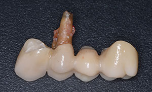 |
| Figure 2. Teeth after crown removal using the laser. | Figure 3. Extraction of tooth No. 5 with removal of the bridgework, retained in the prosthesis. |
 |
 |
| Figure 4. Bridge prepped for reinsertion. | Figure 5. Following the extraction and laser-induced hemostasis. |
My crown-removal technique works on zirconia bi-layered (porcelain layered over zirconia copings), solid monolithic zirconia, IPS Empress, e.max pressed, and e.max CAD/CAM restorations. For crowns, I use the R14 handpiece with a quartz tip. The power is 3.0 W, 200mJ per pulse; the repetition rate is 15 Hz; and the pulse width SSP equals 50 µsec with water spray. These crowns are removed en masse and recemented without the need to remake.
Patients have the need for removal of resin-cemented ceramic crowns without damaging the existing crowns or underlying tooth structure or causing damage to the nerve systems.
CASE REPORT
Diagnosis and Treatment Planning
A 63-year-old female (the patient referred to previously) presented with pain in tooth No. 5 (Figure 1). The tooth had a history of endodontic therapy, including an apicoectomy and a retrograde fill. The treating endodontist had now deemed further surgery to be a futile effort. The 4-unit monolithic zirconia bridge was remade 3 years ago, double abutting the anterior teeth to support tooth No. 5. The remade bridge had been placed using a resin cement. The only treatment option was now extraction, followed by replacing the fixed 4-unit bridge or performing additional sinus lift surgery for single-tooth implants at the Nos. 4 and 5 sites. The patient wanted treatment with an immediate replacement without any additional surgical procedures.
 |
 |
| Figures 6 and 7. After the extraction and laser treatment, the revised bridge was resin cemented. |
 |
| Figure 8. Postoperative photo at 6 months. |
Treatment would consist of removing the resin-bonded, 4-unit zirconia (BruxZir [Glidewell Laboratories]) bridge with a pulsed Erbium laser. Tooth No. 5 would then be extracted. Laser hemostasis would be achieved utilizing a pulsed Nd:YAG laser. A pontic would be made in the restoration and fit into the extraction site, and, finally, the bridge would be luted into place using a self-adhesive resin cement.
Clinical Protocol
Two carpules of 2% Mepivacaine (with 1:20,000 Levordefrin) were infiltrated around surrounding teeth Nos. 3, 5, and 6 for pain control during the extraction. Laser safety and regulatory compliance were observed. I removed the BruxZir abutment restorations from teeth Nos. 3 and 6 using the PowerLase AT laser system with Optical Handpiece Model R14, 8/1.0 Cylindrical Quartz tip. The laser parameters were a wavelength of 2,940 nm, power at 3.0 W (200 mJ), a repetition rate of 15 Hz, a super short pulse of 50 µsec pulse width, and a 3:2 water/air spray. A vacuum tip was used to remove water from the patient’s mouth during this laser procedure.
Only the Erbium laser was used with the tip in contact with outer surfaces of the BruxZir crowns; the focal distance from the fiber tip is 0.7 mm. Using the shortest pulse width (50 µsec) increases the photo acoustic response, and the slow movement of the tip increases the power density. The pulse repetition rate was at a frequency of 15 pulses per second. For each restoration to be removed, the tip was first placed in contact with the occlusal aspect, slowly painting back and forth over the entire occlusal surface, followed by the same for the buccal and lingual surfaces. After laser crown-removal treatment, a scaler or crown-removing pliers are used to remove the existing crown without damage. In this case, after the laser treatment, I was unable to remove the crown from tooth No. 5. I used a tapping bridge remover to remove the loose bridge abutments on teeth Nos. 3 and 6. Tooth No. 5 was extracted during the bridge removal, with the tooth still bonded within the crown abutment (Figures 2 and 3). There was no cement retained internally in the crowns for teeth Nos. 3 and 6. Cement did remain bonded to the prepared tooth structure and the composite resin core material.
Blood was allowed to pool in the extraction site. Hemostasis was then established using the PerioLase II Digital MVP-7 Nd:YAG laser (Millennium Dental Technologies). Laser parameters were set at 3.6 W and 20 Hz, and the pulse width was set to 650 µsec. This free-running, pulsed laser utilizes a 360-µm contact fiber tip. The fiber was placed to the depth of the extraction site and fired as it was gradually moved in a coronal direction. Observation showed color changes in the blood with a thermal clot forming; exposure time was less than a minute. Four minutes after laser therapy, the site was ready for restoration.
Cement was removed from the abutment teeth using 12-fluted composite finishing burs (Midwest). The remaining tooth structure in the No. 5 abutment was shaped into a highly polished bullet-shaped pontic (Figure 4). Air abrasion (50 µm of aluminum oxide) was used to prepare the internal ceramic surfaces prior to cementation (Figure 5). Next, the restoration was bonded into place using a self-adhesive resin cement (RelyX Unicem [3M]) (Figures 6 and 7). The patient was then given post-extraction and oral hygiene instructions and dismissed.
CLOSING COMMENTS
In my private practice, I began using the Nd:YAG for hemostasis following extractions in 1999. My technique was made possible and developed via the utilization of a laser that had more than one pulse width and from knowledge gained during LANAP periodontal laser training offered by Millennium Dental Technologies. These past 20 years, I have used this technique with a wide pulse width following all adult extractions, and it has been instrumental in minimizing cases of alveolar osteitis and after-hours calls from patients with post-extraction bleeding.
In this case, the extraction, laser treatment, and immediate restoration technique minimized treatment duration (Figure 8). The patient was presented with a long-term, aesthetic, and functional restoration in a timely manner. With the use of lasers, the bridge was removed undamaged, and, in addition, the usual challenges that would have otherwise resulted from a lack of hemostasis were easily prevented during the insertion of the appliance.
Dr. Cranska practices full-time laser and family dentistry in Severna Park, Md. He has an Advanced Proficiency Laser Certification from the Academy of Laser Dentistry and a Standard Proficiency and Training Certification from the Institute for Advanced Laser Dentistry (IALD). He can be reached at (410) 975-9331 or via email at familylaserdentistry@outlook.com.
Disclosure: Dr. Cranska has received compensation as a clinical consultant for presenting, lecturing, and training from the IALD, Millennium Dental Technologies, and Fotona LLC.
Related Articles
Use a Laser to Replace a Fractured Solid Zirconia Crown
Use of Lasers to Treat Failing Dental Implants
Laser Removal of All-Ceramic Restorations: Solving a Difficult Clinical Challenge


