As an oral and maxillofacial pathologist, I have the unique perspective of both seeing patients who are referred to me for assessment of oral lesions as well as analyzing biopsy specimens that are sent to me for microscopic diagnosis. With regard to both aspects of what I do, I frequently encounter the term leukoplakia.
To patients, this word is scary and vague, and it often connotes something bad. To clinicians, it represents a lesion that may be challenging to manage and difficult to explain.
The guidelines set forth by the ADA, while thorough, are equally confusing. They recommend periodic followup for a “clinically evident, seemingly innocuous lesion” and biopsy or immediate referral to a specialist for a “clinically evident, suspicious lesion.” How, then, should we go about managing patients in whom we detect a leukoplakia?
What Is It?
By definition, a leukoplakia is a flat, white lesion that cannot be wiped off and cannot be identified as any other entity.1 In other words, it represents a diagnosis of exclusion. Diagnosis may not be the most appropriate word, however, as a leukoplakia is not a single diagnostic entity.
 |
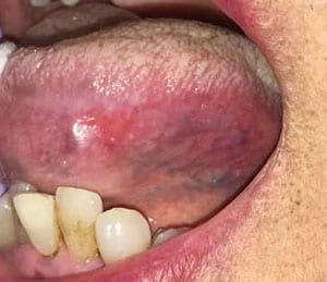 |
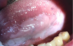
| Figure 2: A thick, relatively homogenous appearing leukoplakia of the ventro-lateral tongue. Biopsies were taken of the anterior, middle, and posterior aspects of the lesion and showed mild epithelial dysplasia. | Figure 3: A “speckled” leukoplakia demonstrating a thick, whiter anterior segment and a thin, red posterior aspect. Biopsy revealed invasive squamous cell carcinoma. |
Microscopically, leukoplakias may be benign (hyperkeratosis and epithelial hyperplasia), pre-malignant (mild, moderate, and severe epithelial dysplasia), or malignant (invasive squamous cell carcinoma). Leukoplakias represent the most common type of oral precancer, accounting for approximately 85% of such lesions. However, epithelial dysplasia or carcinoma is found in only 5% to 25% of all oral leukoplakias.1
What Does It Look Like?
Leukoplakias may all be white patches, but that does not mean all leukoplakias are alike. Lesions may vary in thickness, ranging from thin and homogenous (Figure 1) to thick and irregular (Figure 2). As a general rule, thinner leukoplakias are more likely to be benign or have only mild dysplasia, while lesions that are thicker may have higher degrees of dysplasia.2
This is not always the case, however, and biopsy is required to definitively exclude premalignant or malignant changes. In some cases, leukoplakic lesions may appear “speckled,” meaning that they have both red and white areas (Figure 3). These speckled leukoplakias are also referred to as erythroleukoplakias, and they often show dysplasia or invasive cancer on biopsy.1
Initial Workup and Diagnostic Decisions
Taking a thorough medical and social history is an important first step in all patient care, and it becomes especially critical when evaluating a patient with a leukoplakic lesion. Smoking status and alcohol use still represent the most commonly associated risk factors for developing oral precancer and cancer, though human papillomavirus (HPV) is a known causative agent for base of tongue and oropharyngeal carcinoma.3
When evaluating leukoplakias, one of the first decisions we make as clinicians is establishing a level of clinical concern. In the words of the ADA, is the lesion “seemingly innocuous” or “suspicious”? This can be extremely challenging and often involves at least some degree of personal clinical judgment.
As an oral pathologist, my inherent bias is to view all lesions as suspicious until proven otherwise (via biopsy confirmation), but it is important risk stratify any given lesion. This helps both with patient education and acceptance of treatment protocols, whether that be incisional biopsy, excisional biopsy, or close monitoring. While it is difficult to provide a complete checklist of clinical features that I look for when evaluating a leukoplakia, here are some (hopefully) helpful things to look out for:
- The lateral aspects of the tongue and floor of mouth are the high-risk sites for developing oral cancer. Even if a lesion occurring at these sites appears clinically innocuous, it should be viewed with a higher degree of suspicion.
- Leukoplakias of the occlusal plane on an edentulous ridge often (but not always) represent examples of frictional keratosis secondary to the forces of mastication and food or other irritants rubbing against the area. These are less clinically concerning, especially if they are present bilaterally.
- Put appropriate weight on the patient’s stated social history. A lack of tobacco and alcohol consumption does not necessarily make a leukoplakia less suspicious. Conversely, not all leukoplakias in patients with a history of smoking and drinking will be premalignant or malignant. Similarly, patients may overstate (or understate) the degree of local irritation (such as tongue biting) to a site, especially if asked leading questions.
- Symptoms can be misleading! Lesions that are painful are not always bad, and asymptomatic leukoplakias will not always be innocuous.
- Texture plays an important role in the evaluation of a lesion. Firm or indurated lesions often are clinically worrisome and may be more likely to show epithelial dysplasia or invasive carcinoma.
Biopsy Recommendations
When performing a biopsy, the most critical aspects to consider are site selection and size of the sample. For leukoplakias that are small and uniform in appearance, a single biopsy of the most clinically accessible area can be taken. Leukoplakic lesions that are not homogenous pose more of a challenge. In these cases, the “worst” looking area (clinically indurated, thick, “speckled” in appearance) should be biopsied. If a lesion is large, multiple biopsies can be taken, but the sites from which they are taken should be clearly identified on the specimen containers.
To properly diagnose leukoplakias microscopically, we need to see both surface epithelium and connective tissue. With regard to sample size, the golden rule for adequate depth is to see bleeding clinically. This means that you are through the epithelium and into the underlying connective tissue.
Deciding when to biopsy is sometimes just as difficult, if not more so, than how to biopsy. It is easy to fall into the mindset of not wanting to “hurt the patient” by having to take a biopsy, but much more harm is done by choosing not to biopsy a potentially suspicious lesion.
As a general rule, a leukoplakic lesion that has not resol
ved after two weeks should be biopsied. This does not mean, however, that you need to observe all leukoplakic lesions for two weeks. For those lesions that are at higher-risk sites or are clinically concerning for malignancy, I would recommend that a biopsy be taken as soon as the lesion is identified.
Diagnosis and management of oral premalignant lesions, such as leukoplakias, is one of the most challenging aspects of our job as dental practitioners, but early detection can be life-saving.
References
- Arduino PG, Surace A, Carbone M, et al. Outcome of oral dysplasia: a retrospective hospital-based study of 207 patients with a long follow-up. J Oral Pathol Med 2009; 38:540-4.
- Kumar A, Cascarini L, MaCaul JA, et al. How should we manage oral leukoplakia? Br J Oral Maxillofac Surg 2013; 51:377-83.
- Felier L, Lemmer J. Oral leukoplakia as it relates to HPV infection: a review. Int J Dent 2012; 540561.
Dr. Peters is an assistant professor of oral and maxillofacial pathology at Columbia University College of Dental Medicine. He is a diplomate of the American Board of Oral and Maxillofacial Pathology, a fellow of the American Academy of Oral and Maxillofacial Pathology, and the recipient of the Academy’s prestigious William G. Shafer Award. He also is a member of the Columbia University Oral Diagnostics Biopsy Service and the clinical oral pathology service located at ColumbiaDoctors Midtown. He can be reached at [email protected].
Related Articles
Early Diagnosis of Oral Cancer Can Save Lives
Promote Oral Cancer Awareness This April