


An amazing blend of science and clinical practice, dentistry is constantly evolving to create a better patient experience. Patients perceive technology as an important adjunct to a more comfortable, less stressful dental appointment. As dental technologies evolve, fewer traditional instruments will be utilized, with old-fashioned techniques being replaced by less invasive ones. At the forefront of technology, dental lasers have continued to evolve—from large, boxy units to smaller, user-friendly efficient units. Dental lasers have the ability to replace the old standbys such as handpieces, scalpels, chisels, and more. While these instruments are not obsolete, dental lasers can provide an efficient and less invasive approach to common dental surgical procedures.
As lasers have advanced, the ability to cut hard tissues (tooth structure, bone) with dental lasers has also advanced, and the speed at which lasers can cut has increased dramatically. However, this is only part of the reason dental lasers are replacing conventional instruments and techniques. Other significant advantages include the fact that lasers are less invasive, provide better homeostasis, produce a bactericidal effect, and promote better healing.1 Speed of cutting is only one factor. Many clinicians are surprised at the efficiency with which lasers cut hard tissues.2
However, patient perception is the most important difference between conventional instruments and techniques and the use of lasers.3 When offered the choice between a scalpel or laser for surgical procedures, most patients choose the laser. When offered the choice between a drill and a laser, most patients also choose the laser. Why? Because lasers signify cutting-edge technology and advancements in technique, even if the patient doesn’t understand the details of the technology or procedure.
With fingertip access to the Internet for information, today’s inquisitive patient population is sure to start seeking out newer technologies and the providers that offer them. This, in turn, will drive lasers to become more commonplace in dentistry.
TECHNIQUE FOR ATRAUMATIC OSSEOUS TISSUE REMOVAL AND RECONTOURING
The case report being presented here illustrates the use of an all-tissue laser for successful hard- and soft-tissue surgeries, supplemented by the use of a diode laser for biostimulation. In this clinical case, 2 laser wavelengths were utilized: erbium (all-tissue laser) and diode (soft-tissue laser).
The erbium laser used is an Er,Cr:YSGG, 2,780 nm wavelength laser (Waterlase iPlus [BIOLASE]). This all-tissue laser has the ability to interact with soft tissue, dentin, enamel, and bone. While many dentists recognize the effectiveness of this laser in incising soft tissue, they sometimes question its ability and effectiveness to cut hard tissue. Erbium lasers have advanced significantly in the last 20 years and are capable of cutting tooth structure and bone quite effectively, with significant benefits. While not as quick as a high-speed handpiece in some instances, lasers are clearly more effective for hard-tissue procedures than most clinicians believe.4 The ability of the erbium laser to remove tooth structure without creating heat or microfractures is a clear advantage.5 Similarly, when utilized to remove and recontour bone, the erbium laser generates no heat nor does it create a zone of osseous necrosis.6
The second laser utilized in this clinical technique is a 940-nm diode laser (EPIC [BIOLASE]). Diode lasers have the ability to cut and recontour soft tissues with little or no bleeding despite the fact that diode lasers utilize heat to cut tissue. Here, the diode laser is utilized for a different effect: biostimulation. While not widely known, diode laser wavelengths can reduce bacterial counts, biostimulate tissues on a cellular level and promote increased blood flow.3 All of these factors can lead to better healing and greater patient comfort following surgical procedures.
Following the surgical procedure with the erbium laser, the diode laser was utilized to biostimulate the surgical site. This combination of 2 wavelengths provides efficient cutting and contouring of both soft and hard tissues with minimal bleeding and improved post-op comfort for the patient. The lasers also provide biostimulation, allowing the surgical site to heal more quickly with fewer complications.
CASE REPORT
A 62-year-old male presented with excessive anterior mandibular exostosis (Figure 1). Numerous traumatic ulcerative lesions were present because the patient was wearing an ill-fitting mandibular removable partial denture (RPD) replacing teeth Nos. 19, 20, 23 to 26, 28, and 30. The patient also stated that he had difficulty in placing and removing his RPD, and had trouble with food impaction below the buccal exostosis. The patient’s medical history revealed high blood pressure that was controlled with medication. No other medical issues were noted and the patient appeared to be in good health.
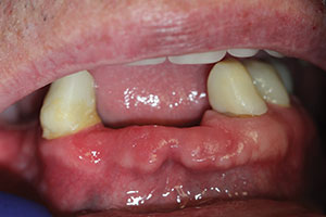 |
| Figure 1. Pre-op: Bony exostosis extended labially from the anterior edentulous mandible. Patient related a history of gingival irritation and an inability to wear a removable partial denture. |
The treatment plan included removing and recontouring the excessive osseous tissues, and then restoring the mandibular arch with a combination of fixed bridgework and posterior implants. By removing and recontouring the exostosis, we planned to create an aesthetic appearance with a better contour for pontic placement with easier cleansability.
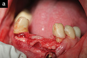
His preoperative blood pressure was 122/82. Following administration of 3.6 cc 2% xylocaine (1:100,000 epinephrine), used to achieve anesthesia in the mandibular region from teeth Nos. 22 to 27, the YSGG laser was utilized to create an incision across the edentulous ridge from teeth Nos. 22 to 27 (laser setting 2.0W, 50Hz, 20% water, 10% air) (Figure 2). The laser was in light contact with the tissue, creating a clean, fine incision with very little bleeding. The gingival tissues were reflected to expose the protruding osseous tissues, which were then evaluated and outlined as to how much tissue would have to be removed in order to create the final ridge appearance.
 |
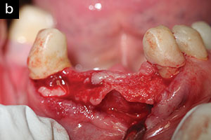 |
| Figures 2a and 2b. Tissue reflected: Following laser incision, the gingival tissue was reflected to expose the osseous tissues with the YSGG laser. |
 |
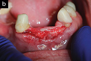 |
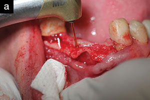
| Figures 3a and 3b. Osseous tissue removed: The final contour of the ridge was created by removing and contouring the osseous tissues with the YSGG laser. |
 |
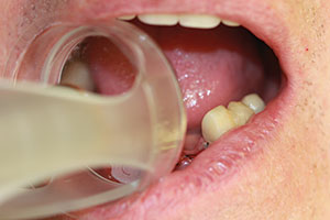 |
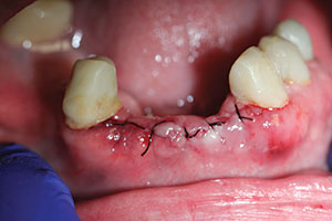
| Figure 4. Tissue sutured: The gingival tissues were re-approximated and sutured with six 4-0 silk sutures (Suture Silk [Henry Schein]) to create a more normal appearance and shape to the anterior ridge. | Figure 5. Biostimulation: Biostimulation with the 940-nm diode laser helped to promote healing and lessened postoperative symptoms. |
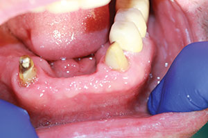 |
| Figure 6. One-week post-op: Sutures removed; results indicated full-tissue coverage with osseous contour that would then facilitate restoration of the missing teeth. |
The YSGG laser (setting 4.0W, 20Hz, 40% water, 40% air) was first utilized to incise the excessive osseous tissues (bulk removal). The laser was then utilized in a defocused mode (approximately 3.0 to 4.0 mm away from the tissues) to recontour and smooth the bone, removing any sharp edges and creating the final ridge contour (Figure 3). By varying the distance of the laser tip from the targeted tissue, the operator controls the amount of tissue interaction and ablation. Near contact results in distinct cutting, while a farther distance from the targeted tissues allows the operator to bevel, smooth, or contour the targeted tissues very precisely.
Following re-approximation of the tissues, six 4-0 silk sutures (Suture Silk [Henry Schein]) were placed (Figure 4) and the patient’s existing RPD was reshaped and relined with a soft liner material (Softline [Henry Schein]). Before final placement of the existing RPD, the 940-nm diode laser was utilized to biostimulate the surgical site and to increase blood flow to the affected tissues. As stated earlier, diode lasers can stimulate tissues on the cellular level, increasing adenosine triphosphate production, mitochondrial activity, and also microcirculation.7 A special attachment (deep-tissue handpiece [BIOLASE]) that focused the diode laser energy to the targeted tissues was used (Figure 5). The setting for this procedure was 3.0W, continuous. The handpiece was held slightly above the tissue for 2 minutes, slowly moving over the surgical site. The patient’s existing RPD was then put in place and used as a temporary prosthesis.
Postoperative instructions included rinsing with warm salt water, over-the-counter nonsteroidal anti-inflammatory drugs, and a soft diet. The patient was asked to report any signs of inflammation, pain, or other symptoms. A postoperative suture removal appointment was set for one week later. A call to the patient that evening revealed slight discomfort with no swelling or other symptoms. The RPD fit well and the patient was able to eat soft foods.
At the one-week postoperative appointment, 6 sutures were removed, and the diode laser was one again utilized to biostimulate the surgical site with the deep-tissue handpiece. The tissue appeared to be healing satisfactorily, and the patient was very happy with the initial results (Figure 6).
DISCUSSION
As dentists, we are taught certain age-old techniques to perform osseous procedures (usually a scalpel and high-speed handpiece). However, it is important to be open-minded to new technology that can provide alternative, often better methods. Sometimes these alternative methods take time to develop or are oversold by the companies selling the product. As clinicians, we must evaluate and learn the true value of these methods. Dental lasers are no different. As laser use in dentistry continues to expand, clinicians will see the value of the use of lasers on a daily basis. In many dental procedures, lasers can provide a better clinical outcome and enhance the patient experience.
Research is showing great progress in dental applications for YSGG lasers as an adjunct in treating periodontal disease,8 disinfection in endodontics,9 placing and exposing implants, and in pediatric applications. Lasers have gone far beyond just providing another “cutting tool.”
IN SUMMARY
In this clinical case report, an alternative method was utilized to perform a common surgical procedure. While there are many different techniques to remove and recontour osseous tissues, lasers show promise in providing a precise, predictable technique, which enhances the patient’s experience through minimal post-op symptoms and faster tissue healing.
References
- Wigdor HA, Walsh JT Jr, Featherstone JD, et al. Lasers in dentistry. Lasers Surg Med. 1995;16:103-133.
- Matsumoto K, Hossain M, Hossain MM, et al. Clinical assessment of Er,Cr:YSGG laser application for cavity preparation. J Clin Laser Med Surg. 2002;20:17-21.
- Walsh LJ. The current status of laser applications in dentistry. Aust Dent J. 2003;48:146-155.
- Chen WH. Why the Er,Cr:YSGG laser is attractive to dental clinicians. US Dentistry. 2006:33-34.
- Hossain M, Nakamura Y, Yamada Y, et al. Effects of Er,Cr:YSGG laser irradiation in human enamel and dentin: ablation and morphological studies. J Clin Laser Med Surg. 1999;17:155-159.
- Wang X, Zhang C, Matsumoto K. In vivo study of the healing processes that occur in the jaws of rabbits following perforation by an Er,Cr:YSGG laser. Lasers Med Sci. 2005;20:21-27.
- Sun G, Tunér J. Low-level laser therapy in dentistry. Dent Clin North Am. 2004;48:1061-1076, viii.
- Hakki SS, Korkusuz P, Berk G, et al. Comparison of Er,Cr:YSGG laser and hand instrumentation on the attachment of periodontal ligament fibroblasts to periodontally diseased root surfaces: an in vitro study. J Periodontol. 2010;81:1216-1225.
- Schoop U, Goharkhay K, Klimscha J, et al. The use of the erbium, chromium:yttrium-scandium-gallium-garnet laser in endodontic treatment: the results of an in vitro study. J Am Dent Assoc. 2007;138:949-955.
Dr. Koceja graduated from Marquette University School of Dentistry in 1986. He served 8 years in the United States Navy Dental Corps, where he completed a periodontal fellowship in San Diego in 1991. He has been in private practice since 1994 and has been actively involved in the use of lasers in his practice since 1999. Dr. Koceja received his laser certification in 2000 and has since achieved Mastership level in the World Clinical Laser Institute (WCLI). He serves on the certification committee for the WCLI. He has written articles and lectured throughout the world on lasers, teaching and promoting their integration into all aspects of dental practice. He can be contacted at (360) 953-8135 or via email at mkoceja@comcast.net.
Disclosure: Dr. Koceja has no financial interest in any of the companies mentioned in this article but did receive compensation from BIOLASE for the writing of this case report.


