INTRODUCTION
Adhesion dentistry is performed in nearly every restorative dental practice today. As early as 1955, Buonocore1 established that adhesion to inorganic enamel could be achieved micromechanically, using weak acids to decalcify with subsequent infiltration with adhesive resins. Dentin, on the other hand, being organic and laden with moisture, proved to be more difficult to adhere to until the mid-1980s. To this day, it presents the greatest clinical challenge in adhesion dentistry due to variability in dentin structure as a result of caries, age and vitality, location within the tooth itself, and the long-term stability of the bond.
Adhesive products have come and gone over the last 40 years with the development emphasis being initially on improving the strength of the bond, commonly expressed as shear bond strengths. Once clinically effective bond strengths were achieved to dentin and enamel, the development emphasis shifted to reducing the complexity of the technique through the use of fewer bottles and chemical agents. While this trend toward simplicity is now the norm, questions still remain with respect to the potential trade-offs that may have occurred regarding bond strengths themselves.
 |
 |
| Before Image. A patient presented with a fractured distal lingual cusp, maxillary left first molar (tooth No. 14). | After Image. The final result. Note the excellent aesthetics achieved in this case. |
Adhesion dentistry is most often practiced with the placement of direct restorations, such as composite resins. In addition, applications exist, including: sealants, desensitization, intraoral repairs to restorations, and their use in conjunction with adhesive resin cements for the placement of indirect restorations. As a result, dentists must generally maintain a portfolio of multiple adhesive products from a variety of manufacturers to address these wide ranging clinical applications. Clinicians must be competent in the use of the products, each with their own specific instructions. Many times, these instructions vary from product to product, despite the fact they often share similar chemistries. It is easy to understand how this could create opportunity for error, negatively affecting final outcomes. Inventories for these products are costly to maintain and store due to upfront costs and the need to monitor their effective shelf lives.
Historical Perspectives on Dentin Bonding: First to Fourth Generation Adhesives
Dental adhesives are often categorized in “generations,” which reflect the mechanism of action for these varying adhesive approaches incorporating adhesive resins, primers, and acids to create bonds to enamel, especially dentin. The earliest approach with the first-generation products relied solely on adhesives with no conditioning of dentin, producing very weak shear bond strengths in the 2 to 3 MPa range. It was characterized by the use of unstable and difficult to handle methacrylates, polyurethanes and cyanoacrylates. These produced disappointing results and failure rates approaching 50%.
Second- and third-generation products were an improvement over the first-generation materials, producing bond strengths up to 16 MPa. Although lower long-term failure rates were reported, they were not sufficient enough to create widespread acceptance into the early 1990s. The adhesive products could be characterized as largely ignoring the dentinal smear layer, using citric acid and ethylmethacylates, polyurethanes, and phosphate esters to achieve low-to-modest shear bond strengths.
With the introduction of fourth-generation systems, bonding took a quantum leap. This approach used 3 separate bottles in what amounted to 3 separate steps. The dentinal smear layer was removed using cleaners such as maleic acid, or with stronger acids such as phosphoric acid and chelating agents like EDTA. They decalcified the enamel and demineralized the dentin in one step, coining the term “total- etch.” Peritubular dentin was exposed to greater depths while maintaining exposed and upright collagen fibrils. The dentin also needed to remain moist to maintain collagen fibrils in the upright position, as their collapse risked loss of adhesion the drier the dentin became. In the second step with this generation of adhesives, volatile primers such as hydrophobic acetone or hydrophilic ethanol were used to enhance adhesion by improving the wettability of the dentin. These primers used a 2-bottle delivery system (bottles A and B) that required dispensing and mixing to activate the primer; the mechanism of action was to increase the surface area for contact for the adhesive resin itself.
In step 3 (bottle C), an unfilled to partially filled resin was applied to infiltrate the collagen fibril network and peritubular dentin. This surrounded the upright collagen fibrils, producing a “hybridization zone” or the adhesive interface. Although complex in terms of application and time required, these fourth-generation systems demonstrated consistently high bond strengths to both enamel and dentin in the 25 to 30+ MPa bond strength range. As such, they are often described as the “gold standard” for adhesion dentistry.2
The application of phosphoric acid to vital dentinal tooth structure raised concerns as to the potential for postoperative sensitivity, which was a reported finding when employing this technique. This was not directly attributable to the phosphoric acid itself, but rather to poor technique with respect to its use, including time of application and improper/incomplete removal. Other factors unrelated to the acid also came into play with this generation of adhesives, such as having the dentin “properly” moist. As a result, solutions were sought to overcome these obstacles without compromising bond strengths themselves.
Single Bottle Systems: Fifth Generation
The fifth-generation systems (2-step systems) were developed using etching as step one. Simultaneous priming and resin infiltration occurs in step 2, using a single-bottle approach by combining the primer with the adhesive. These systems were consistent and reliable clinical performers, with a modest drop-off in bond strengths compared to fourth-generation products.3 Like their fourth-generation counterparts, they relied on proper moisture levels in the dentin for optimal performance. The hope was that, through the elimination of steps and extra bottles, more consistency could be achieved with less opportunity for error. While there was some simplification, postoperative sensitivity was still reported and linked to the use of phosphoric acid, the correct usage of which (time applied, proper and thorough removal, and maintaining a correct residual dentin moisture level for the prescribed product) still presented challenges.
Self-Etch Revolution: Sixth Generation and Beyond
Currently, sixth-generation self-etching adhesives are in widespread use, following the trend to single-bottle systems containing etchant, primer, and resin adhesive. They are designed to simultaneously etch, prime, and bond in one step, in the shortest amount of time. Through the incorporation of acids (that are weaker than phosphoric acid; pH less than 2.5) into the primer/adhesive bottle, these self-etching primers will penetrate the dentinal smear layer, demineralizing surface dentin with neutralization of the acid and with simultaneous resin infiltration in one step (etch-prime-bond). Postoperative sensitivity is purportedly eliminated by keeping the dentinal tubules plugged with the smear layer, thereby reducing the risk of osmotic fluid shifts in the dentinal tubules associated with odontogenic pain.4 Despite less aggressive dentin etching, good dentin shear bond strengths can be achieved.5,6 However, due to the weaker acids used, this can be problematic in terms of bond strengths to enamel, especially uncut enamel; bond strengths to uncut enamel were less than those obtained when using phosphoric acid.7,8 To compensate, “selective enamel etching” with orthophosphoric acid 35% to 37% was recommended. This presents a conundrum of sorts; while effective for enamel, its very use complicates what should be a simplified procedure by adding an additional step. If phosphoric acid should come into inadvertent contact with the dentin, it will diminish bond strengths to it; any part of the dentin smear layer that is removed will negate the effect of the self-etch primers that rely on its presence for optimal bond strengths. The risk exists that outcomes will be compromised with potential for postoperative sensitivity.
Beyond the “Generations”
What should be clear with this discussion is that adhesive bonding is an exercise in “risk tolerance” in terms of efficiency, convenience, and performance. On one hand, bonding has become less complex, but on the other, there are concerns with loss of efficacy with the addition of “work-arounds” to make up for certain deficiencies, creating some confusion and uncertainty.
Would it be possible to develop a truly “universal” adhesive system minimizing risk with no loss of efficacy and with the ability to adhere to all restorative, nontooth substrates? To be termed universal, it would have to be versatile in wide ranging clinical situations, convenient to use, and compatible with other products. By definition it would combine all the desirable attributes of previous adhesive generations with the efficacy of the gold standard 3-bottle, fourth-generation systems. To transcend the generational categorization, the universal adhesive product would be capable of being used in all of the following procedures:
- Direct composite restorations, Classes I to VI
- Indirect restorations (crowns, veneers, inlays, onlays) made from base, precious, nonprecious metals; glass ceramics including feldspathics, leucite reinforced, and lithium disilicates; metal oxides including alumina and zirconia; lab fabricated composite restorations, and nanoengineered ceramics (such as Lava Ultimate Restorative [3M ESPE])
- Desensitization of root surfaces
- Sealants
- Alloys
- Ceramic repairs
- Endodontic posts
- Implant restorations.
And with the versatility to: - Total-etch, selective-etch, self-etch or any combination with comparative effectiveness and virtually no postoperative sensitivity using one bottle with adherence to tooth substrate and virtually all dental restorative materials without using additional chemical solutions and with protocols for application that are identical, regardless of the material to which adhesion is desired.
- Identical protocols for surface treatment of intaglios and tooth substrate with no additional chemical products.
Bonding to Tooth Structure
A recently introduced universal adhesive (Scotchbond Universal Adhesive [3M ESPE]) was developed to meet these criteria, subsequently redefining the adhesive category with a universal product for both direct and indirect restorative applications. This is a single-bottle system that will address all adhesive needs for all direct and indirect materials. This adhesive allows for choice of any etch mode (total-, selective-, self-etch) with consistent bond strengths across the board to both dentin and enamel. When used in the self-etch mode alone, shear bond strengths were 24 MPa to cut enamel and 30 MPa to dentin. When total-etch was employed, values were 27 MPa for both dentin and enamel (3M ESPE internal data). Burgess et al9 also reported mean values of 30 MPa to etched and unetched dentin with similar values to cut enamel at 24 hours under thermocycled conditions, indicating high bond stability, exceeding the 25 Mpa threshold, and regardless of etch mode employed. Significantly, on tooth substrates that typically are more challenging to bond to such as cervical dentin and enamel, immediate bond strengths in independent testing were shown to approach 25 MPa.10
Dentinal moisture conditions (too wet/dry) are often cited as contributory factors to postoperative sensitivity due to bond degradation from microleakage either immediately or throughout time. Perdiga et al11 demonstrated no difference in adhesion performance to moist or dry dentin whether used in total-etch or self-etch mode, though etching enamel with phosphoric acid is still recommended.12 This is advantageous as many dentists prefer to selectively etch enamel, allowing for enhanced enamel bonds without the risk for disruption of dentin bonds whether through over-drying or inadvertent etchant contact with the dentin itself.9
A copolymer consisting of methacylate-modified polyalkenoic acid (Vitrebond [3M ESPE]) was added to this new adhesive. It is responsible for tolerance to dentin moisture imbalances due to its natural bonding affinity to dentin and etched enamel. This adhesive is ethanol-based, with added water to improve hydrophilicity and moisture forgiveness. (This is in contrast to those adhesives containing acetone as a solvent that is far more sensitive to dentinal moisture content.) Hydroxyethyl methacrylate was added to discourage sensitivity and to improve bond strengths, as it can react with collagen due to an ester and hydroxyl group. In a noninterventional study with more than 120 dentist participants, postoperative sensitivity was virtually nonexistent, irrespective of chosen etch mode, indicating this proprietary formulation is effective.13
Bonding to Dental Materials
New criteria have been created to accommodate adhesion to a variety of nontooth substrates such as base to high noble metals, metal oxides (alumina and zirconia), glass ceramics (such as feldspathic, leucite-reinforced porcelains, and lithium disilicate) and alloys, all requiring additional chemicals such as silane and metal primers. Unless one is using a truly universal adhesive product, clinicians must incorporate a variety of strategies and products to match the restorative choice at hand.
Additionally, in these times, we must be able to repair porcelain substrates, bond direct resins and indirect restorations to teeth, secure crowns made of metal to clinically nonretentive preparations, desensitize hypersensitive teeth, and more. Compatibility issues using products from various manufacturers, monitoring the shelf lives of these products, and simply understanding the directions with proper execution create challenges for every dentist today.
Scotchbond Universal Adhesive will bond to any light reactive dental restorative composite/sealant and, with the use of the dual-cure activator (DCA) (3M ESPE) in a 1:1 ratio, it will react with all dual-cure resin and resin cements; this includes self-cure only core build-up materials. Silane has been added to this adhesive, which behaves as a chemical coupler forming covalent bonds with acid etchable glass silicas (feldspathic, leucite-reinforced, or lithium disilicates), resin nanoceramics, and to resin cements used to secure veneers, inlays, onlays, and crowns made of these materials. This universal adhesive has a film thickness of less than 10 µm and is filled 11% by weight, which assures proper restoration seating and maximal adhesion even in cases of endodontic posts and for routine usage in the treatment of dentinal hypersensitivity. Methacryloyloxydecyl dihydrogen phosphate (MDP) is also a component of this adhesive. As an acidic self-etching monomer with a phosphate ester group, it will form a strong chemical bond to metals and alloys as well as nonetchable, nonglass containing metal oxide ceramics such as zirconia and alumina.13,14 Applications would include zirconia and alumina restorations of all types, including precious and nonprecious crowns, implant abutments of titanium or zirconia, and dental amalgam improving retention and marginal seal. Methacryloyloxydecyl dihydrogen phosphate also reacts with the dentin through ionic bonds with surface calcium ions improving bond strengths to it. Due to the hydrolytic stability of MDP, this adhesive has a 2-year shelf life at room temperature.
CASE REPORT
Diagnosis and Treatment Planning
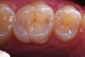
A patient presented with a symptomatic fracture of the distal lingual cusp of tooth No. 14 (upper left first molar) (Before Image). A conservative treatment approach was chosen to replace the cusp, rather than to place a full-coverage crown.
Clinical Protocol
The foundation for success in adhesive dentistry is proper isolation and, if it cannot be assured, a barrier (rubber dam or other isolation device, eg, Isolite) is recommended in most cases. In this case, proper isolation for the procedure could be achieved without the use of a rubber dam.
Tooth preparation was performed to respect the parameters for the chosen restorative material (Lava Ultimate Restorative, a polymer-based [resin] nanoceramic). In this case, the CEREC AC (Sirona Dental Systems) system was utilized for impressioning, restoration design, and in-office fabrication. Tooth preparation involved 2 mm reduction in cuspal areas and 1.6 mm in fissure areas. Once the preparation was completed (Figure 1), a digital impression was taken using the CEREC AC system. The restoration was then designed using the system software, and a shade A3 HT (high translucency) block (Lava Ultimate Restorative) was chosen. Note: These types of restorations can be fabricated either at the chair or through a dental laboratory. Once milled in-office, the restoration was trial-seated for fit verification marginally and proximally (Figure 2).
Next, the restoration was prepolished, and then the intaglio of the restoration was pretreated using air abrasion to create micromechanical retention via surface roughening. Due to the composition of Lava Ultimate Restorative, acid is insufficient to create this roughening effect. Therefore, CoJet Soft (30 µm) (3M ESPE) with a microetcher (Danville Materials) was used to enhance the bond through roughening the internal surfaces (3 bar [35 psi]) for 10 to 15 seconds. (Alternatively, 50 µm aluminum oxide may be used.) The restoration was then set aside.
 |
 |
| Figure 1. The tooth was prepped before imaging with the CEREC AC (Sirona Dental Systems). | Figure 2. The restoration was milled in-office using a Lava Ultimate Restorative (3M ESPE) block (shade A3 HT), and then tried-in for fit. |
 |
 |
| Figure 3. The adhesive resin cement, universal adhesive, applicators, and a dispensing well as set-up prior to the procedure. | Figure 4. The enamel was etched with phosphoric acid. |
 |
 |
| Figure 5. Adhesive (Scotchbond Universal Adhesive [3M ESPE]) was dispensed into the well. The bottle allows for one-handed opening and closing. | Figure 6. Adhesive was applied with a microtipped applicator. |
Since adhesive fixation to the tooth is critical to the long-term performance of Lava Ultimate restorations, our clinical setup included an adhesive resin cement, Scotchbond Universal Adhesive, microapplicators, and a dispensing well (Figure 3). Lava Ultimate is a polymer-based ceramic, requiring the use of silane on the internal surfaces. Since silane is a chemical component of the Scotchbond Universal Adhesive, there was no need for a separate silane treatment.
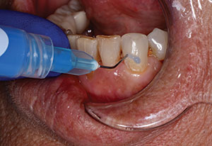
First, in the steps for cementation, the restoration and tooth were conditioned to prepare them for adhesive bonding. Selective etch of the tooth was employed to obtain the best marginal seal in the area of greatest wear (cavosurface) and greatest potential for leakage (cervical areas). Since Scotchbond Universal adhesive has a highly effective self-etch mechanism, any sensitivity issues associated with the use of phosphoric acid (pulpal floors) were avoided. Scotchbond Universal Etchant (3M ESPE) gel (containing 36% phosphoric acid) was precisely applied to the enamel and left in place for 15 seconds. Next, the tooth was rinsed for 10 seconds and then dried for 2 to 3 seconds using oil-free air, leaving the dentin surface “moist” and exhibiting a dull sheen (Figure 4). Using one hand, the cap of the universal adhesive was opened, dispensed (one or 2 drops) and then immediately closed (Figure 5). It was applied with a microtipped applicator, scrubbing it into the intaglio surface for 20 seconds in a single application. Likewise and concurrently, the dentist can also perform the application to the tooth (if isolation is sufficient) with one application using a microtipped applicator for 20 seconds. This allows for optimal workflow, maximizing product, minimizing time, and improving predictability. Both surfaces were air-thinned with moisture-free air until the solvent evaporated and the adhesive no longer moved over the treated surface (approximately 5 seconds) (Figures 6 and 7).
 |
 |
| Figure 7. Adhesive was scrubbed into the intaglio surface of the restoration. | Figure 8. Adhesive resin cement (RelyX Ultimate Adhesive Resin Cement [3M ESPE]) was injected into the preparation. |
 |
 |
| Figure 9. Cement flowed evenly from the margins upon seating. | Figure 10. Excess was cleaned with a mini-sponge. |
With all of its attributes, what really sets this adhesive apart from the others is its ease of use. Tooth and material surfaces are handled identically to indirect restorations, with the only variables being choice of etching mode and the need for a DCA (none needed if using RelyX Ultimate Adhesive Resin Cement [3M ESPE]). After immediate dispensing, application to any surface takes less than 30 seconds. There is no need for the use of desensitizers or disinfectants with Scotchbond Universal adhesive, and they are not recommended.
An adhesive resin cement (RelyX Ultimate Adhesive Resin Cement) was then injected into the preparation, covering all the surfaces (Figure 8). This cement contains a DCA that will react with Scotchbond Universal adhesive to create an auto- and light-cured reaction, ensuring maximum polymerization in any areas that light may not be able to penetrate. Note: If using a resin cement with dual-cure capabilities from another manufacturer, a DCA should be added to the Scotchbond Universal to enable the autocure feature of the alternative cement. Next, the restoration was seated, noting even cement flow from all the margins (Figure 9). A mini-sponge (3M ESPE) was used to clean away the excess, and then the restoration was cured with an LED light (Bluephase [Ivoclar Vivadent]) (minimum 1,100 mW output) using five 10-second cycles, for 60 seconds total (Figure 10).
The occlusion was checked and adjusted with a 20-µm diamond (Neodiamond [Microcopy]), and final polish was then immediately imparted after equilibration using a fine diamond paste (Diashine Intraoral diamond polishing paste [VH Technologies]) using a latch grip, soft bristle prophy brush (Crescent Dental). The very aesthetic final restoration can be seen in the After Image.
CLOSING COMMENTS
In the author’s opinion, the adhesive system described in detail and used in this case report will once again redefine adhesive dentistry. Although new to the market, extensive prior research done by the manufacturer, along with independent testing, corroborates my anecdotal evidence of more than one year of predictable clinical success with virtually no complications for any task requiring an adhesive approach.
References
- Buonocore MG. A simple method of increasing the adhesion of acrylic filling materials to enamel surfaces. J Dent Res. 1955;34:849-853.
- Soderholm KJ, Soares F, Argumosa M, et al. Shear bond strength of one etch-and-rinse and five self-etching dental adhesives when used by six operators. Acta Odontol Scand. 2008;66:243-249.
- Al-Ehaideb A, Mohammed H. Shear bond strength of “one bottle” dentin adhesives. J Prosthet Dent. 2000;84:408-412.
- Brannstrom M. The hydrodynamic theory of dentinal pain: sensation in preparations, caries, and the dentinal crack syndrome. J Endod. 1986;12:453-457.
- Hegde MN, Manjunath J. Bond strength of newer dentin bonding agents in different clinical situations. Oper Dent. 2011;36:169-176.
- Knobloch LA, Gailey D, Azer S, et al. Bond strengths of one- and two-step self-etch adhesive systems. J Prosthet Dent. 2007;97:216-222.
- Erickson RL, Barkmeier WW, Kimmes NS. Bond strength of self-etch adhesives to pre-etched enamel. Dent Mater. 2009;25:1187-1194.
- Barkmeier WW, Erickson RL, Kimmes NS, et al. Effect of enamel etching time on roughness and bond strength. Oper Dent. 2009;34:217-222.
- Burgess J, Shah S, Cakir D, et al. Shear bond strength to restorative materials and tooth structure. Presented at: AARD/CADR Annual Meeting and Exhibition; March 23, 2012; Tampa, FL. Abstract 636.
- Maeno M, Akiyama S, Ogawa S, et al. Bonding performance of recent all-in-one adhesive systems to abrasion-lesion dentin. Presented at: AARD/CADR Annual Meeting and Exhibition; March 24, 2012; Tampa, FL. Abstract 1307.
- Perdigao J, Sezinando A, Monteiro P. Evaluation of a new universal adhesive using different bonding strategies. Presented at: AADR Annual Meeting; March 21, 2012; Tampa, FL. Abstract 18.
- Guggenberger R, Cerny B, Thalacker C, et al. Postoperative sensitivity with a new universal adhesive. Presented at: IADR General Session; June 20, 2012; Iguaçu Falls, Brazil. Abstract 186.
- Blatz MB, Zbaeren C, Mante F. Bond of a new self-etch adhesive to alumina and zirconia. Presented at: AADR Annual Meeting; March 23, 2012; Tampa, FL. Abstract 710.
- Burgess J, Shah S, Cakir D, et al. Shear bond strength to restorative materials and tooth structure. Presented at: AADR Annual Meeting; March 23, 2012; Tampa, FL. Abstract 636.
Dr. Poticny maintains a private, general restorative practice in Grand Prairie, Tex, and is an adjunct associate clinical professor at the University of Michigan School of Dentistry, department of cariology, endodontics, and restorative sciences. He is a graduate of the Ohio State University, Baylor College of Dentistry, and a Fellow of the Academy of CAD/CAM Dentistry. He is a member of the ADA, the International Association for Dental Research, and the International Society of Computerized Dentistry. He is internationally recognized for his dedication, work, and peer education to the disciplines of digital restorative dentistry, material sciences, their clinical applications, and original research. He is widely published on these topics, including numerous peer reviewed journals and publications and has presented extensively for the same. He also serves as a consultant to numerous dental manufacturers. He can be reached at djpoticny@earthlink.net.
Disclosure: Dr. Poticny has received honoraria from 3M ESPE.