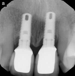
Throughout the last 50 years, dental research and clinical studies have documented numerous new concepts and treatments for periodontal disease. In an era in which we now have a better understanding of the systemic effects associated with this disease process, it is our responsibility as oral care practitioners to both educate and provide our patients with the care they require to enhance the quality of their lives beyond the oral cavity. It is the intent of this article to expand the reader’s knowledge and to provide insight into additional therapies that can contribute superior results to their periodontal therapy.
CHOOSING THERAPIES FOR THE BEST QUALITY TREATMENT
The dilemma that we all face is deciding which pathways and modalities to choose in order to enhance treatment outcomes for our patients. The clinical modalities that are offered within our own practice in caring for our patients’ periodontal needs are as follows: traditional ultrasonic scaling, which includes magnetostrictive and piezoelectric wavelengths; hand instrumentation; localized antibiotic and antimicrobial administration (ARESTIN [OraPharma], PerioChip [Dexcel Pharma], and Periostat [Colla-Genex]); 980 nm-wavelength lasers (Siro-Laser [Sirona] and GENTLEray [KaVo]; CO2 lasers (Spectra [LUTRONIC] and DEKA); and oral antibiotic prescriptions. Some additional modalities include Nd:YAG lasers (Neodymium: Yttrium Aluminum Garnet), and 810 nm and 940 nm diode lasers.
The question now becomes how to utilize such therapies in order to maximize results, yet keep costs within reason while minimizing side effects such as antibiotic resistance and pain. Within our group, the hygienists perform 99% of all soft-tissue therapy. The therapy consists of multiple pathways in order to achieve periodontal success. Each and every patient is screened with the following procedures to ensure the greatest possibility of success: a full review and understanding of the patient’s medical history and medications; a full review of the patient’s dental history; an understanding of the familial dental history; and yearly full-periodontal and x-ray examinations. The periodontal examination consists of full-walking probings, mobility, fremitus, attachment level, and furcation readings. These readings, plus appropriate vertical or horizontal bite-wings, are all required for a proper diagnosis. Additional occlusal and related issues are reviewed yearly. Our goal is to utilize all of this information to develop a diagnosis and to present a treatment plan. Treatment can be as basic as a limited occlusal adjustment, or as complicated as full rehabilitation.
DIAGNOSIS IS ESSENTIAL
The most common periodontal problem that we encounter in our own practice concerns patients who present with localized probing depths more than 5 mm. The first issue to confirm is whether bleeding is present upon probing. Though subjective, bleeding upon probing is an indication that inflammation and/or infection are present within the pocket. A nonbleeding 5 mm pocket requires a review of the patient’s medical and clinical history. This may further guide the provider to ideal care. For example, if the tissue has been stable for years and pocketing is due to passive eruption, then traditional ultrasonic therapy may be adequate to maintain the area. Given the same 5 mm pocket in an area that has subgingival restorative margins and a long history of periodontal maintenance, one may want to do routine laser decontamination to maintain the area or place a localized antimicrobial on a periodic basis. Pockets with bleeding on probing and a depth ≥ 5 mm may require additional soft-tissue treatment beyond traditional debridement therapy.
Diagnosis becomes essential and the clinician must keep some key questions in mind. Are the bleeding pockets a result of poor restorative work, lack of home care, high gingival architecture, vertical or horizontal bone loss, loss of clinical attachment, trauma, occlusion, and/or a result of a systemic condition and/or medication (calcium antagonists are an example)? These issues have to be considered each and every time, along with other possibilities, in order to deal effectively with the clinical situation.
A typical example would be faulty margins causing the inflammation; in this case, success will depend on treating the restorative situation along with the periodontal issues. The problem will recur if treating the pocket inflammation is all that is done, since the cause (restorative) of the problem has not been dealt with. In the case of faulty crowns, temporary crowns are fabricated along with concomitant soft-tissue therapy and the final impressions may be taken weeks later. Successful tissue management creates a natural harmony that integrates with the eventual restorations, while ensuring effortless final impressions. In many of the situations discussed here, combined clinical care may involve a variety of supportive modalities to treat the areas of localized inflammation and infection. Two such therapies are the use of localized antibiotics and soft-tissue lasers.
ULTRASONIC INSTRUMENTATION IN PERIODONTAL THERAPY
Traditional ultrasonic instrumentation is the standard of care for patients in our practice. When using this instrumentation, the essence of successful therapy is to debride the involved root surfaces, along with comprehensive removal of the bio-film. With 5-mm pockets, visibility and access are often not an issue, and our hygienists can remove most of the involved biofilm as the first step of therapy. As we will discuss later, removing biofilm when treating 6- to 9-mm pockets becomes far more challenging and sequential therapy is then the recommended approach. Sequential therapy is based on the fact that, if biofilm is left after the first visit in these deep areas, the hygienist will need additional visits to effectively remove it. And, depending on the patient type and response to therapy, many will need to be referred to a periodontist as possible surgery candidates. In traditional half-mouth scaling and root planings, it is very difficult for hygienists to fully access each and every diseased root surface, which reduces the likelihood of a positive outcome.
COMBINING TREATMENT MODALITIES TO OPTIMIZE RESULTS
Comprehensive treatment through the initial and recare administration of additional modalities, such as lasers and localized antibiotics, has been proven clinically effective in numerous research studies and clinical trials. The key to using these and other modalities is to understand that they are far more successful when used during biofilm removal. In fact, they are complementary to the entire process. In the case of localized antibiotics, such as minocycline HCL 1 mg Microspheres (ARESTIN [OraPharma]), the additional therapeutic benefits include both antibacterial effects for up to 30 days, and the reduction of collagen degradation resulting from tissue-destructive enzymes.1-3 Chemotherapeutics such as ARESTIN work by modulating the inflammatory response, along with the suppression or elimination of pathogenic bacteria.4 In treating localized 5-mm pockets in which periodontal inflammation is the primary cause of the problem, simple placement concurrent with biofilm removal has been shown to reduce bleeding upon probing, increase clinical attachment levels, and decrease probing depths in comparison to traditional scaling and planing.1
LASERS
The use of lasers has demonstrated many therapeutic effects as a supportive treatment to remove diseased epithelial tissue (a second bio-film) along with reduction of the associated bacteremias within the pocket. Studies have shown that con-comitant use of lasers, ultrasonic therapies, and locally applied antibiotics can produce enhanced results regarding clinical attachment gains, along with reductions in bacterial counts and gingival bleeding.5,6 Within the context of the periodontal pocket, the laser light energy is absorbed by the inflamed tissue resulting in cell wall lysis, and ultimately bacterial death. Different wavelengths are utilized by the 3 main types of laser: diode (810, 940, and 980 nm), Nd:YAG, and CO2. Lasers within this segment work because the laser-light energy wavelengths are absorbed by water (high levels are present in inflamed tissue) and/or melanin (present in blood and certain bacterial species). As the low-level energy is absorbed, the bacteria within the pocket are effectively vaporized, resulting in the removal of the diseased tissue. Diode and Nd:YAG lasers work in contact mode, which means they utilize a fiber to directly contact the tissue. CO2 lasers work in a noncontact mode (no direct fiber-to-tissue interaction) within the pocket, and allow the operator to very efficiently treat the inflamed and infected tissues. In the earlier example of treating 5 mm bleeding pockets, a laser is inserted into the pocket immediately before or after ultrasonic therapy. This procedure decontaminates and removes the diseased epithelial tissue, concurrently stimulating new-tissue growth. The patient would then be reevaluated at his or her next recall or sequential periodontal therapy (SPT) visit.
Sequential Therapy
Sequential therapy is performed when more advanced pocketing is observed. For example, a patient presents with quadrants of pockets ranging from 5 to 9 mm. The approach of sequential therapy is to target the deeper pockets (6 to 9 mm) first, then to reinstrument these same pockets at subsequent visits before proceeding to the less-involved areas (those with 5- to 6- mm depths). The rationale behind this approach is that if a hygienist or doctor has to treat 8-mm pockets, full biofilm (tooth and tissue) removal and complete tissue debridement are highly unlikely in one visit. In such cases, the practitioner may need to re-enter the deeper pockets 2 to 3 times to ensure full debridement and biofilm removal. Supportive treatments are then employed according to each individual case.
 |
 |
| Figure 1. Preoperative radiograph of abutment tooth No. 11. |
Figure 2. Preoperative (lingual) view of teeth Nos. 11 and 12. Periodontal treatment (SRP), along with CO2 laser and locally applied antibiotic is initiated to stabilize the area prior to restorative work. Tooth No. 11 (prep) will be used as an abutment for a planned removable bridge from teeth Nos. 6 to 11. |
 |
 |
|
Figure 3. Scaling is performed in deep pockets (7 and 8 mm) to mechanically remove bulk plaque and calculus. |
Figure 4. After scaling, a CO2 laser with a perio tip is used to remove diseased and excess epithelial tissue and to restore proper contour. |
 |
 |
|
Figure 5. Immediately following SRP and laser treatment, a locally applied antibiotic is administered into periodontal pockets to provide targeted, site-specific treatment and promote rapid and complete healing with a resultant reduction in pocket depth. |
Figure 6. The 3-week postoperative photo shows significant healing of gingival tissue around teeth Nos. 11 and 12. Tooth No. 11 has stabilized sufficiently to work as an anchor for the bridge. |
Additionally, in complex casework, the 2 therapies can work together to keep pathologic bacteria levels down, along with facilitating the removal of diseased epithelial tissue (Figures 1 to 6 show a case example). The use of systemic antibiotics can often be minimized for many of our patients and this can help reduce the level of resistant bacteria. Systemic antibiotics should be reserved for use in late-stage therapy in complex casework where they may still be beneficial. It is essential to realize that complete biofilm removal is required even with the use of systemic antibiotics, as these medications often cannot penetrate complex biofilms when they are left intact.
CONCLUSION
In our practices as general practitioners, we are faced each and every day with on-going periodontal issues. Successful treatment requires a full understanding of the potential underlying health issues, along with a comprehensive look at the possible etiologies involved. Treatment options have expanded over the last decade to allow supportive procedures in addition to traditional ultrasonic therapy. Lasers and localized antibiotics are 2 such modalities that can augment our abilities and provide superior, conservative care.
References
- Goodson JM, Gunsolley JC, Grossi SG, et al. Minocycline HCl microspheres reduce red-complex bacteria in periodontal disease therapy. J Periodontol. 2007;78:1568-1579.
- Golub LM, Lee HM, Greenwald RA, et al. A matrix metalloproteinase inhibitor reduces bone-type collagen degradation fragments and specific collagenases in gingival crevicular fluid during adult periodontitis. Inflamm Res. 1997;46:310-319.
- Oringer RJ, Al-Shammari KF, Aldredge WA, et al. Effect of locally delivered minocycline micro-spheres on markers of bone resorption. J Periodontol. 2002;73:835-842.
- Greenstein G, Tonetti M. The role of controlled drug delivery for periodontitis. The Research, Science and Therapy Committee of the American Academy of Periodontology. J Periodontol. 2000;71:125-140.
- Gutierrez T. Diode laser for bacterial reduction and coagulation: an adjunctive treatment for periodontal disease. Contemporary Oral Hygiene. 2005;5:20-21.
- Coluzzi DJ, Convissar RA. Lasers in clinical dentistry. Dent Clin North Am. 2004;48:xi-xii.
Dr. Graham is extensively involved in lecturing and continuing education, focusing on incorporating current clinical advancements through “conservative dentistry.” He is the co-founder of Dental Team Concepts, a continuing education company that makes use of contemporary, interactive formats to integrate time-proven conservative dentistry with 21st century materials and techniques. His courses emphasize diagnosis, evidence-based treatment, dental materials, adhesion and cosmetic dentistry, customized approaches to per-iodontal care, implants, and laser dentistry. He is in private practice in Chicago, Ill, and also holds a part-time faculty position at the University of Chicago. He can be reached at (773) 684-5702 or lgrahamdds@aol.com.
Disclosure: Dr. Graham received an honorarium for lectures and articles.


