Advances in surgical techniques, such as sinus grafting and implant placement, in conjunction with the advances in restorative materials and techniques, allow patients to regain the aesthetics and function of their original dentition—perhaps even better. The key to the successful and predictable use of advanced surgery and materials is being able to identify the critical diagnostic variables, and to organize them in a manner that will create a treatment plan (and/or alternative plan) that addresses the patient’s chief complaint and dental health. The goal of this article is to assist the dentist in identifying and organizing all the variables of treatment that are responsible for creating predictable outcomes.
 |
|
|
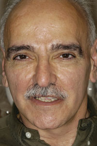
Figure 1. Preoperative full-face view.
|
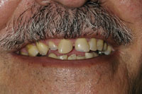
Figure 2. Preoperative smile.
|
CASE REPORT
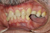
This 56-year-old patient (Figures 1 and 2) was referred with the chief complaints of an unattractive smile, inability to chew his food, and a fear of a lengthy treatment time. Due to the severity of his presenting condition, a complete prosthodontic examination was performed.1 A complete series of photographs was taken, along with panoramic and cephalometric radiographs. Physical records, including diagnostic impressions and casts and face-bow transfer with interocclusal records were accomplished. A thorough review of the patient’s medical and dental history preceded a complete oral exam and charting. The patient was reappointed for a treatment consultation.
|
|
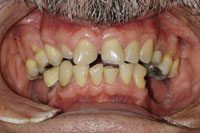
| Figure 3. Maximum intercuspation. (Note the severe wear). |
Figure 4. Protrusive position. |
|
|
|
Figure 5. Right-lateral view.
|
Figure 6. Left-lateral view.
|
|
|
|
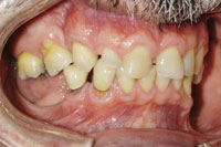
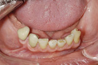
Figure 7. Mandibular occlusal view.
|
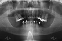
Figure 8. Panoramic radiograph.
|
|
|
|
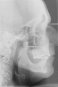
Figure 9. Cephalometric radiograph. (Note severe cant of the posterior maxillary molars and angle of the occlusal plane).
|
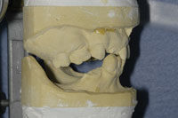
Figure 10. Mounted diagnostic casts at the proposed OVD and CR position. (Note the supra-eruption of teeth Nos. 2 and 3 along with the expansion of the right maxillary tuberosity).
|
|
|
|
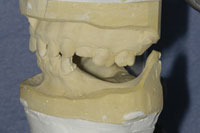
Figure 11. Left view of the diagnostic casts demonstrating the supra-eruption of Nos. 14 and 15.
|
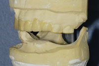
Figure 12. Left-lateral view showing the mandibular anterior cross section and the anterior angulation of the mandibular incisors. The maxillary occlusal plane has been diagnostically corrected, along with the maxillary incisal edge position.
|
|
|
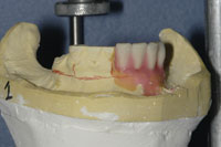
Figure 13. Anterior diagnostic tooth set-up, done to establish the mandibular incisal edge postion.
|
DIAGNOSTIC WORK-UP AND TREATMENT PLANNING CONSIDERATIONS
Examination of the photographs and radiographs (Figures 3 to 9), along with the diagnostic casts (Figures 10 to 12), revealed a grossly malaligned occlusal plane. This misalignment was aggravated by the supraeruption of the maxillary posterior teeth, extensive wear of the maxillary anterior teeth, and the subsequent supraeruption of the mandibular anterior teeth and alveolus. Additional laboratory diagnostic procedures were performed, including cast modification and diagnostic waxing (Figure 13).
A review of the cephalometric image revealed that the patient exhibited an occlusal vertical dimension (OVD) within normal limits; and this was also clinically demonstrated by examination of his lip support and facial profile, as well as speech evaluation. Though the anterior teeth were significantly shortened, the OVD would not allow for an increase to achieve restoration of the maxillary anterior teeth to proper form and length. Though many times, especially in Class II skeletal relationships, the OVD can be modified to achieve restorative space, an increase was not indicated for this patient. However, the maxillary crown length could be adjusted by repositioning the mandibular incisal position. This could be accomplished through a combination of periodontal crown lengthening, endodontic therapy, and prosthodontic preparation of the teeth to create the required space for the upper teeth. Additional possible therapies would include orthodontic intrusion, orthognathic surgery, or extraction.
Cast modifications and diagnostic waxing were valuable tools used in evaluating the possible treatment options outlined above. For this patient, the first step was to establish an ideal maxillary incisal edge position with a pleasing tooth form and gingival interface. Next, the mandibular incisal edge position was established. A corrected occlusal plane was then created with cast surgery and diagnostic waxing. At this point, the diagnostic work-up revealed that the projected position of the mandibular incisors could not be accomplished with endodontics and periodontal crown lengthening at the proposed occlusal plane. This is extremely valuable information to have prior to beginning any treatment on the patient.
| Table 1. Keys to Diagnostic Records |
- Establish the occlusal vertical dimension (OVD)
- Establish the horizontal occlusal dimension-centric relation/occlusion
- Establish the maxillary anterior incisal edge position
- Establish the mandibular anterior incisal edge position
- Establish the occlusal plane
|
| Table 2. Parameters of Care |
- Indications for care
- Therapeutic goals
- Risk factors affecting the quality of treatment
- Standards of care
- Performance assessment criteria
- Known risks and complications
|
Since a chief goal of the patient was limited treatment time, orthodontic therapy, orthognathic surgery, retention of the mandibular teeth with adjunctive endodontics, and periodontal surgery were eliminated as treatment alternatives. Further diagnostic work was then centered on developing a treatment plan utilizing extraction of the remaining mandibular teeth, alveolar reduction in preparation for immediate implant placement, and temporary restoration (Table 1). To organize all the data obtained from the records, the data was formatted in the Parameters of Care for Partial Edentulism by the American College of Prosthodontists2 (Table 2).
|
|
|
Figure 14. One week post operatively.
|
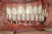
Figure 15. Final restorations: anterior view at maximum intercuspation.
|
|
|
|
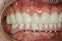
Figure 16. Right-lateral view of final restorations.
|
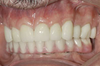
Figure 17. Left-lateral view of final restorations.
|
|
|
|
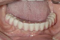
Figure 18. Occlusal view of the mandibular final restoration.
|
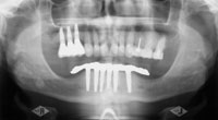
Figure 19. Postoperative panoramic radiograph.
|
|
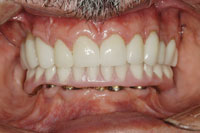
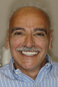
| Figure 20. Final, full-face smile. |
INDICATIONS AND GOALS FOR TREATMENT
The indications for care are worn and chipped anterior maxillary teeth, missing mandibular posterior teeth, and a severely compromised occlusal plane from the supraeruption of the maxillary premolars and molars. In addition, supraeruption of the mandibular anterior teeth and alveolar bone increased the occlusal plane discrepancy, causing the aesthetic compromise of the anterior mandibular incisal edges by being positioned well above the oral commisure.
The therapeutic goals were as follows: to aesthetically restore the maxillary anterior teeth to function; to replace the posterior mandibular teeth while restoring function and correcting the occlusal plane; to increase the ability to chew a normal diet; to create an oral environment that minimizes the need for further future restorative care; and to facilitate the ease of oral hygiene procedures.
RISKS, PERFORMANCE ASSESSMENT CRITERIA, AND STANDARD OF CARE
Risk factors that the patient presented with were reviewed. Severe attrition of the maxillary anterior teeth might require endodontic therapy as well as periodontal crown lengthening with subsequent aesthetic compromises. Coronoplasty of the maxillary molars might require endodontic therapy if pulpal compromise or sensitivity occurred. The loss of functional anterior occlusion, posterior occlusion and changes in the patient’s occlusal vertical dimension could trigger temporomandibular dysfunction (TMD) symptoms during treatment, as well as postoperatively. The severely misaligned posterior maxillary teeth, along with the supraeruption of the mandibular anterior teeth and alveolus, might limit the amount of correction possible surgically and/or prosthetically. In addition to these presenting anatomic risks, a thorough discussion of the risks for all the different treatment options were covered. Known risks include those of surgical interventions, of implant failure, of endodontic failure, paresthesia, material fractures, and the patient’s pain response/tolerance.
Performance assessment criteria were established and discussed. In order to achieve a successful outcome, the anterior maxillary teeth would need to be restored to form and function, along with the replacement of all missing teeth. An increase in phonetics and aesthetics would be necessary along with ease of oral hygiene procedures. Finally, patient satisfaction would be vital in achieving success.
The standards of care necessary for this treatment are outlined in the ACP Parameters of Care for Partial Edentulism for a Class IV Patient according to the Prosthodontic Diagnostic Index of the American College of Prosthodontists.3 The diagnosis for this patient is Class IV Partial Edentulism based on 4 key factors: the lack of anterior guidance; reduced alveolar bone height in both the maxilla and mandible; the lack of a functional occlusal plan; and the etiology in all quadrants.
PRESENTING THE TREATMENT PLAN TO THE PATIENT
The total treatment plan presented to the patient was divided into 2 parts, preprosthetic and final prosthodontic. Based on the diagnostic findings and work-up, the following preprosthetic treatment plan was discussed with him:
To correct the supraeruption of teeth Nos. 2 and 3, and to correct the expansion of the right maxillary tuberosity and sinus in preparation for implant reconstruction, these 2 teeth would be extracted. This would be done in conjunction with a tuberosity reduction and a sinus elevation with bone grafting. Teeth Nos. 14 and 15 would require coronoplasty and/or occlusal reduction, including elective endodontics if necessary, prior to restoration with complete crowns to correct the supraeruption and occlusal plane discrepancy. Prior to restoration with a complete crown, tooth No. 7 would have elective endodontics followed by a cast post and core. Teeth Nos. 22 to 28 would be extracted, along with an alveolectomy of the residual alveolar ridge. These procedures would allow for the proper vertical positioning of the mandibular implants in order to achieve the proposed occlusal scheme. In addition, it would create the necessary autogenous graft material for the sinus graft. Placement of 6 mandibular implants between the mental foramina would be followed by placement of 3 right maxillary posterior implants after graft maturation.
The final prosthodontic plan presented included porcelain-fused-to-metal (PFM) crowns for teeth Nos. 2 to 4. A pink porcelain gingival façade would be incorporated with these crowns to equalize the gingival contours. (PFMs were recommended because they would allow for the use of a pink porcelain gingival façade as well as screw retention for later retrieval and maintenance). Pressed all-ceramic crowns (IPS Empress Esthetic [Ivoclar Vivadent]) were recommended for teeth Nos. 5 to 11 to minimize tooth preparation requirements, and to minimize any periodontal maintenance complications sometimes associated with subgingival margins. These would be fabricated by the dental technician utilizing the layering technique (as opposed to the press and stain technique) in order to achieve optimal anterior aesthetics. Due to a higher strength, all-ceramic crowns made of lithium disilicate (e.Max Press [Ivoclar Vivadent]), using a compatible aesthetic layering porcelain (e.Max Ceram [Ivoclar Vivadent]), were recommended as single-unit restorations for the maxillary left bicuspids and molar (teeth Nos. 12 to 14) due to their higher strength. The preparations would be designed without subgingival margins to assist in long-term periodontal health. The final mandibular restoration would be a metal/acrylic fixed-hybrid implant bridge restoring teeth Nos. 19 to 30.
TREATMENT BEGINS: SEQUENCING
To meet the patient’s goal of treatment immediacy and minimal postoperative prosthetic adjustment, the following treatment sequence was accomplished in a single day.
Teeth Nos. 5 to 14 were anesthetized and prepared for complete restorations with a reduction guide made from the diagnostic wax-up. Custom acrylic transitional crowns were fabricated based on the diagnostic wax-up and cemented.
Mandibular anesthesia was initiated and the extraction of teeth Nos. 22 to 28, along with the proposed alveolectomy to prepare the implant sites and to harvest autogenous bone for the sinus elevation and graft, was performed. Mandibular soft-tissue flaps were lightly sutured for protection until implant placement.
Right maxillary anesthesia was initiated. Teeth Nos. 2 and 3 were extracted, along with a vertical reduction of the posterior right tu-berosity. A lateral-wall sinus access was accomplished for the sinus elevation and graft. This required a secondary reconstruction of the sinus membrane with an alloplastic membrane fortified with plate-let-rich plasma. Utilizing the harvested autogenous bone and additional alloplastic graft material, the augmentation was completed and the soft tissues were primarily closed by suture.
Mandibular anesthesia was augmented and 6 endosseous implants were placed according the diagnostic work-up. Final closure of the soft tissues was accomplished with careful suturing. The immediate denture was then adapted and converted to a fixed transitional acrylic bridge as described by Balshi4 and inserted with screw retention. Normal postoperative care was provided following this procedure.
PLACEMENT OF FINAL RESTORATIONS: SEQUENCING
At 12 weeks following the surgery, final restorations were accomplished on teeth Nos. 5 to 14 along with the final mandibular restoration. At 16 weeks after sinus grafting, the right maxillary posterior implants were placed in a single-stage protocol. Six months later, final prosthodontic restoration of the right maxillary implants was completed according to the treatment plan. The patient was given a detailed post-treatment maintenance schedule.
SUMMARY
This patient’s treatment involved a complex diagnostic challenge, as well as a challenging clinical sequence due to the utilization of immediate implant placement and restoration after extraction along with immediate prosthodontic restoration with sinus elevation with bone grafting. The inability to have diagnostic patient wax try-ins required a detailed and exacting diagnostic work-up which included significant laboratory diagnostic wax-ups. The utilization of the ACP Parameters of Care for Partial Edentulism for a PDI Class IV patient provided a framework in which care could be planned and executed with confidence. The availability of various reconstructive materials and techniques to create a seamless restorative result is essential to the success of this type of advanced treatment.
References
- American College of Prosthodontists. Parameters of Care for the Specialty of Prosthodontists. J Prosthodont. 2005;14(4 suppl 1):18-25.
- McGarry TJ, Nimmo A, Skiba JF, et al. Classification system for partial edentulism. J Prosthodont. 2002;11:181-193.
- Prosthodontic Diagnostic Index. American College of Prosthodontists Web site.http://www.prosthodon-tics.org/membership/pdi.asp. Accessed October 7, 2008.
- Balshi T, Wolfinger G. Immediate placement and implant loading for expedited patient care: a patient report. Int J Oral Maxillofac Implants. 2002;17:587-592.
Dr. McGarry received his DDS degree in 1975 from UMKC School of Dentistry. He was awarded his certificate in prosthodontics in 1978 from the Veterans Administration Hospital in San Francisco, CA, and became a Diplomate of the American Board of Prosthodontics in 1991. He is a Fellow in the American College of Prosthodontists and has served as president of the College. He currently holds an appointment as an associate professor at the University of Oklahoma School of Dentistry, and is also an adjunct assistant professor at the University of Illinois School of Dentistry. He maintains a full-time private practice limited to prosthodontics and is the clinical director for the McGarry Implant Institute. He can be reached at mcgarry@implantassociates.net or (405) 755-7777.
Disclosure: Dr. McGarry has no financial interest in any of the companies mentioned in this article.