INTRODUCTION
Early detection of potentially premalignant oral mucosal abnormalities is essential in the battle against oral cancer because even small, asymptomatic lesions can have significant malignancy potential. When early diagnosis is made and appropriate intervention and treatment is rendered, morbidity and mortality rates improve. Since conventional oral cancer screening exams are basic to dental school curricula, it is concerning that oral cancer survival rates have only improved about 5% since 1974, fluctuating around 55%. Several adjunctive visual screening devices and minimally invasive sampling techniques have been introduced for assisting clinicians in finding suspicious lesions in early stages of development. Since lesions detected in these preneoplastic stages have unclear prognoses, significant controversy exists over whether or not they contribute to overtreatment. The clinician’s choice of whether to utilize adjunctive visual screening devices or not should be based on objective ethical reasoning.
The art and science of dentistry has substantially improved through technology since the introduction of the high-speed handpiece in the 1950s.1 There has been a paradigm shift from paternalistic management of obvious problems to a medical model of dental care, which includes prevention as well as management of dental disease. The discovery of the relationship between oral cancers and systemic diseases such as the human papillomavirus (HPV)2 and periodontal disease,3 as well as the understanding that approximately 75% of oral cancers are related to the popular social behavior of smoking while drinking alcohol,4 has raised awareness to the importance of oral cancer screening examinations. Technology has advanced rapidly in the area of oral cancer screening, offering a variety of adjunctive visual aids (Table 1) to the practitioner to assist in identification and early diagnosis of potentially premalignant oral lesions.5 It is incumbent upon each practitioner, as a member of an ethical profession, to be aware of current oral cancer screening technologies and to make educated, ethical, and rational choices about which, if any, technologies to employ for the most ethical management of their patients.
|
Table 1. Currently Available Adjunctive Visual Screening Devices |
||||||||||||||||||||
ADJUNCTIVE VISUALIZATION AIDS
|
Recognition of the importance of optimal oral health and rapid advancements in technology may lead the practitioner to be overzealous in treatment. A comprehensive plan of care that is “right” for the patient is one that includes application of ethical principles in the development, acceptance, and implementation of diagnostic techniques and appropriate management. The science of dentistry provides opportunities to render improved care, but the “art of dentistry” includes appropriate communication with the patient and application of ethical principles in making treatment recomendations.6 The ADA’s Principle of Ethics and Code of Professional Conduct7 lays the groundwork; how the Code is actualized reflects the dentist’s individual values.
Different approaches to prevention may yield similar outcomes. In fact, it has been argued that properly performed conventional oral cancer screening exams alone are adequate for recognizing problematic soft-tissue lesions,8 which is generally accepted as the standard of care. However, this theory has been challenged in a recent retrospective study that suggested that direct tissue fluorescence imaging with the VELscope Vantage (LED Dental) was effective in identifying dysplastic lesions that had not been observed in the same low-risk patient population examined with conventional oral cancer screening exams only the year previously.9 Although techniques for palpation and incandescent light visual examination have been taught in dental schools for decades, the overall 5-year survival rates for oral cancer have only improved about 5% since 1974, fluctuating around 55%.10 Since professional responsibility includes acting in a manner that promotes “good” for the patient, ethical principles should affect all choices made in care management.11
Because dentistry is a moral profession that is guided by normative principles,12 dentists are obligated to choose diagnostic modalities that allow them to be “…caring and fair in their contact with patients.”7 Although patient autonomy should be the overwhelming decision-making principle as to whether or not treatment of early soft-tissue lesions is performed, the dentist is obliged to maintain competency in recognition of soft-tissue abnormalities. In fact, the court system has supported this statement by consistently ruling in favor of patients who have sued for negligence in oral cancer screening, stating that defendant dentists should have but did not recognize lesions early enough to prevent malignant transformation of dysplastic lesions.13 Preservation of the profession of dentistry and the self-policing autonomy that it enjoys necessitates adherence to the normative picture.14 The community at large, as well as members of the profession of dentistry, expects dentists to act ethically7 according to certain norms: autonomy, justice, veracity, nonmaleficence, beneficence, and veracity (Table 2).
|
Table 2. Ethical Decision Making in Dentistry is Determined by the Integration and Understanding Core Ethical Principles |
| SUMMARY OF ETHICAL PRINCIPLES • Autonomy (self-governance). The dentist is free to determine appropriate care to his or her patients but has a duty to respect the patient’s rights to self determination and confidentiality. The patient must be appropriately informed in order to make decisions that are in his or her best interest. • Nonmaleficence (do no harm). The dentist has a duty to refrain from harming the patient, which requires the dentist to maintain a current working knowledge and basic skill set necessary to recognize and render appropriate clinical management of oral disease. • Beneficence (do good). The dentist has a duty to promote the patient’s welfare and to promote public health. • Justice (fairness). The dentist has a duty to treat people fairly in accordance with generally acceptable clinical practices. • Veracity (truthfulness). The dentist has a duty to communicate truthfully with colleagues and with patients, based on appropriate supporting evidence and to avoid disparaging commentary based on difference of opinion rather than fact. |
Autonomy includes the right of a patient to select and/or to refuse treatment and the right of the dentist to decide upon appropriate management for patients under his or her care. This means that the dentist must inform the patient of atypical soft-tissue findings, follow accepted diagnostic protocols,15 and discuss appropriate treatment options, which may include referral to appropriate specialists. The patient must be involved in the treatment decision, necessitating that the dentist be an educator, informing the patient about the etiology of his or her findings and potential outcomes of treatment or deferral of treatment. Autonomy includes the decision making of the patients as well as decision making for the dentist. The right to refuse treatment, to refer for treatment, or to render treatment is inherent in the principle of autonomy for the practitioner.16 However, wise practitioners understand that they may legally share responsibility for treatment rendered or not rendered by clinician to whom they refer.13
Justice requires that the clinician not be judgmental or condescending, understanding that patients are extremely fearful of cancer and that preneoplasia and dysplasia are not cancer. Patients trust that dentists are current with their working knowledge of skills of modern dental techniques and practices. This is why minimal continuing education is mandated in every state. It is also an ethical obligation of the dental profession. Referral of the patient to more qualified clinicians is expected if treatment is needed beyond the skill of the primary dentist. However, the assumption that dental specialists have more knowledge or skills than general dentists may be incorrect in certain incidences because they may “…be at a disadvantage” by not having adjunctive screening technologies in their practices. For example, one particular malpractice carrier has suggested that their surgeons should not feel obligated to perform biopsies of lesions discovered with adjunctive screening technologies, nor should they feel obliged to purchase them.17 In such a case, the primary dentist would probably be more qualified to perform the biopsy than a resistant specialist or he/she should reconsider the choice of referral.
Veracity includes judgment about what information is necessary for patients to make informed choices about their care. For example, when a suspicious oral lesion is discovered, it is ethically responsible for the clinician to inform the patient that something atypical has been discovered while exerting caution in avoiding stimulation of unnecessary fear and anxiety. It would not be appropriate to give the patient a diagnosis until a biopsy has been performed. It is, however, entirely ethical and appropriate to discuss sexual practices with patients in the context of medical history review in light of the documented links between HPV and dysplasia.18
Beneficence and nonmaleficence refer to acting in a manner that promotes the good of the patient while avoiding unnecessary harm to the patient or to society. For example, the decision not to incorporate adjunctive visual screening technologies into the oral cancer screening exam may be made based on reports that they result in an unacceptable number of false positives thus yielding overtreatment by unnecessary biopsies.17 However, it could be argued that such a position leads to delay in diagnosis when early management may save lives. Cases have been described in the literature where dysplasias have been found by adjunctive visualization aids that are not visible to the naked eye under white light alone.19 Furthermore, one device has been reported to be more indicative of dysplastic margin delineation than toluidine blue.20
Ethical decision making reduces the tendency toward overtreatment. The following case report illustrates the ethical rationale for implementing adjunctive visual screening technology into the oral cancer screening exam process. Although the VELscope Vantage was the screening device of choice in this particular case, the rationale would be similar for any adjunctive visual screening tool.
CASE REPORT
Subjective
A 45-year-old female patient of record presented for routine recare. She was a nonsmoker with a history of elective hysterectomy due to cervical epithelial dysplasia. She reported a history of multiple male sexual partners. Currently, she was in a monogamous, long-term relationship with a partner with no known history of sexually transmitted disease. Otherwise her medical history was unremarkable.
|

Objective
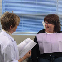
During the conventional oral exam, a raised, pink sessile lesion was noted on the left posterior tonsillar pillar, a highrisk area for squamous cell carcinoma. The lesion appeared to have regular borders. There was minimal gag reflex and there was no history of pain or “scratchy” throat. The lesion was soft and raised and firm upon palpation (Figure 1).
Under direct tissue fluorescence imaging, orange fluorescence suggesting significant bacterial colonization prompted re-evaluation under 3.8x magnification with HiRes Plus loupes (Orascoptic) and enhanced white lighting with a Feather Light LED headlamp (Ultralight Optics). The suspicious area did not blanch under moderate pressure applied with a periodontal probe. Loss of fluorescence appeared to be confined to less than one mm from the base of the lesion circumferentially (Figure 2).
Assessment
Papilloma. The patient opted not to have HPV typing performed on the specimen due to financial considerations. The biopsy reported that the margins of the excision were clean.
Plan
The patient was informed that “a suspicious area” was found during my examination in a high-risk site. She was evaluated 14 days later. When the lesion appeared unchanged at the follow-up appointment, the patient opted for excisional biopsy and a sample was obtained and submitted to Tufts Oral Pathology Services. The VELscope Vantage was used to assist in surgical margin determination, and the lesion was removed, extending to a tissue depth of 2 mm and extending approximately 2 mm into tissues that exhibited normal tissue fluorescence. Educational literature and counseling about HPV transmission was provided to the patient, along with educational information about safer oral sex practices, and the patient was seen for follow-up evaluation of the excision site 7 days later. The healing of the tissue was unremarkable.
Ethical Rationale
• Autonomy: The clinician chose to utilize direct tissue fluorescence imaging as an adjunctive screening device because it was the system most supported by published literature at the time of the examination. From his own clinical experience, it was the most patient-friendly and easiest screening system to implement routinely into his practice. The patient was clearly informed about the lesion immediately via instantaneous digital photo documentation using a MagnaVu PSII (Magnified Video Dentistry), as described elsewhere.21 The patient was carefully educated about the possible etiology of her papilloma, the possibility of conversion to carcinoma if untreated, and the risk of possible HPV transmission to sexual partners.
• Veracity: Care was given not to rush to premature conclusions based on the screening exam alone. The patient was informed that adjunctive technology was used to enhance the thoroughness of the oral exam. The patient was informed that cytology was a secondary screening modality and was not diagnostic. The findings of cytological testing were used for patient education as described literature.22
• Nonmaleficence: Although it may be argued that adjunctive screening technologies lead to unnecessary false positives, the clinician in this case considered the risk of a false positive to be negligible to the risk of a false negative results from conventional screening alone, based on his own research and experience.9 An appropriate protocol to rule out inflammation and to minimize false positives15 was followed. Although the general understanding is that oral papillomas are usually benign, biopsy confirmed that this lesion was not normal tissue, and therefore, not a false positive.
• Benevolence: The clinician believes strongly that early detection of premalignant lesions saves lives through early intervention. Even though the patient opted not to have her lesion HPV typed, intervention through education may reduce the chance of propagation of HPV to her sexual partners, which is in the best interest of the community. The choice to remove the lesion was made in her best interest because of the unusual clinical appearance and borders of the lesion in a high risk zone in a patient who has multiple risk factors.
• Justice: The clinician met the legal obligation to the patient by discovering a potentially premalignant lesion, educating the patient thoroughly, and by making an appropriate and well-founded referral to a skilled and educated colleague for definitive diagnosis.
CLOSING COMMENTS
In the current age of rapidly advancing technology in the field of oral cancer early detection and seemingly aggressive marketing practices of the vendors of these technologies, clinicians need to make the decision whether or not to implement adjunctive technology based on ethics and supporting acceptable literature and experiences of trusted colleagues. The reality is that conventional oral cancer examinations alone have not improved survival rates substantially throughout the past 50 years,5 and any tool that allows cancer screening to be performed better should be welcomed, based on every ethical principle of the dental profession.
References
- A millennium of dentisty: a look into the past, present and future of dentistry. Academy of General Dentistry. knowyourteeth.com/infobites/abc/article/?abc=a&iid=305&aid=1255. Accessed October 22, 2010.
- Syrjänen S. Human papillomaviruses in head and neck carcinomas. N Engl J Med. 2007;356:1993-1995.
- Tezal M, Sullivan MA, Hyland A, et al. Chronic periodontitis and the incidence of head and neck squamous cell carcinoma. Cancer Epidemiol Biomarkers Prev. 2009;18:2406-2412.
- Kahn M. Embracing technology to save lives: a review of oral cancer screening techniques and new technologies. Course lecture presented at: Ohio Academy of General Dentistry Master Track Program, The Ohio State University College of Dentisty; October 3, 2008; Columbus, OH.
- Lingen MW, Kalmar JR, Karrison T, et al. Critical evaluation of diagnostic aids for the detection of oral cancer. Oral Oncol. 2008;44:10-22.
- Huff K, Huff M, Farah C, et al. Ethical decision-making for multiple prescription dentistry. Gen Dent. 2008;56:538-547.
- American Dental Association. Principle of Ethics and Code of Professional Conduct. ada.org/sections/about/pdfs/ada_code.pdf. Accessed October 22, 2010.
- Patton LL, Epstein JB, Kerr AR. Adjunctive techniques for oral cancer examination and lesion diagnosis: a systematic review of the literature. J Am Dent Assoc. 2008;139:896-905.
- Huff K, Stark PC, Solomon LW. Sensitivity of direct tissue fluorescence visualization in screening for oral premalignant lesions in general practice. Gen Dent. 2009;57:34-38.
- American Cancer Society. Cancer Facts & Figures 2005. Atlanta, GA: American Cancer Society; 2005.
- Ethics Handbook for Dentists. Gaithersburg, MD: American College of Dentists; 2004. acd.org/ethicshandbook.htm. Accessed on October 22, 2010.
- Windhorn RJ, Cuenin MF. An implant versus a conventional fixed prosthesis: a case report. Gen Dent. 2007;55:44-47.
- Lydiatt DD. Medical malpractice and head and neck cancer. Curr Opin Otolaryngol Head Neck Surg. 2004;12:71-75.
- Weinstein BD. Dental Ethics. Philadelphia, PA: Lea & Febiger; 1993.
- Huff, K. Enhanced oral cancer screening exams improve quality of care. SPEAR. In press.
- Nichols PS, Winslow GR. What patients need versus what they want. Gen Dent. 2003;51:503-504.
- MONITOR. Questions about oral cancer screening systems. MONITOR. 2008;19(1).
- Huff KD. The challenges of implementing oral cancer screening technologies. Dental Economics. 2009;99:54.
- Huff KD, Garren KC, Huff MS. A novel, minimally invasive approach to managing mild epithelial dysplasia. Gen Dent. 2010;58:126-129.
- Poh CF, Zhang L, Anderson DW, et al. Fluorescence visualization detection of field alterations in tumor margins of oral cancer patients. Clin Cancer Res. 2006;12:6716-6722.
- Huff KD. Photography: an integral component of oral cancer screening. Dent Today. 2009;28:100.
- Huff KD. Brush cytology…a great patient education tool. Dental Economics. 2009;99:17.
Dr. Kevin Huff is a general dentist in Dover, Ohio, and is a clinical instructor in the department of comprehensive care at the Case School of Dental Medicine in Cleveland, Ohio. He is the coordinator of oral medicine at the Mercy Medical Center GDR program and is also a visiting faculty member at Spear Education in Scottsdale, Ariz. He serves as a senior clinical consultant at the Imaging Center at Case in Cleveland, Ohio, and is a mentor for Spear Education in Scottsdale, Arizona. Dr. Huff lectures regularly on the topic of oral cancer screening technologies and has received honoraria from LED Dental, for some of his courses. He can be reached via e-mail at dr@doctorhuff.net.
Disclosure: Dr. Kevin Huff has received sponsorship for lecturing from VELscope but has not received compensation for writing this article.
Dr. Marlene Huff is an associate professor in the College of Nursing at The University of Akron in Akron, Ohio. She can be reached at via e-mail at mhuff@uakron.edu.
Disclosure: Dr. Marlene Huff reports no disclosures.