


INTRODUCTION
The range of direct resin-based restorative products available in the field has expanded greatly in recent years.1-3 In addition to the classic universal composites, the enormous rise in patients’ aesthetic expectations has resulted in the launch of a large number of so-called “aesthetic composites” on the market, which are characterized by composite materials in a sufficient number of different shades and different grades of translucency and opacity.4 Opaque dentin shades, translucent enamel shades, and, if required, body shades make it possible to achieve highly aesthetic direct restorations using the multicolored layering technique. They are practically indistinguishable from the hard dental tissue and can even rival the aesthetics of all-ceramic restorations. Some of these composite systems consist of more than 30 different versions of shades and opacities. However, it is essential to have appropriate experience in the handling of these materials, which are primarily used in the anterior region with a layering technique employing 2 or 3 different opacities and translucencies.4,5
 |
 |
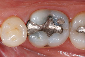
| Figure 1. Pre-op photo showing the previously placed amalgam restoration (tooth No. 19). | Figure 2. Amalgam filling was removed. |
 |
 |
| Figure 3. After excavation, the cavity was finished and isolated with a rubber dam. | Figure 4. Demarcation of the cavity with a sectional matrix. |
Background on Layering Versus Bulk-Fill Techniques
Due to their polymerization properties and limited depth of cure, light-curing composites are generally used in a layering technique with individual increments of no more than 2.0 mm in thickness. Each individual increment is polymerized separately, with exposure times from 10 to 40 seconds, depending on the power of the curing light and color/translucency of the composite material.6 With the materials available up until recently, thicker composite layers resulted in insufficient polymerization of the composite resin and thus offered inferior mechanical and biological properties.7-9
Applying the composite in 2.0-mm increments can be a very time-consuming procedure, especially in large posterior cavities. Consequently, there is considerable demand in the market for composite-based materials that are more simplistic, quicker to use, and therefore more economical, for this range of indications.10 To satisfy this demand, bulk-fill composites have been developed during recent years that, given a sufficiently powerful curing light, can be placed more quickly in the cavity, using a simplified application technique, in layers 4.0 to 5.0 mm thick and with short increment curing times of 10 to 20 seconds.6,11-14 Taken literally, “bulk-fill” means that they can be used to fill the cavity in a single-step technique without the need for layering.15 With plastic restorative materials, this is currently only possible with cements and chemically activated or dual-curing core build-up composites. However, the former do not possess adequate mechanical properties for restorations that are clinically stable in the long term in the masticatory load-bearing posterior region of the permanent dentition. Consequently, they are only suitable for use as interim restorations or long-term temporaries.16-18 The latter are neither approved as restoratives nor suitable for such indications from a handling perspective (eg, shaping of occlusal surfaces). The bulk-fill composites currently available for the simplified filling technique in the posterior region are not actually “bulk” materials in the true sense, when examined more precisely, as the approximal extensions of clinical cavities, in particular, are generally deeper than the maximum depth of cure specified for these materials (4.0 to 5.0 mm).19,20 This said, it is possible to fill cavities with depths of up to 8.0 mm in 2 increments if a suitable material is selected, and this covers the majority of defect dimensions encountered in routine clinical practice.
 |
 |
| Figure 5. Selective enamel etching with 35% phosphoric acid (Vococid [VOCO]). | Figure 6. The tooth was carefully rinsed of all acid gel and dried. |
 |
 |
| Figure 7. A bonding adhesive (Futurabond M+ [VOCO]) was applied to the enamel and dentin using a microbrush. | Figure 8. The solvent was dried from the adhesive system with a gentle stream of air. |
 |
 |
| Figure 9. The bonding adhesive was then light-cured for 10 seconds. | Figure 10. Once the adhesive was applied, the entire sealed cavity exhibited a shiny surface. |
Introducing ORMOCER-Based Restoratives
Most composites contain organic monomer matrices based on conventional methacrylate chemistry.21 Silorane technology22-27 and ORMOCER (“organically modified ceramics”) chemistry28-35 present alternative approaches. ORMOCERs are organically modified, nonmetallic, inorganic composites.36 ORMOCERs can be classified between inorganic and organic polymers and possess both an inorganic and an organic network.35,37,38 This group of materials was developed by the Fraunhofer Institute for Silicate Research (ISC), in Würzburg, and marketed for the first time as a dental restorative material in 1998 in collaboration with partners in the dental industry.33,34 Since then, there has been considerable further development of the ORMOCER-based composites for this range of applications. However, the use of the ORMOCERs is not limited to dental restoratives. These materials have been successfully employed for years in fields such as electronics, microsystems technology, plastic refining, preservation, anticorrosion coatings, functional coatings for glass surfaces, and highly resistant scratch-proof protective coatings.39-41
ORMOCER-based dental restorative materials are currently available from 2 dental companies. In the dental ORMOCER products of the past, additional methacrylates were supplemented to the pure ORMOCER chemistry (in addition to initiators, stabilizers, pigments, and inorganic fillers) in order to improve workability.42 Therefore, it is better to speak of ORMOCER-based direct restoratives here.
According to the manufacturer of the product to be demonstrated in this case report article, the new bulk-fill nano-ORMOCER Admira Fusion x-tra (VOCO) (launched in 2015) no longer contains any conventional monomers in addition to the ORMOCERs in the matrix. It features a nanohybrid filler technology with an inorganic filler content of 84% by weight. It is available in a universal shade and displays polymerization shrinkage of just 1.2% by volume and simultaneously low shrinkage stress. Admira Fusion x-tra can be applied in layers of up to 4.0 mm, with each increment being cured in 20 seconds (curing light power > 800 mW/cm2). The malleable consistency and the material data of Admira Fusion x-tra allow the dentist to restore cavities using the bulk-fill technique using a single material. As a result, an occlusal covering layer with an additional composite (as required when flowable bulk-fill composites are used) is no longer necessary.
CASE REPORT
Diagnosis and Treatment Planning
A 47-year-old patient presented at our clinic requesting to have his remaining amalgam fillings replaced gradually throughout time with tooth-colored restorations. In the first treatment session, the old amalgam filling in his mandibular right first molar (tooth No. 19) was replaced (Figure 1). The tooth responded normally to a cold test without delay, and the percussion test was also normal. Having been informed of the possible treatment alternatives and their costs, the patient elected to have a direct restoration with the nanohybrid ORMOCER recommended (Admira Fusion x-tra) using the bulk-fill technique.
 |
 |
| Figure 11. The first increment of Admira Fusion x-tra (VOCO) filled the mesial area of the cavity, up the approximal wall to the level of the marginal ridge. | Figure 12. The restorative was light-cured 20 seconds. |
 |
 |
| Figure 13. The tooth after removal of the matrix. | Figure 14. The second increment of Admira Fusion x-tra completely filled the cavity. |
 |
 |
| Figure 15. The occlusal anatomy was then shaped, functional but uncomplicated. | Figure 16. Next, the mesial-occlusal restoration was fully light-cured. |
 |
| Figure 17. The completed, highly polished restoration. Function and aesthetics of the tooth were successfully restored. |
Clinical Protocol
Treatment started with thorough cleaning of the tooth with a fluoride-free prophylaxis paste and a rubber cup to remove external deposits. As Admira Fusion x-tra is only available in a universal shade, there is no need for detailed determination of the tooth shade. After administration of local anesthesia, the amalgam was carefully removed from the tooth (Figure 2). Following excavation, the cavity was finished with a fine-grit diamond bur and a rubber dam placed to isolate the tooth (Figure 3).
The rubber dam separates the operating site from the oral cavity, facilitates clean and effective working, and guarantees that the working area remains clean of contaminating substances such as blood, sulcus fluid, and saliva. Contamination of the enamel and dentin would result in considerably poorer adhesion of the composite to the dental hard tissue and would endanger the long-term success of a restoration with optimal marginal integrity. Additionally, the rubber dam protects the patient from irritating substances such as the adhesive system. The rubber dam is thus an essential aid in ensuring quality and facilitating work in the adhesive technique. The minimal effort required in applying the rubber dam is also compensated by avoiding the changing of cotton rolls and the patient’s requests for rinsing.
The cavity was then demarcated with a sectional matrix made of metal (Figure 4). The universal adhesive Futurabond M+ (VOCO) was chosen for the adhesive pretreatment of the dental hard tissue. Futurabond M+ is a modern one-bottle adhesive, which is compatible with all conditioning techniques: the self-etch technique and the phosphoric acid-based conditioning techniques (selective enamel etching or complete etch and rinse pretreatment of enamel and dentin). In this case, we chose the selective enamel etching technique, applying 35% phosphoric acid (Vococid [VOCO]) along the enamel margins and allowing it to work for 30 seconds (Figure 5). The acid was then rinsed off for 20 seconds with the compressed air and water jet, and excess water carefully removed from the cavity with compressed air (Figure 6). Figure 7 shows the application of a generous amount of the universal bonding agent Futurabond M+ on enamel and dentin with a microbrush. The adhesive was thoroughly rubbed into the dental hard tissue with the applicator for 20 seconds. The solvent was then carefully dried off with dry, oil-free compressed air (Figure 8) and the bonding agent light-cured for 10 seconds (Figure 9). The result was a shiny cavity surface, evenly covered with adhesive (Figure 10). This should be carefully checked, as any areas of the cavity that appear matte are an indication that insufficient adhesive was applied to those sites. In the worst case, this could result in reduced bonding of the restoration in these areas and, at the same time, in reduced dentin sealing, which may lead to postoperative sensitivity. If such areas are found in the visual inspection, additional bonding agent is again selectively applied to them.
In the next step, the cavity measured in advance with a periodontal probe (6.0 mm deep from the floor of the box to the occlusal marginal ridge), was filled with Admira Fusion x-tra in the area of the mesial box until a residual depth in the whole cavity of no more than about 4.0 mm remained. At the same time, the mesial approximal surface was built up completely to the level of the marginal ridge (Figure 11). The restorative material was cured by a polymerization lamp (light intensity > 800 mW/cm2) for 20 seconds (Figure 12). The buildup of the mesial approximal surface converted the original Class II cavity into an “effective” Class I cavity, and then the matrix system was removed, as it was no longer required (Figure 13). This facilitates access to the cavity with hand instruments for shaping the occlusal structures in the further course of the treatment and, thanks to the improved visibility of the treatment area, allows improved visual control of the material layers subsequently applied. The second increment of Admira Fusion x-tra filled the residual volume of the cavity completely (Figure 14). Following the shaping of a functional but uncomplicated occlusal anatomy (Figure 15)—which also helps to ensure rapid finishing and polishing—the restorative material was cured again for 20 seconds (Figure 16).
After removal of the rubber dam, the restoration was carefully finished with rotary instruments and abrasive discs, and the static and dynamic occlusion adjusted. Diamond-impregnated silicone polishers (Dimanto [VOCO]) were then used to give the surface of the restoration a smooth and shiny finish. Figure 17 shows the finished, direct nano-ORMOCER restoration, which reproduces the original tooth shape with an anatomically functional occlusal surface, physiologically shaped approximal contact, and aesthetically acceptable appearance. Finally, a foam pellet was used to apply the fluoride liquid varnish (Profluorid L [VOCO]) to the teeth.
CLOSING COMMENTS
The place for the use of direct resin-based restorative materials will continue to grow in the future. These materials are capable of providing high-quality permanent restorations for the masticatory load-bearing posterior region, whose reliability has been documented in the literature. The results of an extensive review have shown that the annual loss rate for composite restorations in the posterior region (2.2%) is not statistically different from amalgam restorations (3.0%).43 Ever-increasing economic pressure in the healthcare sector creates a need for a simpler, faster, and thus more cost-effective basic treatment as an alternative to more time-consuming layered composite restorations. For some time now there have been composites with optimized depths of cure on the market for this purpose, which can be used to make clinically and aesthetically acceptable posterior restorations with a procedure that is more cost-effective compared with traditional hybrid composites.44,45 In addition to the bulk-fill composites with classic methacrylate chemistry, the range of products available within the field of resin-based direct restorative materials with an increased depth of cure has now been expanded with a nanohybrid ORMOCER version.
References
- Ferracane JL. Resin composite—state of the art. Dent Mater. 2011;27:29-38.
- Kunzelmann KH. Komposite – komplexe Wunder moderner Dentaltechnologie. Teil 1: Füllkörpertechnologie. Ästhetische Zahnmedizin. 2007;10:14-24.
- Kunzelmann KH. Komposite – komplexe Wunder moderner Dentaltechnologie. Teil 2: Matrixchemie. Ästhetische Zahnmedizin. 2008;11:22-35.
- Manhart J. Charakterisierung direkter zahnärztlicher Füllungsmaterialien für den Seitenzahnbereich. Alternativen zum Amalgam? Quintessenz. 2006;57:465-481.
- Manhart J. Direkte Kompositrestauration: Frontzahnästhetik in Perfektion. ZWP Zahnarzt-Wirtschaft-Praxis. 2009;15:42-50.
- Ilie N, Stawarczyk B. Bulk-Fill-Komposite: neue Entwicklungen oder doch herkömmliche Komposite? ZMK. 2014;30:90-97.
- Caughman WF, Caughman GB, Shiflett RA, et al. Correlation of cytotoxicity, filler loading and curing time of dental composites. Biomaterials. 1991;12:737-740.
- Ferracane JL, Greener EH. The effect of resin formulation on the degree of conversion and mechanical properties of dental restorative resins. J Biomed Mater Res. 1986;20:121-131.
- Tauböck TT. Bulk-Fill-Komposite: wird die Füllungstherapie einfacher, schneller und erfolgreicher? Bavarian Zahnärzteblatt. 2013;16:318-323. doi: 10.5167/uzh-87909.
- Burtscher P. Von geschichteten Inkrementen zur Vier-Millimeter-Bulk-Fill-Technik – Anforderungen an Komposit und Lichthärtung. DZW Die Zahnarzt Woche. 2011;6-8.
- Czasch P, Ilie N. In vitro comparison of mechanical properties and degree of cure of bulk fill composites. Clin Oral Investig. 2013;17:227-235.
- Finan L, Palin WM, Moskwa N, et al. The influence of irradiation potential on the degree of conversion and mechanical properties of two bulk-fill flowable RBC base materials. Dent Mater. 2013;29:906-912.
- Manhart J. Neues Konzept zum Ersatz von Dentin in der kompositbasierten Seitenzahnversorgung. ZWR Das Deutsche Zahnärzteblatt. 2010;119:118-125.
- Manhart J. Muss es immer Kaviar sein? — Die Frage nach dem Aufwand für Komposite im Seitenzahnbereich. ZMK. 2011;27:10-15.
- Hickel R. Neueste Komposite—viele Behauptungen. BZB Bayerisches Zahnärzteblatt. 2012;49:50-53.
- Frankenberger R, Garcia-Godoy F, Kramer N. Clinical performance of viscous glass ionomer cement in posterior cavities over two years. Int J Dent. 2009;2009:781462. doi: 10.1155/2009/781462. Epub: Feburary 22, 2010.
- Hickel R, Ernst CP, Haller B, et al. Direkte kompositrestaurationen im seitenzahnbereich—indikation und lebensdauer. Gemeinsame Stellungnahme der Deutschen Gesellschaft für Zahnerhaltung (DGZ) und der Deutschen Gesellschaft für Zahn-, Mund- und Kieferheilkunde (DGZMK) aus dem Jahr 2005. [Joint Statement of the German Society of Operative Dentistry (DGZ) and the German Society of Oral and Maxillofacial Surgery (DGZMK).] Deutsche Zahnärztliche Zeitschrift. 2005;60:543-545.
- Lohbauer U. Dental glass ionomer cements as permanent filling materials?—Properties, limitations and future trends. Materials. 2010;3:76-96.
- Barfuß A, Frankenberger R, Biffar R, et al. Die richtige Basisversorgung—Expertenzirkel. Dent Mag. 2012;30:12-24.
- Frankenberger R, Vosen V, Krämer N, et al. Bulk-Fill-Komposite: Mit dicken Schichten einfacher zum Erfolg? Quintessenz. 2012;65:579-584.
- Peutzfeldt A. Resin composites in dentistry: the monomer systems. Eur J Oral Sci. 1997;105:97-116.
- Guggenberger R, Weinmann W. Exploring beyond methacrylates. American Journal of Dentistry. 2000;13:82-84.
- Ilie N, Hickel R. Silorane-based dental composite: behavior and abilities. Dent Mater J. 2006;25:445-454.
- Ilie N, Hickel R. Macro-, micro- and nano-mechanical investigations on silorane and methacrylate-based composites. Dent Mater. 2009;25:810-819.
- Lien W, Vandewalle KS. Physical properties of a new silorane-based restorative system. Dent Mater. 2010;26:337-344.
- Weinmann W, Thalacker C, Guggenberger R. Siloranes in dental composites. Dent Mater. 2005;21:68-74.
- Zimmerli B, Strub M, Jeger F, et al. Composite materials: composition, properties and clinical applications. A literature review. Schweiz Monatsschr Zahnmed. 2010;120:972-986.
- Hickel R, Dasch W, Janda R, et al. New direct restorative materials. FDI Commission Project. Int Dent J. 1998;48:3-16.
- Manhart J, Hollwich B, Mehl A, et al. Randqualität von Ormocer- und Kompositfüllungen in Klasse-II-Kavitäten nach künstlicher Alterung. Deutsche Zahnärztliche Zeitschrift. 1999;54:89-95.
- Manhart J, Kunzelmann KH, Chen HY, et al. Mechanical properties and wear behavior of light-cured packable composite resins. Dental Materials. 2000;16:33-40.
- Wolter H. Kompakte Ormocere und Ormocer-Komposite. Fraunhofer-Institut für Silicatforschung (ISC)—Tätigkeitsbericht 1995. 1995;56-63.
- Wolter H, Storch W. Neuartige Silanklasse—werkstoffe für formkörper. ISC-Tätigkeitsbericht 1992. 1992;61-72.
- Wolter H, Storch W, Ott H. Dental filling materials (posterior composites) based on inorganic/organic copolymers (ORMOCERs). Macro Akron. 1994;503.
- Wolter H, Storch W, Ott H. New inorganic/organic copolymers (ORMOCERs) for dental applications. Materials Research Society Symposia Proceedings. 1994;346:143-149.
- Wolter H, Storch W, Schmitzer S, et al. Neue biokompatible Dentalwerkstoffe auf Ormocer-Basis. In: Planck H, Stallforth H (eds). Tagungsband Werkstoffwoche 1998, Band 4, Symposium 4: Werkstoffe für die Medizintechnik. Weinheim, Germany: Wiley-VCH Verlag GmbH & Co; 1998:245-248.
- Greiwe K, Schottner G. ORMOCERe: Eine neue Werkstoffklasse. FhG-Berichte. 1990;2:64-67.
- Moszner N, Gianasmidis A, Klapdohr S, et al. Sol-gel materials 2. Light-curing dental composites based on ormocers of cross-linking alkoxysilane methacrylates and further nano-components. Dent Mater. 2008;24:851-856.
- Moszner N, Völkel T, Cramer von Clausbruch S, et al. Sol-gel materials 1. Synthesis and hydrolytic condensation of new cross-linking alkoxysilane methacrylates and light-curing composites based upon the condensates. Macromol Mater Eng. 2002;287:339-347.
- Ciriminna R, Fidalgo A, Pandarus V, et al. The sol-gel route to advanced silica-based materials and recent applications. Chem Rev. 2013;113:6592-6620.
- Schmidt H, Wolter H. Organically modified ceramics and their applications. Journal of Non-Crystalline Solids. 1990;121:428-435.
- Wolter H, Schmidt H. Isolationsschichten auf der grundlage organisch modifizierter keramiken und deren applikationen [Insulation layers on base of organic modified ceramics and their application]. DVS Berichte. 1990;129:80-85.
- Ilie N, Hickel R. Resin composite restorative materials. Aust Dent J. 2011;56 Suppl 1:59-66.
- Manhart J, Chen H, Hamm G, et al. Review of the clinical survival of direct and indirect restorations in posterior teeth of the permanent dentition. Oper Dent. 2004;29:481-508.
- Burke FJ, Palin WM, James A, et al. The current status of materials for posterior composite restorations: the advent of low shrink. Dent Update. 2009;36:401-402.
- Manhart J, Chen HY, Hickel R. Three-year results of a randomized controlled clinical trial of the posterior composite QuiXfil in class I and II cavities. Clin Oral Investig. 2009;13:301-307.
Dr. Manhart received his DDS from the Dental School of the Ludwig-Maximilians University in Munich, where he is also a professor. He offers training sessions and practical workshops in aesthetic-restorative dentistry (composite, all-ceramic, veneers, adhesive root posts, and aesthetic treatment planning). He can be reached at manhart@manhart.com or via the website manhart.com.
Disclosure: Dr. Manhart reports no disclosures.


