What is required to generate consistently 3-D obturated root canal systems?
As the purpose and possibilities of the gutta-percha cone fit are explored, it is appropriate to ask why both lateral condensation and vertical compaction of warm gutta-percha have continued to grow in popularity over the last 50 years. In addition, more recently, carrier-based endodontic obturation techniques have gained in acceptance. Restorative dentists and endodontists have discovered confidence, control, and predictability in these techniques that have as their foundation a master cone.
 |
 |
| Figure 1a. Tapered gutta-percha cone. | Figure 1b. Gutta-percha cone formed into various shapes. |
In this article, the master cone will be referred to as the cone. The fact that the gutta-percha cone is a tailor fit and then compacted into the root canal system is a major reason for the popularity of these time-tested obturation techniques (Figures 1a and 1b). In carrier-based obturation, a “verifier” is utilized to confirm shape after rotary use and before obturation. Almost all other methods of root canal obturation have failed to measure up to the gutta-percha cone fit standard. Over the decades, multiple endodontic obturation systems have come and gone. Many mechanical devices have been extremely technique sensitive or do not produce the intended result. Endodontic obturation techniques that utilize the dynamic and physical advantages of the gutta-percha cone medium have given dentists confidence to produce consistent, 3-D endodontic fillings.
THE CONE: FIRST CRITICAL DISTINCTION
The fit of the gutta-percha cone is the last step in successful cleaning and shaping of the root canal. When the cone fits, the root canal system is ready to be packed. In restorative dentistry, this would be analogous to saying when the crown fits, it is ready to cement. Cementation of a crown or obturation of a root canal system is a simple process when the receptacle is properly prepared. Without this known shape and without the goal of specific mechanical objectives, we are simply creating volume inside teeth with no scientific principles for the exact desired shape.1 The engineering of the warm gutta-percha technique allows for significant lateral and vertical pressures to obturate both discovered and undiscovered portals of exit.2
The Critical Distinction of the Cone Has 5 Major Advantages:
 |
 |
| Figure 2a. Radiograph of gutta-percha cone fit. | Figure 2b. Line diagram of round gutta-percha cone short of asymmetric foramen. |
 |
 |
| Figure 2c. Vertical plugger molding warm gutta-percha apically into asymmetric constriction. | Figure 2d. Symmetric gutta-percha cone takes on the asymmetric shape of the apical constriction. |
 |
 |
| Figure 2e. Gutta-percha cones being demonstrated in various multiple planes. | Figure 2f. Gutta-percha cones related to the apical gauging instruments in terms of length, diameter, and contour. |
 |
 |
| Figure 3a. Gutta-percha cone fit. | Figure 3b. Gutta-percha is molded 0.5 mm apically, developing endodontic hydraulics. |
(1) Verify the shape. The gutta-percha cone can be de-scribed and formed in the same multiple planes as the prepared canal, which is dictated by the original shape and dimension of the root canal itself (Figures 2a to 2f). The advantage of the cone shape is first to create space with the introduction of vertical pluggers, which develop obturation hydraulics. The shaping also facilitates cleaning because walls are accessible to files and irrigants. In addition, this continuously tapering cone allows for elimination of hidden cul-de-sacs that occur in tortuous and complex areas of the root canal system. The gutta-percha cone is fit to the radiographic terminus of the root canal system. The cone is then cut back short of the radiographic terminus, which allows for molding into the portal of exit. This sequence maximizes the obturation of the foramen by gutta-percha and minimizes the interface between the gutta-percha and sealer (Figures 3a and 3b).
 |
 |
| Figure 4a. Silver cone retreatment. | Figure 4b. Gutta-percha cone tracing the endodontic failure. |
 |
 |
| Figure 4c. Gutta-percha cone fit 1.0 mm from radiographic terminus. | Figure 4d. Down-pack film demonstrating significant and successful hydraulics as well as easy apical movement. |
 |
 |
| Figure 5a. First instrument to radiographic terminus of a maxillary first premolar. | Figure 5b. Gutta-percha cone fit. Note that the cone is fit closer to the radiographic terminus due to the curvature. |
 |
| Figure 5c. Deepest point of compaction radiograph note the preservation of the portal of exit position along the side of the root. |
Portals of exit may be defined as any communication of the internal root canal system with the surrounding attachment apparatus (foramina, resorptions, and perforations). The appropriate cutback guidelines are based on length, diameter, and curvature of the root canal system. As the shaped root canal becomes shorter, wider, and straighter, the easier it is to move and form gutta-percha apically (Figures 4a to 4d). A 1.0 mm (+/-) apical movement, for example, is easily and safely accomplished. As the shaped root canal becomes longer, as the diameter of the apical portal of exit becomes smaller, and as the shaped canal curvature becomes greater, then the cone is fit closer to the radiographic terminus; 0.5 mm or less is often appropriate (Figures 5a to 5c).
 |
 |
| Figure 6a. The gutta-percha cone can be cut back to mimic literally any apical dimension. | Figure 6b. ProTaper gutta-percha cones designed for exact fit of finishing files. |
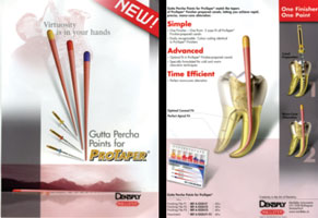
Therefore, the cone fit advantage in warm gutta-percha techniques is an opportunity for continuous clinical judgment. There is no absolute cutback length. It depends on the above factors and good clinical judgment. The tendency for beginners in warm gutta-percha techniques is to cut the gutta-percha cone back too far. The cone can be cut back in an infinite number of cross-sectional dimensions, making the gutta-percha cone versatile and adaptive to any clinical situation, from extremely open apices to curved root canal systems (Figure 6a). Precutback gutta-percha cones such as ProTaper gutta-percha cones (DENTSPLY/Tulsa) make the cone fit easier, and the full length of the geometries better replicate the geometries of the metals of the finishing files that created those shapes (Figure 6b).
 |
| Figure 6c. Example of lateral canal obturation during the down-pack heat wave. |
(2) Vehicle for conduction. The presence of a cone allows for a warm wave of compaction moving apically and then a warm wave of compaction moving back coronally (Figure 6c). The heat wave, which is the second critical distinction of warm gutta-percha techniques, will be described in a future article. Without this conduction vehicle, there would be little chance to eliminate foramina or portals of exit predictably along the walls and in the apical third of the root canal system. In the past, chemicals have been used to soften the gutta-percha, but excessive shrinkage often oc-curred. Friction can be used to mold gutta-percha, but the clinician experiences less control.
(3) Control. Considerable control is achieved by carefully fitting the cone, then warming and pressing it in repetitive cycles until obturation is complete. The appropriate shape of the cone also ensures the correct geometric relationship between itself and the shaped canal; ie, the cone fits in the apical third of the root canal system and then has a diminishing taper toward the middle and coronal thirds of the root canal space.
(4) Peace of mind. The cone fit gives the clinician exceptional confidence because it is known what the outline form of the down-pack and subsequent finish-pack films will look like. This takes all of the stress out of endodontic obturation. The only question is how many foramina will be discovered that were previously not known to exist. The basic shape, however, is no surprise, and there is virtually no concern about the apical position of the packing being significantly too short or too long. Confidence of 3-D obturation is achieved, and the first prerequisite for a predictable result is met.3
(5) Choices. The Rodale Synonym Finder defines choice as selection, discretion, selectivity, differentiation, druthers, option, possibility, answer, a way out, and solution. Perhaps the most significant contribution that the cone fit makes is to offer more solutions and more opportunities to finesse the obturation with greater precision. Each shaped canal is unique in itself. The cone allows us to adapt to the characteristics of each situation, which in turn gives the clinician a greater dimensional flexibility and more latitude in judgment. Obturation of root canal systems is analogous to squeezing a round peg into a square hole. The dimensions of the gutta-percha cone must change to fit the dimensions of the foraminal constriction. This provides freedom in treatment, which translates into confidence and a state of well-being during treatment. The process of blending the components of the gutta-percha cone and the root canal shape is the foundation for mastering clinical endodontics and achieving a sense of enjoyment and energy as well as excellence. Endodontic success then be-comes more a matter of choice than chance.
Guidelines for Cone Fit
(1) Establish the vision. Know the shape you desire to design so the cone will fit correctly.4
(2) If the cone does not fit, then fit a second one by cutting back at a slightly different cross-sectional diameter than previously. If the second cone does not fit, then put down the cones and pick up the files again because the shape needs improvement. The cone is simply matching to a shape, and if the cone does not fit, then the appropriate shape is not yet accomplished.
(3) Fit the cone to the radiographic terminus and then cut back 0.5 to 1.0 mm (+/-) depending on the length, width, and curvature of the root canal. Use quality, sharp Irsis scissors or a straight surgical blade such as a No. 11 Bard-Parker.
(4) Be sure to cut back the cross-sectional diameter of the gutta-percha cone equivalent to the cross-sectional diameter and length of the last finishing instrument, which gauges the diameter of the radiographic terminus but is held there by the foraminal constriction. Verify with a radiograph. After the cutback for packing, verify with a radiograph again.
(5) Freeze the shape of the gutta-percha cone by submerging it in 70% alcohol with cotton pliers and then forming the canal curvature replication by pulling the cone through a 2-x-2 gauze pad. This step creates a shape that mimics the flow of the shaped root canal preparation. A template of the canal is therefore made, and controlled packing can be accomplished. The cone can be stored if packing will be accomplished at a later visit.
(6) Before fitting the cone, an important step is to dry the canal. Then, in order to prevent the accumulation of possible dentin mud from along creases of the root canal system, confirm patency of the foramen with a file smaller than the largest gauging instrument that fits easily to the radiographic terminus. Many portals of exit are blocked without this last and very important step.
(7) Coat the apical third of the gutta-percha cone with a thin layer of sealer and gently follow the cone to place in preparation for obturation.
CONCLUSION
Incorporating the guidelines discussed in this article and understanding the significance of the gutta-percha cone fit in relationship with proven endodontic biologic principles will consistently create obturation excellence and produce results that are increasingly satisfying for the patient and clinician.
In a later issue, the author will discuss the second critical distinction of warm gutta-percha techniques: the heat wave.
References
1. Schilder H. Vertical compaction of warm gutta-percha. In: Gerstein H. Techniques in Clinical Endodontics. Philadelphia, Pa: WB Saunders Co; 1982:84-90.
2. Cohen J. A Quantitative Analysis of the Forces and Pressures Generated Within the Root Canal System During Vertical Condensation of Warm Gutta-Percha [thesis]. Boston, Mass: Henry M. Goldman School of Dental Medicine; 1974.
3. West J. The Incidence of Underfilled Foramina in Endodontic Failures [thesis]. Boston, Mass: Henry M. Goldman School of Dental Medicine; 1975.
4. Schilder H. Cleaning and shaping the root canal. Dent Clin North Am. 1974;18:269-296.
Dr. West is founder and director of the Center for Endodontics Pioneering New Possibilities in Endodontics in Tacoma, Wash. He currently serves as a trustee of the American Association of Endodontists (AAE) Foundation, has served on the educational affairs committee and committee on dental care and clinical practice for the AAE, and is an international lecturer and teacher. He is a member of the American Academy of Esthetic Dentistry and the International College of Dentists, an editorial board member of The Journal of Esthetic and Restorative Dentistry, and scientific editor for Boston Universitys Communiqu. He is an affiliated associate professor at the University of Washington School of Graduate Endodontics and clinical instructor at Boston University Henry M. Goldman School of Dental Medicine. He was senior author of Cleaning and Shaping the Root Canal System in Cohen and Burns 1994 and 1998 editions of Pathways of the Pulp, is a contributing author to the 1995 edition of Goldstein and Garbers Complete Dental Bleaching, and co-authored Obturation of the Radicular Space with Dr. John Ingle in Ingles 1994 and 2002 editions of Endodontics. He maintains a private practice in Tacoma and can be reached at (866) 900-7668 or johnwest@centerforendodontics.com.


