


Drawing blood in the dental office is a regular occurrence. It’s unavoidable, really. Gums bleed when we nick them, floss them, and move them around during procedures. Occasionally, the tongue will bleed when it’s pinched by a suction, or a cheek will bleed when it’s scraped by a dry angle. But now, it’s becoming common practice for blood to be drawn at the dental office intentionally! Enter PRP, PRF, and PRGF.
Platelet-rich plasma is PRP.
Platelet-rich growth factor is PRGF.
Platelet-rich fibrin is PRF.
If these acronyms “promote and accelerate wound healing, regulate inflammation, and improve soft and hard tissue regeneration,” then, which one might be the best option? Let’s look at how they are the same, how they are different, which one is used for what, and which is the best option depending on one’s need.
How Are They the Same?
PRP, PRGF, and PRF all require the obtaining of an autologous blood sample to extract the platelet-rich layer used for accelerated healing in oral surgical procedures. In other words, they all require taking a blood sample from the patient to aid in their healing.
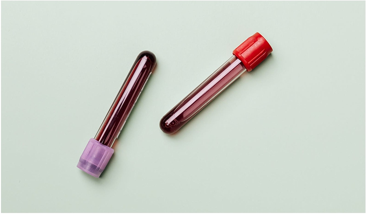
The samples in each case are spun in a centrifuge to separate the blood into three layers. The heaviest red blood cells sink to the bottom. The middle platelet-rich layer is suspended above that. The very light platelet-sparse layer is at the top.
The speed at which a blood sample is spun will yield a different concentration of certain blood components in these layers. The gel-like layer of platelets suspended in plasma in the middle is collected and injected or applied into the site of injury (surgery) to accelerate the patient’s healing following a surgical procedure.
How Are They Different?
With so many acronyms, it’s easy to feel lost. Many times, the terms might be used synonymously when in fact they’re each a little different.
In simplest terms, PRP and PRGF can be thought of as more of a gel-like product and PRF as a more clot-like product. But just for fun, lets dive in a little deeper.
PRP, the older approach, hails from the “dark ages” of 1997. (I kid—sort of.) It is primarily used for soft-tissue regeneration rather than osteogenesis and requires more blood than PRF. PRP is spun at a higher speed, making all the heavy white blood cells and stem cells sink to the bottom of the tube, where they are not collected in the sample.
Additional research resulted in the discovery that a higher concentration of platelets with the inclusion of white blood cells and stem cells in the sample would be even more therapeutic. Enter PRF. It turns out the inclusion of white blood cells aided in avoiding postoperative infection, and the presence of stem cells had obvious regenerative capacity.
Since PRF is spun at a lower speed, a higher concentration of white blood cells, stem cells, and platelets remains in the middle plasma layer, protected from the mechanical damage that happens when spun at a higher speed (like PRP) along with a higher platelet concentration.
PRF ends up producing about twice as many platelets as found in the body compared to PRP. This fact is important to point out since platelets are responsible for releasing growth factors that repair and help regrow tissue and, combined with fibrin, clot the blood, forming a plug that seals up blood vessels to prevent the loss of blood.
It makes sense that a higher platelet concentration results in a greater yield of growth factor. The growth factor in PRF is slowly released over about seven to 10 days due to its fibrin network, whereas PRP releases growth factor immediately in the absence of a dense fibrin network.
Unlike PRF, PRP sometimes requires the initial addition of an anticoagulant to prevent the blood sample from clotting too much and too quickly. The most commonly used anticoagulant in PRP is acid citrtate dextrose (ACD), which “binds calcium and prevents the coagulation proteins from initiating the clotting cascade.” PRF doesn’t need an anticoagulant.
After being spun, PRP is collected and usually mixed with bovine thrombin or calcium chloride. Both are coagulants that activate a PRP sample, prompting fibrinogen to convert to fibrin, resulting in a more gel-like product. Centrifugation for PRF results in a clot that is plucked out of the test tube after it is spun.
Finally, PRGF offers growth factor and blood clotting at the site of wound healing, but it is the most costly and does not yield a lot of fibrin. PRGF is very similar to PRP in the way that it is collected and prepared but does not require animal thrombin for coagulation. However, it still requires the addition of calcium chloride as a post centrifugation fibrinogen activator (coagulator) like PRP and is also a more gel-like product.
Which One Is Used for What?
All in all, PRP and PRF overlap all over the place in their applications. But again, it’s hard to avoid the fun of diving in a little deeper.
As mentioned, after collection, PRP is mixed with a coagulating activator, prompting fibrinogen to convert to fibrin, resulting in a gel-like product. The PRP can then be used at extraction sites and is often used in cases where the surgical site can be reapproximated or covered easily with the patient’s own gum tissue since it is not as dense as PRF. Sometimes, it is used to hydrate bone graft or coat implants.
PRGF, like PRP, is a liquid product that requires an activator coagulant (calcium chloride) to elicit the conversion of fibrinogen to fibrin to achieve a gel-like consistency and is then used commonly in the same way that PRP is without the addition of an anticoagulant at the start. PRGF is not the superior option for hard-tissue healing since it lacks the stem cells found in PRF but is still good for coating an implant before it is placed.
PRF, a clot product, is most often used for guided tissue and bone (both hard and soft tissue) regeneration. Because PRF can be used as a membrane or plug, it is often used in root coverage procedures, socket and soft-tissue grafting procedures, bone grafting and management around implants, and recession cases. It can even be used in cosmetic facial surgeries and in the form of “I-PRF” in the case of temporomandibular joint (TMJ) pain.
Wait… There Are Sub-Acronyms, Too?
Oh yes.
Let’s close this out with a note on three slight variations of PRF used depending on the application:
- L-PRF: leukocyte and platelet-rich fibrin
- A-PRF: advanced platelet-rich fibrin
- I-PRF: injectable platelet-rich fibrin
First, to simplify, these slight variations of PRF are really just the result of different speed and time of spin in the centrifuge, yielding a higher percentage of different cell types over the others. A-PRF and L-PRF are both clot-like results, and I-PRF is a liquid gel.
L-PRF is great for decreasing the chances of postoperative infection, given it is rich in white blood cells, leukocytes to be more specific, which helps decrease inflammation. It is great for extraction sites by minimizing bone loss, and it can even be used to revitalize the pulp of a tooth to avoid the need for root canal or extraction in some cases.
A-PRF is spun for the same amount of time as the traditional PRF sample but at a slower speed, lending a more uniform spread of growth factor in the clot. It also is used for the same applications as PRF. The slow spin generally yields a greater red blood cell inclusion, as you can imagine.
I-PRF is the injectable form of PRF. It can be used as treatment for TMJ pain by being injected directly into the joint! It makes delivering stem cells directly into the joint possible because it is liquid as it is being injected and clots after the fact. It helps enhance vascularity. It’s useful in bone grafting where it can hydrate the bone initially. As it firms up, the graft is easier to place because the tiny bone particles can no longer flake out.
So, Which Is the Best Option?
PRF results in a greater concentration of platelets. With that comes a greater concentration of growth factor and fibrin and, therefore, more efficacious healing—not to mention the growth factor is released more slowly in PRF, so the product keeps working well beyond the time of surgery. Stacked up against PRP and PRGF, PRF appears to be winning.
Looking at the brief overview of which one is used for what, we can see a lot of overlap between applications. PRF is easier to collect and prepare. It also does not require as much blood or any additives. Plus, it can be used to heal of both hard and soft tissue while stimulating new bone and new blood vessel formation.
So, PRF appears to reign supreme when it comes to autologous platelet-rich therapies in dentistry. It is simply a choice between using the everyday PRF form or the L-PRF, A-PRF, or I-PRF applications, depending on the procedure.
All in all, it appears that drawing blood at the dental office isn’t going away and, in fact, may be beneficial for optimal healing!
References
- Giannini S; Cielo A; Bonanome L; Rastelli C; Derla C; Corpaci F; Falisi G, European review for medical and pharmacological sciences [Eur Rev Med Pharmacol Sci], ISSN: 2284-0729, 2015; Vol. 19 (6), pp. 927-30; Publisher: Verduci; PMID: 25855914, Database: MEDLINE Complete PubMed(2)
- The International Cellular Medical Society. (n.d.). Guidelines for the Use of Platelet Rich Plasma. The International Cellular Medical Society. http://www.cellmedicinesociety.org/attachments/206_ICMS%20-%20Guidelines%20for%20the%20use%20of%20Platelet%20Rich%20Plasma%20-%20Draft.pdf. (3)
- Kobayashi E;Flückiger L;Fujioka-Kobayashi M;Sawada K;Sculean A;Schaller B;Miron RJ; (2016). Comparative release of growth factors from PRP, PRF, and advanced-PRF. Clinical oral investigations. https://pubmed.ncbi.nlm.nih.gov/26809431/. (6)
- PRF vs PRP: PRF stands out as superior compared TO PRP. (2020, August 09). Retrieved April 05, 2021, from https://www.dentaliceberg.com/prf-prp-prgf-which-one-is-the-best/#:~:text=PRF%20stands%20out%20as%20a,the%20blood%20into%20distinct%20layers. (1)
- Rehan, K. (2020, April 20). A Quicker Way to Heal? PRP and PRF in Dentistry. constituent. https://www.agd.org/constituent/news/2020/04/20/a-quicker-way-to-heal-prp-and-prf-in-dentistry. (5)
- Team, I. P. (n.d.). The benefits and limitations of using platelet-rich fibrin for implants: Implant Practice US. Implant Practice US – Dental Publication. https://implantpracticeus.com/ce-articles/a-comprehensive-clinical-review-of-platelet-rich-fibrin-prf-and-its-role-in-promoting-tissue-healing-and-regeneration-part-2/. (4)
Dr. Schneider is the dental director at Delta Dental of Arizona and has more than 19 years of experience in dental administration, dental education, and clinical dentistry.
Related Articles
Empower Patients to Screen Themselves for Oral Cancer
Teledentistry Can Help Us Solve Dentistry’s Access to Care Challenges
Proactive Collaboration Between Dentists and Physicians Is Key to Improving Care


