
INTRODUCTION
Prosthetic management of partial edentulism can be challenging with the presence of limited interocclusal space. Over time, edentulous areas that are not restored may lead to drifting, tipping, rotation, and supraeruption of neighboring and/or opposing teeth.1-3 The extrusion of opposing teeth in combination with the alveolar extrusion of the edentulous areas reduce the space needed for fabricating a removable or fixed prosthesis when the edentulous areas are present in maxilla.4 Regaining the lost interocclusal space is a requirement for a successful prosthetic treatment for these cases.2-6 Also, interarch distance dimension must be clearly visualized prior to implant placement.7 Several approaches, such as no treatment, restoration with a shortened prosthesis, intrusion of the extruded teeth, posterior maxillary alveoloplasty, or the reduction of the extruded teeth (which may require endodontic treatment and periodontal surgery) have been proposed for the extension of interocclusal space.2-4,8,9 The clinical situations in combination with the desires of the patient are the critical factors in selecting the appropriate treatment option.3
The combined surgical and prosthetic treatment of a partially edentulous case with limited interocclusal space will be presented in the following clinical case report.
CASE REPORT
A 45-year-old female patient presented to department of prosthodontics, Istanbul University, for prosthodontic treatment of the missing maxillary left posterior region (Figure 1). Clinical photos (Figures 2a and 2b), radiographic (Figure 3) examination, and mounted diagnostic casts (Figure 4) showed severe alveolar extrusion of the maxillary left posterior segment and a slightly reduced vertical dimension of occlusion (VDO).
 |
| Figure 1. Partially edentulous maxilla. |
 |
 |
| Figure 2a. Preoperative smile. | Figure 2b. Intraoral view of the reduced interocclusal space. |
 |
 |
| Figure 3. Panoramic radiograph prior to treatment. | Figure 4. Reduced interocclusal space, as observed in the mounted casts. |
The treatment was started by increasing the VDO by adjustment of the occlusal plane of the mandibular teeth. A Chromascope Shade Guide (Ivoclar Vivadent) was used to select an appropriate shade, and then the worn mandibular right posterior teeth were prepared with supragingival shoulder margins for ceramic onlays. Preparations were made using an inlay/onlay preparation set (Inlay Preparation Set 4261 [KOMET/Brasseler]). Next, the mandibular left posterior teeth were prepared for ceramic crowns and a definitive impression of the tooth preparations was made using a vinyl polysiloxane (VPS) impression material (Brecision [Bredent]). The impression of the opposing arch was made with an irreversible hydrocolloid (Xantalgin [Heraeus Kulzer]). Three ceramic onlays (Figure 5) and a 3-unit metal ceramic bridge with metal occlusal surfaces (in order to gain interocclusal distance) were fabricated in the dental laboratory. After checking the occlusion and aesthetics, the ceramic onlays were cemented using dual-cure resin cement (NX3 Nexus Third Generation [Kerr]) and the 3-unit metal ceramic was cemented using a polycarboxylate cement (Poly F Plus [DENTSPLY DeTrey GmbH]). After the restoration of the mandibular teeth, the prosthetic treatment of the left maxillary edentulous area was planned. It was not possible to restore the left maxillary edentulous area with a fixed or removable prosthesis due to missing interocclusal space. Therefore, insertion of 3 dental implants after regaining the adequate interocclusal space by a maxillary alveoloplasty would be performed.
 |
 |
| Figure 5. Fabricated ceramic onlays shown on the cast. | Figure 6. Alveoloplasty, before the insertion of implants. |
 |
 |
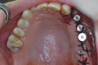
| Figure 7. Intraoral view of the implants immediately after surgery. | Figure 8a. View of the regained interocclusal space. |
 |
 |
| Figure 8b. Panoramic radiograph after the insertion of implants. | Figure 9. The implant positions shown on the cast. |
 |
 |
| Figure 10a. Final view of the patient. | Figure 10b. Cemented ceramic onlays. |
A crestal incision throughout the maxillary left edentulous area and vertical releasing incisions were made. A mucoperiosteal flap was elevated buccally and lingually. Following the alveoloplasty (Figure 6), 3 dental implants (Swissplus [Zimmer Dental]) were place
d according to manufacturer’s instructions. One stage surgery was chosen; the implants were supplied with healing abutments, and mucoperiosteal flaps were sutured with a close adaptation of the wound margins to the implant shoulders (Figure 7).
Four months later (Figures 8a and 8b) the gingiva formers were unscrewed and a definitive impression was made using VPS material (Brecision). Three separate metal ceramic crowns were fabricated on the model (Figure 9) and cemented on the 3 maxillary implants.
The patient was instructed in maintenance and hygiene procedures associated with the fixed dentures. The routine recall appointments were scheduled on a 6-month basis and no complications occurred during the 4-year follow-up period (Figures 10a and 10b).
DISCUSSION
The prosthetic rehabilitation of partially edentulous patients can engender a challenge for the clinician when there is reduced and inadequate interocclusal space. The occlusal scheme may be deformed due to early loss of teeth when the opposing dentition supraerupts towards the edentulous space.4 The extrusion of opposing teeth and/or the alveolar extrusion of the edentulous areas reduce the space needed for fabricating the partial denture.4 Before any prosthetic reconstruction can commence, lost intermaxillary space must be regained.3 Restoring the accurate plane of occlusion can be accomplished by periodontal, orthodontic, conservative restorative, and surgical procedures. Conservative treatment options like no treatment, intruding the mandibular posterior teeth, or periodontal surgery in combination with endodontic treatment were not chosen for the case presented in this article. Since sufficient bone for alveoloplasty was available in the maxillary edentulous area, the plane of occlusion in the mandible was corrected by a conservative approach.10 The mandibular right posterior teeth were restored with ceramic onlays, a relatively conservative treatment option, when compared with the alternative choice of full-coverage crown restorations.11 The mandibular left posterior teeth were restored with a ceramic bridge because the teeth had been prepared prior to treatment in our clinic.
The use of dental implants to replace missing teeth has become the standard of care for edentulous spaces. With the presence of restricted interocclusal clearance, screw-retained restorations have been proposed because it may not be possible to develop adequate retention to retain restorations on implants with cement.5,12 Screw-retained restorations can be secured to implants with as little as 4 mm of space from the surface of the implant to the opposing occlusion.12 However, if the crown length is too short, it may negatively affect the aesthetics; especially when the crowns are in the smile line as in the patient presented in this report. The main reason why a screw-retained implant restoration was not necessary here was the sufficient coronal distance in the maxilla gained by doing the alveoloplasty. Alveoloplasty was performed before the insertion of the implants in the same surgical session without a requirement for a second surgery.
CLOSING COMMENTS
Limitation in interocclusal space is a common problem in prosthetic dentistry. Several approaches have been proposed to solve this problem. The treatment presented here involved a combined prosthetic and surgical approach in order to gain interocclusal distance allowing a functional as well as aesthetic result. A 4-year follow-up period showed the treatment chosen had provided for a successful and stable result.
References
- Mopsik ER, Buck RP, Connors JO, et al. Surgical intervention to reestablish adequate intermaxillary space before fixed or removable prosthodontics. J Am Dent Assoc. 1977;95:957-960.
- Lee HE, Lee KT, Tseng YC, et al. Interdisciplinary management of unfavorable posterior intermaxillary space. Br J Oral Maxillofac Surg. 2008;46:413-415.
- Chun YS, Row J, Yang SJ, et al. Management of extruded maxillary molars to accommodate a mandibular restoration: a clinical report. J Prosthet Dent. 2000;83:604-606.
- Chen CM, Tseng YC, Huang IY, et al. Interdisciplinary management of dental implant patient: a case report. Kaohsiung J Med Sci. 2004;20:415-418.
- Armellini D, Bilko S, Carmichael RP, et al. Screw-retained prosthesis for Straumann implant sites with limited interocclusal clearance. J Prosthodont. 2006;15:198-201.
- Chaimattayompol N, Arbree NS. Assessing the space limitation inside a complete denture for implant attachments. J Prosthet Dent. 2003;89:82-85.
- Alsiyabi AS, Felton DA, Cooper LF. The role of abutment-attachment selection in resolving inadequate interarch distance: a clinical report. J Prosthodont. 2005;14:184-190.
- Melsen B, Fiorelli G. Upper molar intrusion. J Clin Orthod. 1996;30:91-96.
- Alessandri Bonetti G, Giunta D. Molar intrusion with a removable appliance. J Clin Orthod. 1996;30:434-437.
- Bittner N, Lal K, Neurohr J. Fabrication of a custom abutment for a wide-diameter implant in a situation with limited interocclusal space. J Prosthet Dent. 2008;100:474-477.
- Sorensen JA, Choi C, Fanuscu MI, et al. IPS Empress crown system: three-year clinical trial results. J Calif Dent Assoc. 1998;26:130-136.
- Chee W, Jivraj S. Screw versus cemented implant supported restorations. Br Dent J. 2006;201:501-507.
Dr. Geckili earned his DDS from the Marmara University School of Dentistry (1999). He also holds a PhD in prosthodontics (2007) from the School of Dentistry, Istanbul University, Department of Prosthodontics, where he is currently a research assistant. He is a member of Academy of Prosthodontics and Gnathology Society, Turkish Society of Oral Implantology, and European Academy of Osseointegration. He is experienced in implantology and prosthodontics, has taught for more than 10 years, is a proficient lecturer, and has published more than 15 papers in peer-reviewed professional journals. He can be reached at [email protected].
Disclosure: Dr. Geckili reports no disclosures.
Dr. Bilhan earned his DDS in 1993; has a postgraduate certificate in oral surgery and implantology from the School of Dentistry, Karl-Franzens University of Graz, Austria (1994); and holds a PhD in oral surgery from the School of Dentistry, Zurich University, Department of Oral and Maxillofacial Surgery (1996). In 2004, he received a PhD in prosthodontics from School of Dentistry, Istanbul University, Department of Removable Prosthodontics, where he is currently a research assistant. He is a member of Academy of Prosthodontics and Gnathology Society, Turkish Society of Oral Implantology, a Fellow of the International Team for Implantology, a Fellow of International Congress of Oral Implantology, and a master of Implant Prosthodontics Section. His experience with implantology, prosthodontics, and oral surgery includes extensive clinical experience with more than 10,000 successfully performed oral surgeries. He has taught for more than 14 years and has published more than 34 papers in peer-reviewed professional journals. He has extensive international lecture experience with multilingual capabilities (fluent in Turkish, German, English, and intermediate level French). He can be reached via e-mail at [email protected].
Disclosure: Dr. Bilhan reports no disclosures.
Dr. Mumcu holds a DDS from Istanbul University School of Dentistry (1998) and a PhD in Prosthodontics (2006) from School
of Dentistry, Istanbul University, Department of Removable Prosthodontics (2006), where he is also a research assistant. He holds memberships in the International Association for Dental Research, Academy of Prosthodontics and Gnathology Society, Turkish Society of Oral Implantology, European Academy of Osseontegration, International Team for Implantology, the Assocication of Prosthodontics, American College of Prosthodontists, Association of Prosthodontics of Canada, and the European Association for Osseointegration. He is experienced in implantology, prosthodontics, and temporomandibular disorders. He is a proficient lecturer, has taught for more than 12 years, and has published more than 32 papers in peer-reviewed professional journals. He can be reached via e-mail at [email protected].
Disclosure: Dr. Mumcu reports no disclosures.

