


Beth Groenke, DDS, MS, has received the INfORM Samuel F. Dworkin Predoctoral Award from the International Association for Dental Research for her research into the use of MRIs in oral health diagnosis. She is a graduate student in the University of Minnesota School of Dentistry’s Division of TMD and Orofacial Pain.
“I was always intrigued by TMD and orofacial pain,” Groenke said. “Though I worked with excellent faculty, many of them couldn’t give me satisfactory answers on managing my TMD patients.”
Citing a poor understanding of the field, Groenke said, she decided she wanted to learn more. She joined the University of Minnesota’s Orofacial Pain program “because of its excellent mix of clinical and research opportunities,” she said, which enabled her to learn the clinical aspects of pain management and use her undergraduate engineering skills again.
After completing a master’s degree in dentistry, Groenke has continued researching with the dental MRI development team. She is now working with Donald Nixdorf, DDS, MS, to explore the use of MRI as a diagnostic tool. In particular, her study on detecting vertical root fractures was the focus of her award-winning presentation at the virtual International Network for Orofacial pain & Related disorders Methodology (INfORM) conference.
“In orofacial pain, we see patients who have had root canals who then present with pain perceived in the tooth,” she said.
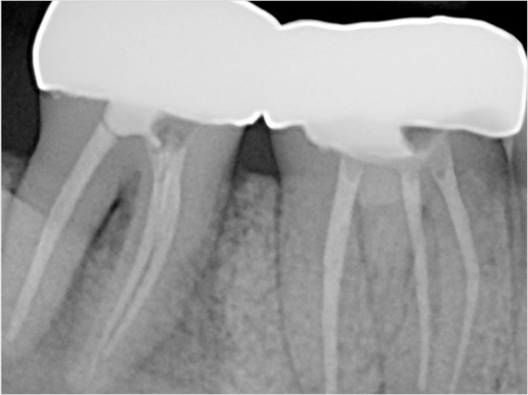
One potential cause of that pain is a vertical root fracture, which extends from the tip of the root toward the dental crown. Early and specific detection is key for this kind of pain, the school said. If dentists can detect a vertical root fracture, they know to extract or amputate part of the tooth and eliminate the pain. But if the pain stems from another cause like neuropathy or TMD, that surgery can make the problem worse.
That’s where Groenke’s research comes in, the school said, as the most common imaging tools used by dentists such as CBCT scans can often miss these small fractures.
“Our research aimed to explore if dental MRI could detect these very small fractures,” Groenke said, as well as evaluate the sensitivity and specificity of MRI to detect fractures compared with more frequently used tools like a CBCT.
In a study of 115 extracted teeth, Groenke found that the MRI performed similarly to the CBCT scan in terms of sensitivity and specificity and was able to identify smaller fractures than those previously reported for CBCT scans.
“This is encouraging for the future of dentistry because based on this research, MRI is performing at an equivalent level to existing technologies,” Groenke said, even though the MRI’s use in dentistry is in its early stages.
Groenke said she is grateful to have received the Samuel F. Dworkin Award for this work.
“Within the orofacial pain field, the INfORM network is well known to be comprised of many preeminent researchers from all over the field,” she said. “I was extremely honored to receive the award.”
For Groenke, the award is confirmation of what she already knows: this field, and this lab, are where she is meant to be.
“I love that it’s cutting edge,” she said. “Only a handful of other groups in the world are working to develop MRI for dentistry, and I don’t believe any of them are pushing the boundaries of technology quite like our group is.”
As part of the award, Groenke will be a judge in next year’s presentations.
Related Articles
Vertical Root Fractures Occur More Frequently When a Post Is Present
A More Precise Way to Re-Treat Root Canals
A Short Case Study: The Second Time’s the Charm


