


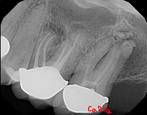
A patient came in with pain on tooth No. 15. The diagnosis was necrotic pulp with a large periapical radiolucency (Figure 1).
The size of the lesion has nothing to do with the healing potential. Some providers may assume it’s a cyst and jump into surgery or an extraction and implant.
I treated the case with root canal therapy in two visits using calcium hydroxide (Figure 2). The patient returned asymptomatic, and I completed the case with bioceramic sealer and gutta-percha (Figure 3).
The patient returned nine months later for an evaluation (Figure 4). The area showed very nice osseous repair due to the large size of the lesion.
 |
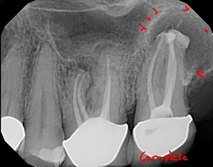 |
| Figure 2. Treatment involved two visits with root canal therapy and calcium hydroxide. | Figure 3. Bioceramic sealer and gutta-percha was used to complete treatment. |
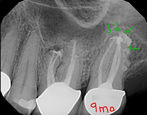 |
| Figure 4. Nine months later, there was very nice osseous repair. |
As you can tell, I’m passionate about this stuff! Did you know:
- You can’t diagnose a cyst on a radiograph. It needs a biopsy with histology (White et al 1994 OOO).
- There is a statistically significant higher proportion of cysts found in the premolar than in the molar region (Mortensen et al EJOS 1970)
- Fistulas occur more often in connection with granulomas than with cysts (Mortensen et al EJOS 1970)
- 85% of all periapical lesions are granulomas. The others are cysts or other non-odontogenic pathologies (Nair 2004 OOO).
- Radicular cysts are considerably less frequent and occur in two distinct histological categories: apical true cysts and apical pocket cysts (Nair 1996 OOO).
- Radicular true cysts are entirely enclosed by epithelium (Nair 2004 OOO).
- Granulomas and pocket cysts may heal after non-surgical root canal therapy, whereas true cysts are self-sustaining and usually require apical surgery (Simon 1990 JOE).
- Metastases of cancer in the mouth are extremely rare, comprising 1% of all malignant mouth neoplasms. Because of its rarity, the diagnosis of metastatic lesions in the oral cavity is very often missed (Sanchez 2005 MOPO).
- Metastatic cancers are located in the mandible in 80% to 90% of cases (Sanchez 2005 MOPO).
- It’s very rare, but sometimes metastatic lesions in jaws may show vague pain and be misdiagnosed as pathologic entities of dental original such as pulpal or periapical disease (Khalili 2010 JOE).
Dr. Short attended the Medical College of Georgia School of Dentistry to attain a DMD degree in 1999. In 2002, he earned his post-doctorate degree in endodontics from Nova Southeastern University and then became a Diplomate of the American Board of Endodontics in 2009. Dr. Short is an expert consultant in endodontics to the Georgia Board of Dentistry, author, speaker, and assistant clinical professor at the Dental College of Georgia in Augusta. His private practice, Apex Endodontics PC, is located in Smryna, Ga. He can be reached at dr.short@yahoo.com.
Related Articles
Retreatment Vs Extraction and Implant: Making Sound Clinical Treatment Decisions
A Short Case Study: Everything Including the Kitchen Sink
A Short Case Study: Separated Instruments? Perforation? Relax!


