


INTRODUCTION
Origin and Features of the Nitinol Alloy
In 1958, scientists at the United States Naval Ordinance Laboratory were looking for a metal that could be used to improve missile nose cones. They wanted a metal that could resist fatigue, heat, and the force of impact. They ultimately discovered what we know today as NiTi, a mixture of nickel and titanium alloys.
NiTi is an exotic metal in that it does not behave like normal alloys. Normal alloys become more ductile when they are heated and eventually melt when a certain temperature is reached. NiTi is a metal that does the opposite. At high temperatures, NiTi transitions to a cubic molecular structure which is more robust and resistant to torsional stress; this is known as the Austenite, or parent, phase (A-phase) (Figure 1a). At cooler temperatures, NiTi transitions to a more complicated molecular structure known as the Martensite, or daughter, phase (M-phase) (Figure 1b). In this M-phase, the metal is ductile and highly resistant to cyclic fatigue (think of a wet noodle). The temperature at which NiTi transitions from the M-phase to the A-phase can be altered with proprietary processes. During the manufacturing process, NiTi can be formed into a desired parent state (A-phase) shape. At lower temperatures, the metal can be manipulated; however, once exposed to a predefined (warmer) temperature, it will transition to the A-phase and morph into the predetermined shape.
 |
 |
| Figure 1. (a) Austenitic (A-phase) NiTi crystalline structure exhibits shape memory and forms at higher temperatures. This structure provides more strength and is torque resistant. (b) The Martensitic (M-phase) crystalline structure exhibits control memory and is very ductile and flexible. However, it is not ideal for cutting, since it is torque-sensitive. |
 |
| Figure 2. Several conventional NiTi files in cross section. These files cut around a central axis, creating a round cross-sectional shape during rotary or reciprocating motion. |
Evolution of NiTi File Designs
Setting metallurgy aside, there are more than 30 different NiTi file designs available on the market today. Many articles and textbooks attempt to categorize these files into generations (first, second, third, and so on); however, there is no standardized or universally agreed upon lexicon for categorizing these instruments. For simplicity’s sake, we can categorize all NiTi file designs into one of the following 3 categories:
1. Conventional NiTi Files—This includes any NiTi file design that features a symmetric cutting axis. The pitch of the flutes can be consistent or inconsistent, but every cutting flute is designed to touch the canal walls. These files bore a perfectly round hole and require a higher torque setting because the cutting flutes do not disengage. These are the original batch of NiTi files on the market, and some popular file brand names in this category include the following: ProFile, ProFile GT, ProTaper, ProTaper Gold, K-3, Quantec, V-Taper, Vortex, Vortex Blue, and Edge Files (Figure 2).
2. Eccentric NiTi Files—This category of file represents any of the more modern NiTi file designs that feature an asymmetric cutting axis. An eccentric NiTi file can cut slightly outside of its central axis of rotation and requires lower torque because the cutting flutes can disengage along the axis of the canal, and all cutting edges are not engaged simultaneously. Some popular brand names in this category include EndoSequence, ESX, ProTaper Next, and TruShape.
3. Expanding NiTi Instruments—This category is the newest category of NiTi instrumentation, and it includes any NiTi file design that features an instrument that can expand in the canal to adapt to the canal’s natural morphology (oblong canals, etc). As a result, these instruments are considered to possess adaptive cores, where the envelop of motion is greater than the file diameter itself. To date, there are only 3 instruments that fall into this category: the self adjusting file (SAF), XP-3D Shaper (Brasseler USA), and XP-3D Finisher (Brasseler USA) (Figure 3).
 |
 |
| Figure 3. Three adaptive files so far have been introduced onto the market. (a) The original was the self adjusting file (SAF). (b) The 2 new ones are the XP-3D Shaper and Finisher (Brasseler USA). |
 |
| Figure 4. Because of their round cutting in cross sections, traditional NiTi rotary and reciprocating files are unable to clean the oval areas of the root canal adequately. |
 |
 |
| Figure 5. The centric axis of rotation of the file imposes a great amount of torque on traditional rotary instruments. Excessive torque with round cross section files (a) that creates round holes in canals (b) can over torque the instrument leading to unwinding and possible file separation. |
 |
| Figure 6. To reduce torsional and flexural fatigue, instrumentation must be done with serially larger files, increasing the number of instruments needed to prepare a root canal. |
Disadvantages of Conventional and Eccentric NiTi Files
Despite the improved efficiency associated with NiTi instrumentation, all conventional and eccentric NiTi files have the same universal disadvantages, as follows:
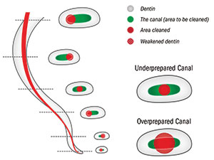
1. Nonanatomic Shaping—The root canal system is highly complex. Most canals have an irregular anatomic shape. Despite significant advancements, traditional files can only make round shapes and cannot reach oval areas of the canal during treatment (Figure 4).
2. Excessive Torque and Fatigue—Traditional NiTi files are prone to file deformation and potential separation, and they apply unwanted stress to the tooth. This is because of the excessive torque that is applied due to a centric axis of rotation (Figure 5).
3. Requiring the Use of Multiple NiTi Files—To reduce cyclic and torsional fatigue, multiple file sequencing techniques are utilized. Furthermore, in multirooted teeth, different master file sizes may be required. This adds cost, complexity, and chair time to the procedure of shaping canals for obturation (Figure 6).
Expanding NiTi Instrumentation
Most clinicians would agree that the ideal root canal cleaning instrument should deliver the following benefits:
- Simplicity—One to 2 instruments is ideal
- Safety—The instrument should not separate, and if it does, it should be easy to remove
- Efficiency—Excellent debris removal (cleans as quickly as possible while maintaining safety)
- Anatomical—Adaptation to canal irregularities (cleaner is always better)
- Minimally invasive—Saving as much tooth structure as possible (while achieving endodontic success)
- Gentle, nonaggressive treatment—Avoid dentinal microcracks due to excessive torque
- Minimize patient discomfort—Avoid extrusion of debris and irrigants which can cause pain and failure.
3-D INSTRUMENTATION
The XP-3D Shaper attempts to address the shortcomings of traditional NiTi instrumentation, ushering in a new biologic standard of care in endodontic instrumentation. Once a minimum size 15/02 to 15/04 space has been scouted, and a path has been created in the root canal, the XP-3D Shaper’s patented MaxWire NiTi technology allows the instrument to expand inside this space at body temperature. As the instrument rotates, its orbit expands and contracts to abrade the broad and narrow aspects of the canal. This intuitive micromechanical debridement allows the practitioner to utilize a single instrument to safely clean and enlarge the canal while respecting the original canal morphology.
MaxWire Adaptive NiTi Technology
The MaxWire alloy has been designed to transition from a M-phase to an A-phase at body temperature (Figure 7). Therefore, the instrument is in its ductile and malleable M-phase structure at room temperature and becomes stiffer and assumes its programmed shape when it’s introduced into the root canal in vivo (at body temperature). This turns a very flexible instrument into an expanding instrument that is stiff enough to cut dentin upon rotation.
 |
| Figure 7. The MaxWire alloy has been designed to transition from a M-phase to an A-Phase at body temperature. This change in shape inside the root canal expands the file’s envelop of motion to touch a greater amount of root structure. |
Adaptive Core Technology
The central core of the XP-3D Shaper is a size No. 30 (0.3 mm) tip with a 1.0% taper. This thin MaxWire core allows for maximal flexibility and resistance to cyclic fatigue. When introduced to body heat, the Shaper expands its virtual core of motion to the equivalent of an 8.0% taper. The instrument responds to the resistance from feedback from the root canal wall. This is like a spring with the file’s virtual core exerting gentle outward pressure on the root canal wall in all directions inside the envelop of motion of the instrument. When engaged at 800 to 1,000 rpm, the Shaper efficiently debrides the root canal walls while respecting the original anatomy. In other words, this file will not create round shapes inside oval canals; instead, it cleans the entire oval to a larger size. Due to its off-axis rotation at the tip, the Shaper’s tip can contact a much larger diameter than its solid core diameter of size No. 30, reaching canal widths as wide as 30 or more. However, it will not expand the size of the apex unless the canal is naturally smaller than a size No. 30 (Figure 8). The adaptive core pulsates in the canal, dislodging dentinal debris and causing significant turbulence.
 |
 |
 |
| Figure 8. (a) Due to its off-axis rotation at its tip, (b) the XP-3D Shaper (Brasseler USA) can contact a much larger diameter than its solid core diameter of size 30 (0.3 mm), (c) thus reaching much greater canal widths than its own diameter. |
 |
 |
| Figure 9. Unlike the XP-3D Shaper, the XP-3D Finisher (Brasseler USA) is intended to clean a prepared canal and will not change the shape of the canal. This instrument can be used after conventional instrumentation to remove packed debris without significantly shaping the canal. | Figure 10. EndoSequence BC Sealer (Brasseler USA) has material properties like a filler, allowing pooling to take place without shrinkage or washout. This makes it an ideal cement for obturating natural shapes where pooling of cement is possible. |
The XP-3D Finisher
Like the XP-3D Shaper, the XP-3D Finisher utilizes MaxWire technology to adapt to the canal’s natural anatomy. This instrument has a sickle shape at body temperature and is incredibly flexible.1-7
Unlike the Shaper, the Finisher is intended to clean a prepared canal and will not change the shape of the canal (Figure 9). The Finisher has a larger expansion capacity than the Shaper and can reach upwards of 6.0 mm in diameter. The size No. 30 Finisher has been shown to provide superior final irrigation/cleaning compared to ultrasonics alone. The Finisher can also be used for retreatment and for cases with larger abnormal anatomy (eg, internal resorptions, immature teeth). The Finisher is available in sizes No. 25 and No. 30 with a a zero percent taper.
Premixed Bioceramic Bonded Obturation
In the era of modern endodontics, shaping is no longer dictated by the limitations of obturation materials. The introduction of nonshrinking bonded obturation (EndoSequence BC Sealer and BC Points [Brasseler USA]) allows practitioners to embrace the new adaptive NiTi instrumentation. Unlike traditional sealers, BC Sealer does not shrink, and it bonds to dentin and BC Points, so it is not necessary to condense gutta-percha to minimize the sealer interface (Figure 10). Therefore, some pooling in the oval areas of the canal are well tolerated, since BC Sealer is a filler, rather than a sealer, in its traditional terminology. With BC Sealer, the function of gutta-percha is simply to take up space, provide a path for retreatment, and provide for hydraulics/delivery of the sealer.
 |
 |
 |
| Figure 11. The hydration and precipitation reactions of BC Sealer showcase its biocompatibility, antimicrobial, and hydrophilic properties. The material properties of bioceramics produce an actual cement filler that, (a) due to its hydrophilic flow characteristics, (b and c) can fill anatomical spaces in the oval areas of the canal beyond the main gutta-percha cone and set without shrinkage, where there is significant pooling. |
During the BC Sealer setting/hydration reaction (Figure 11), the calcium silicate picks up water molecules from the dentin and forms calcium hydroxide. The calcium hydroxide combines with the calcium phosphate to form hydroxyapatite.
The material properties of bioceramics produce an actual cement filler that, due to its hydrophilic flow characteristics, can fill anatomical spaces in the oval areas of the canal beyond the main gutta-percha cone and set without shrinkage where there is significant pooling. The material properties of this cement overcome historical limitations of resorbable and shrinkable resin and zinc oxide eugenol-based cements that were the inspiration for the modern warm- and cold-condensation techniques (Figure 12).
 |
 |
 |
| Figure 12. (a) The pre-op and post-op radiographs of a maxillary second molar using the advanced XP-3D protocol, (b) showcasing the adaptive file technology offered by the XP-3D Shaper to (c) produce 4 different-sized master cones from a single Shaper instrument based upon the anatomical shape of the original root canal. |
The following are the steps of the XP-3D Obturation Technique:
Step 1. Confirm the apical diameter with a gutta-percha point, paper point, or standard file (in fingers).
Step 2. Coat the canal walls with BC Sealer.
Step 3. Coat the appropriate gutta-percha point with BC Sealer and place to working length.
Step 4. Sear off gutta-percha at the canal orifice and vertically compact with the appropriate size plugger.
CASE REPORT
To illustrate the expansion capacity of a single XP-3D Shaper file to accommodate different canal diameters and results into different master cone sizes, we can look at the clinical case in Figure 12a.
This necrotic maxillary second molar was treated endodontically in a single visit using the advance XP-3D protocol and obturated with different sized master cones (Figures 12b and 12c). Following access and initial coronal enlargement using an orifice opener and some hand files, each of the 4 canals were instrumented to a size No. 15 hand file. Additional taper was then provided by enlarging the canals to a size 15/04 with an ESX Scout (Brasseler USA). Following this tapered No. 15 preparation, the XP-3D Shaper file was introduced in each canal to working length. Once the working length was reached, 2 cycles of 10 long strokes, separated by irrigation and rinsing out the generated debris, were performed. Each of the constant tapered BC Cones were then fitted in each canal, starting with a size 30/04 and going up in size serially until a cone that was fitted to working length was determined for each canal. What’s interesting is that the same file provided different sizes for each of the 4 root canals (Figure 12c). Obturation with BC Sealer showed adequate enlargement appropriate for each canal.
The anatomical shaping possible with MaxWire’s expanding file technology allows a single file to provide anatomical shaping based on the natural shape of the root canal being treated. Here, the palatal root was naturally wide and, therefore, a size No. 50 was fitted after cleaning. A No. 40 in the mesiobuccal 1 (MB1), a No. 35 in the distobuccal, and a No. 30 in MB2 show that a single file can adapt to the walls and provide final shapes appropriate for the original natural shapes. This is, historically, a remarkable advancement in how we have gauged root canals and why anatomical shaping using adaptive file technology can potentially expedite the root canal process by saving the number of files and the guess work required in final master file determination in multirooted teeth.
CLOSING COMMENTS
The recent advancements in NiTi metallurgy and bioceramic nanotechnology are premitting clinicians to embrace more minimally invasive techniques for endodontic debridement that combine anatomic shaping with safe instrumentation. These technologies will prove to make for easier, more efficient, and predictable clinical outcomes. More importantly, these revolutionary technologies are increasing the potential for long-term endo-restorative success by combining the bonding power of bioceramics with the minimally invasive root canal preparations offered by NiTi wire technology.
References
- Bao P, Shen Y, Lin J, et al. In vitro efficacy of X-endo Finisher with 2 different protocols on biofilm removal from apical root canals. J Endod. 2017;43:321-325.
- Keskin C, Sariyilmaz E, Sariyilmaz Ö. Efficacy of XP-endo Finisher file in removing calcium hydroxide from simulated internal resorption cavity. J Endod. 2017;43:126-130.
- Wigler R, Dvir R, Weisman A, et al. Efficacy of XP-endo Finisher files in the removal of calcium hydroxide paste from artificial standardized grooves in the apical third of oval root canals. Int Endod J. June 8, 2016 [Epub ahead of print].
- Alves FR, Marceliano-Alves MF, Sousa JC, et al. Removal of root canal fillings in curved canals using either reciprocating single- or rotary multi-instrument systems and a supplementary step with the XP-endo Finisher. J Endod. 2016;42:1114-1119.
- Sanabria-Liviac D, Moldauer BI, Garcia-Godoy F, et al. Comparison of the XP-Endo Finisher file system and passive ultrasonic irrigation (PUI) on smear layer removal after root canal instrumentation effectiveness of two irrigation methods on smear layer removal. Journal of Dental and Oral Health. 2017;4:1-7.
- Leoni GB, Versiani MA, Silva-Sousa YT, et al. Ex vivo evaluation of four final irrigation protocols on the removal of hard-tissue debris from the mesial root canal system of mandibular first molars. Int Endod J. 2017;50:398-406.
- Azim AA, Aksel H, Zhuang T, et al. Efficacy of 4 irrigation protocols in killing bacteria colonized in dentinal tubules examined by a novel confocal laser scanning microscope analysis. J Endod. 2016;42:928-934.
Dr. Nasseh received his master’s in medical sciences degree and certificate in endodontics from the Harvard School of Dental Medicine in 1997. He received his DDS in 1994 from Northwestern University Dental School (Evanston, Ill). He maintains a private endodontic practice in Boston (msendo.com) and holds a staff position at Harvard’s postdoctoral endodontic program. He has done research in the areas of bone biochemistry and has lectured internationally on endodontic diagnosis, anesthesia and sedation, treatment planning, efficiency of care, and microsurgery. He is the endodontic editor for several dental journals and periodicals and serves as the alumni editor of the Harvard Dental Bulletin. He is the CEO and president of RealWorldEndo. He can be reached at anasseh@me.com or by visiting the website realworldendo.com.
Disclosure: Dr. Nasseh is the CEO and president of RealWorldEndo.
Related Articles
Apicoectomy: The Misunderstood Surgical Procedure


