


INTRODUCTION
Management of the soft tissues during restorative procedures is often not discussed but can have a profound effect on the long-term success of a case. Since many dental materials have a resin-based component to the procedure (adhesive, composite, or cement), it is imperative to keep the operating field free of contamination from either blood or saliva to ensure an optimal result. Using acid-etch (total etch) procedures, placing matrix bands for restoration of a Class II composite, or inserting a retraction cord into the gingival sulcus to make a master impression are only a few of several dental procedures that can result in bleeding, even when the gingival tissues appear to be healthy.
This article will explore several clinical situations where it is necessary to control contamination (ie, bleeding and intracrevicular fluid) and will present different techniques to help ensure a dry operating field during the placement of direct/indirect dental restorations and when taking master impressions.
The 2-Cord Technique for Indirect Impression Making
A 2-cord impression technique is an extremely predictable way to capture quality master impressions for full-coverage (circumcoronal) and partial-coverage restorations with either intracrevicular or equi-crevicular margins (at the free gingival margin). For this technique, a small-diameter cord is placed at the base of the sulcus and a larger diameter cord is placed at the level of the restorative margin. Remember, the master impression must capture not only the entire margin, but also about 0.5 mm of the tooth/root surface apical to the margin. The goal of retraction is to create a space in which to inject light-bodied impression material around the preparation (Figures 1 to 4).
 |
 |
| Figure 1. Placement of the #00 retraction cord (Ultrapak [Ultradent Products]) is shown using a PFI A-6 plastic filling instrument (Hu-Friedy). This cord was placed at the base of the sulcus and should not overlap at the ends. | Figure 2. The top retraction cord, #1 (Ultrapak), was placed at the level of the preparation margin. It should be visible 360° around the preparation. |
 |
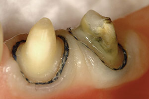
 |
| Figure 3. The facial margins of the #1 cord were gently teased out of the sulcus with the tyne of an explorer. (Note the space created and the absence of blood or saliva—the perfect setting for the master impression.) | Figure 4. An occlusal view of the preparations after removal of the #1 retraction cord. The #00 remained in the sulcus to be removed after the provisional restoration was cemented, and also to aid in cement removal. Note the 360° patency of the sulcus, which was ready for the placement of the light-bodied impression material. |
 |
 |
| Figure 5. The super-pulsed diode laser (Gemini 810 + 980 Dual Wavelength Diode Laser [Ultradent Products]) was used to create some space in this thick periotype case prior to the placement of gingival retraction. | Figure 6. A marginal correction was performed prior to the placement of the #1 retraction cords. |
 |
 |
| Figure 7. A facial view after cord placement for a 10-unit porcelain veneer case prior to making the master impression. Because all of the top (blue) cord was visible, full sulcus patency with no bleeding or moisture was present prior to injection of the light-bodied impression material (or digital scanning). | Figure 8. A view of a completed 10-unit master impression (Thermo Clone [Ultradent Products]) for all-ceramic (IPS e.max [Ivoclar Vivadent]) veneers. |
 |
 |
| Figure 9. A postoperative photo of the completed reconstruction at 5 years. | Figure 10. The site around the implant healing abutment is contoured using a diode laser (Gemini) to help simulate a gingival papilla and allow the implant abutment to more closely resemble the emergence profile of the natural tooth it is replacing. |
 |
 |
| Figure 11. A diode laser (Gemini) is shown being used to remove excess tissue around the implant platform prior to seating the custom implant abutment. | Figure 12. The custom implant abutment was placed, and the margins were fully exposed to allow for complete seating during the cementation process. |
Diode Lasers: An Essential Tool in Soft-Tissue Management
Diode lasers are used as an effective adjunct for the management of the gingival tissues in many areas of restorative dentistry. Before the development of diode lasers, electrosurgery had been used exclusively for some of the same soft-tissue management applications and in many dental practices as well. While electrosurgery can remove a lot of tissue quickly, it can cause an extensive amount of tissue damage (necrosis). Also, electrosurgery devices cannot be used safely around metallic dental restorations or dental implants and are strictly contraindicated. The main objection that clinicians have had in the past when using many of the older diode technologies is that they are slow. As a result, these devices are typically used in continuous mode to cut faster, but the constant output of laser energy can actually result in more tissue damage (necrosis). It is important that the clinician thoroughly understand the nuances of periodontal tissues (attached gingiva vs movable mucosa, keratinized tissue vs nonkeratinized tissue, soft tissue biotype, and alveolar crestal bone position relative to restorative margin position), before performing any laser soft-tissue procedure. Better control of cutting and less peripheral tissue damage are the clinical goals as advances in technology become incorporated into the newest diode lasers on the dental market.1
The Latest Technology in Diode Lasers
One of the first of this generation of super-pulse diode lasers, specifically designed for dentistry, is the Gemini 810 + 980 Dual Wavelength Diode Laser (Ultradent Products). Current diode lasers have either 810-nm or 980-nm wavelengths. The chromophores that these lasers photons target in tissue are hemoglobin (pigment) and water. The 810-nm-wavelength diodes are better at coagulation (hemostasis), while 980-nm-wavelength diodes are better at tissue ablation (cutting). The Gemini combines 810-nm and 980-nm wavelengths in one unit to enhance the coagulation and ablation performance above that of previous dental diode lasers. However, an important difference seen by combining wavelengths in one unit is the ability to achieve 20 W of peak power over an extremely short duty cycle. As a result, the amount of thermal relaxation that occurs between pulses of laser energy allows the soft tissue to cool to such a degree that tissue damage (necrosis) is drastically reduced. Cleaner cuts (with no charring) can be made, resulting in faster healing and less patient discomfort. The degree of collateral thermal damage can be decreased by a factor of about 2 to 3 times when using a super-pulsed laser as opposed to a traditional diode laser used in continuous mode. To summarize, optimal clinical results with less risk of collateral thermal damage (necrosis) can be accomplished with short pulses (duty cycles) at the highest peak power possible using super-pulsed diode laser technology. And, since super-pulsed lasers have such a low heat output, they are extremely safe to use around implant components and to uncover implants prior to the impression phase of implant prosthetic treatment.2
Diode Lasers: Making Restorative Dentistry Easier and More Predictable
When dentists are asked why they’ve added diode lasers into their practices, the most common reason given is to avoid the placement retraction cord. However, the clinician should evaluate the gingival tissues around each preparation prior to doing a troughing procedure. Some of the biologic considerations are as follows: (1) Is there enough attached gingiva present? (2) What is the horizontal thickness of the gingival tissue prior to troughing? (Is the biotype or periotype thin, normal, or thick? What is the level of keratinization present?) (3) What is the proximity of the mucogingival junction to the base of the sulcus to be troughed? And (4) where is the crest of bone relative to the free gingival margin? As long as there is a normal to thick biotype (or periotype) present, adequate thickness of gingival tissues in both horizontal and vertical dimensions, and a normal or high crestal alveolar bone position, troughing can be a useful tool to expose crown preparation margins for both conventional and digital impressions.
 |
 |
| Figure 13. The completed restoration of the case seen in Figure 12. The ceramic restoration on tooth No. 8 is implant-borne. The ceramic restoration on tooth No. 9 was cemented on a natural abutment. | Figure 14. An ovate pontic site (lateral incisor, tooth No. 10 position) was prepared with a diode laser (Gemini) for a 2-tooth cantilevered implant bridge restoring teeth Nos. 9 and 10. The tissue at the base of the ovate site measured 3.0 mm from the bony crest, which was verified by sounding. |
 |
 |
| Figure 15. A 5-year post-op view of the cantilevered 2-unit implant bridge. | Figure 16. A Class V abfraction lesion is seen on the facial surface of tooth No. 5. |
 |
 |
| Figure 17. A diode laser (Gemini) was used to expose the cervical extent of the abfraction to make the area supragingival and easier to restore with composite resin. | Figure 18. The completed Class V composite restoration on tooth No. 5. Note the flawless gingival margin of the restoration and the minimal amount of tissue necrosis as a result of using a super-pulsed diode laser (Gemini). |
 |
 |
| Figure 19. An aesthetic gingivectomy was performed above tooth No. 9 to equalize the zenith position with tooth No. 8. Note that biologic width was not violated, as there was at least 1.0 mm of gingival sulcus remaining after the gingivectomy procedure was performed. | Figure 20. A 2-year post-cementation photo shows teeth Nos. 8 and 9 (shown in Figure 19) with porcelain veneer restorations. Note that the symmetry of the gingival zenith positions has remained after the diode laser procedure performed at the preparation appointment more than 2 years prior to when this photo was taken. |
CASE REPORT
Diode lasers can be useful tools to facilitate placement of retraction cords in cases where a high degree of keratinization of the gingival tissues exists, making it difficult to place the cords.
Figure 5 shows a diode laser being used to open up the sulcus to allow for easy placement (not packing) of a retraction cord to further expand the sulcular space and to compress any tissue tags that may remain after troughing. Using a PFI A-6 (Hu-Friedy) as a gingival retraction instrument to protect the soft tissue, a course cylinder diamond (5855.018 [Komet USA]) was used to refine the margins of the preparation (Figure 6). After verifying the fit of the impression tray, the top cords were placed and ready for the impression procedure (Figure 7). Figure 8 shows the completed master impression (Thermo Clone [Ultradent Products]) of the restorative margins and 0.5 mm of tooth or root surface apical to the restorative margins to ensure accurate reproduction of the emergence angle in the restoration. Figure 9 shows the 5-year postoperative photo of a rehabilitation case where this impression technique was utilized.
DISCUSSION
One way to improve upon gingival aesthetics for fixed bridge pontics and around implant abutments is to use a diode laser to create an ovate pontic site in the edentulous area (or around the circular implant abutment). This helps form an ovoid root shape and simulates gingival papillae, helping to create an anatomic emergence profile for the restoration (Figures 10 to 13). The result is an aesthetic pontic that appears to erupt from the gingival tissues with simulated papillae formed interproximally between the pontic and the adjacent abutment teeth (Figures 14 and 15). It is important to mention, when creating an ovate pontic site with a diode laser in an edentulous area, that there must be a minimum depth of 3.0 mm of soft tissue, which is determined by sounding to the crestal bone with a periodontal probe after the application of local anesthesia. After 4 to 6 weeks of healing, master impressions for the definitive restoration can usually be made.
In operative dentistry, there are many soft-tissue procedures that can be done with a diode laser that can help make routine restorative procedures proceed more smoothly and efficiently. In today’s world of adhesive dentistry, the 2 main enemies of adhesive resin procedures are moisture (saliva) and blood. When doing Class II, Class III, or Class V direct-composite restorations, the gingival tissues often come into play, and, most of the time, this can have a negative impact on the procedure if the operating field cannot be controlled (from bleeding). By using a diode laser to reduce marginal gingival tissue, the restorative margin supragingival can be moved (only as biologic width perimeters allow) and bleeding stopped, prior to matrix and restorative material placement; in this way, the chances of successful adhesion and long-term success of the definitive restoration can increase substantially (Figures 16 to 18).
Another useful procedure easily accomplished with a diode laser is to remove redundant (excess) gingival tissue tags that may grow over the preparation margins during the provisional phase of treatment when the provisional restoration has an ill-fitting (ie, open) margin. This procedure eliminates the possibility of trapping this excess tissue between the preparation and restorative margins, creating a gap for the ingress of bacteria and potential recurrent decay. This same procedure is also useful when placing a cementable implant restoration. Since implant crowns usually fit tightly and have little space between the restoration and abutment, hydraulic pressure from excessive amounts of cement can be a potential barrier to full seating of the restoration. The use of a diode laser to expose the platform (margin) of the implant abutment will facilitate full seating of the implant restoration. It is important to emphasize again that while diode lasers are safe to use around implant components, the use of electrosurgery is not.
Aesthetic gingivectomies performed to balance gingival zenith contours and cervico-incisal heights on teeth in the aesthetic zone (smile zone) can be easily done using a diode laser as long as the clinician has a thorough understanding of biologic width and tissue biotypes and if there is enough free gingiva (unattached) present that will leave a minimum remaining sulcus depth of 1.0 mm. This number is based on the original work by Gargiulo et al3 in 1961. Kois4 and others use this information as part of their “magic numbers,” whereas the measurement from the free gingival margin to the crest of bone should be 3.0 mm on the facial aspect of a crown preparation. Diode lasers afford the clinician a controlled way to artistically provide these procedures with minimal necrosis, maintaining the physiologic contour of the marginal gingiva without fear of gingival recession (Figures 19 and 20).
CLOSING COMMENTS
Management of the gingival tissues, or lack thereof, during routine dental restorative procedures can have a profound influence over the clinical outcomes of the cases. With modern restorative materials that require absolute control of gingival position and the ability to eliminate hemorrhage in the operative area, a diode laser is an essential tool to simplify the procedure and to accomplish these goals. Super-pulse technology is the latest step in the evolution of diode lasers, allowing for more accurate and less traumatic interactions with the gingival tissues. For the applications in restorative dentistry discussed above, this represents a quantum leap in dental laser technology and soft-tissue management.
REFERENCES
- Goharkhay K, Moritz A, Wilder-Smith P, et al. Effects on oral soft tissue produced by a diode laser in vitro. Lasers Surg Med. 1999;25:401-406.
- Borchers R. Comparison of diode lasers in soft tissue surgery using CW and superpulsed mode: an in vivo study. International Journal of Laser Dentistry. 2011;1:17-27.
- Gargiulo AW, Wentz FM, Orban B. Dimensions and relations of the dentogingival junction in humans. J Periodontol. 1961;32:261-267.
- Kois J. Altering gingival levels: The restorative connection, Part 1: biologic variables. J Esthet Dent. 1994;6:3-7.
Dr. Lowe received his DDS degree from the Loyola University School of Dentistry in 1982. He previously taught at the Loyola University School of Dentistry while building a private practice in Chicago. He currently maintains a full-time practice in Charlotte, NC. He can reached via email at boblowedds@aol.com.
Disclosure: Dr. Lowe has received honoraria from Ultradent Products.
Related Articles
Putting Material Science to Work: Use of a Self-Polishing, Universal Nanohybrid Composite
A One-Visit Option: An Alternative to Traditional Ceramic Restorations
“Smart” Class V Preparation Design for Direct Composites


