


Exposure to ultra-high-strength MRI may release mercury from amalgam fillings in teeth, according to researchers at Akdeniz University and Kirikkale University in Turkey. However, the effect was not seen in the lower-strength and more commonly used 1.5-Tesla (T) MRI. Amalgam is approximately 50% mercury.
“In a completely hardened amalgam, approximately 48 hours after placing on teeth, mercury becomes attached to the chemical structure, and the surface of the filling is covered with an oxide film layer,” said lead author Selmi Yilmaz, PhD, a dentist and member of the Department of Oral and Maxillofacial Radiology in the Faculty of Dentistry at Akdeniz University. “Therefore, any mercury leakage is minimal.”
Previous research has found that exposure to the magnetic fields of MRI could cause mercury to leak from amalgam fillings. This concern has been heightened by the recent arrival of ultra-high-strength 7-T scanners in the clinic. Their stronger magnetic field yields more anatomical detail, but its effects on amalgam dental fillings have not been studied.
The researchers evaluated mercury released from dental amalgam after 7-T and 1.5-T MRI in teeth that had been extracted from patients for clinical indications. While the Food and Drug Administration approved 7-T MRI in 2017, the technology has extremely limited availability. The lower-strength 1.5-T MRI is widely available and commonly used for patient exams.
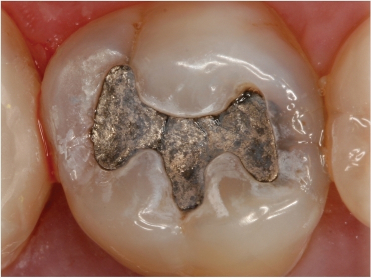
The researchers opened two-sided cavities in each tooth and applied amalgam fillings. After nine days, two groups of 20 randomly selected teeth were placed in a solution of artificial saliva immediately followed by 20 minutes of exposure to 1.5-T or 7-T MRI. A control group of teeth was placed in artificial saliva only.
When the researchers analyzed the artificial saliva, the mercury content in the 7-T, 1.5-T, and control group was 0.67 ± 0.18, 0.17 ± 0.06, and 0.14 ± 0.15 parts per million, respectively. At 0.67 ppm, the mercury content in the 7-T group was approximately four times the levels found in the 1.5-T group and the control group.
“In our study, we found very high values of mercury after ultra-high-field MRI,” Yilmaz said. “This is possibly caused by phase change in amalgam material or by formation of microcircuits, which leads to electrochemical corrosion, induced by the magnetic field.”
The researchers note that an important point of discrimination concerning safety and hazard to human health is the amount of mercury absorbed by the vital tissues.
“It’s not clear how much of this released mercury forms is absorbed by the body,” Yilmaz said.
Further studies may be warranted, Yilmaz added, to evaluate the relationship between high-field MRI and release of mercury from dental amalgam. The researchers have three ongoing projects focused on phase and temperature changes of dental amalgam across different magnetic fields. As no evidence of harmful effects was found in the 1.5-T group, patients with amalgam fillings should not be concerned about having an MRI exam, the researchers said.
The study, “Ex Vivo Mercury Release from Dental Amalgam after 7.0-T and 1.5-T MRI,” was published by Radiology.
Related Articles
Berlin Declaration Shows Amalgam Has Entered Its Twilight Era
What Every Dentist Needs to Know About the New Amalgam Regulations
Coalition Calls for an End to Dental Amalgam in the United States


