This case report will discuss the history and treatment of a patient who suffered from a severe malocclusion and was iatrogenically placed into another even worse malocclusion. Success in this case rests, as it always should, on proper evaluation and diagnosis as well as pretesting
for prognosis.
A 38-year-old female presented to our office; she was referred by one of our patients who was aware that our office treats occlusion-related problems. She presented complaining mainly of jaw pains, earaches, and only 2 teeth touching in the back of her mouth, only on her right side. Her medical history was normal, and she had no medical problems relevant to her dental state. Her dental history was long.
CASE HISTORY
 |
 |
|
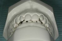
Figure 1. Patient had a deep overbite and no canine-to-canine contact, therefore no anterior guidance. The orthodontist provided us with the preoperative unmounted models, so the true arch-to-arch relationship can only be guessed. |
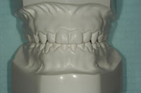
Figure 2. Everything seems fine immediately postoperative (ortho-surgery). But note that models are unmounted. This is therefore not the true arch-to-arch relationship. |
Two years prior to her appointment with us, she had been to her dentist and complained of frequent headaches. She was an Angle Class II with severe anterior overbite (Figure 1). Her dentist discussed the fact that her occlusion might be the cause of her headaches and referred her to an orthodontist. The orthodontist explained that it was possible the occlusion could be the causative factor, and in turn explained that not only orthodontics was needed but also maxillo-facial surgery. The oral surgeon confirmed that occlusion might be the problem, but insisted that no promise could be made as to end results.
The patient accepted the treatment plan because nothing else she had tried had helped. A complete orthodontic treatment was done combined with impaction of the maxilla and saggital split advancement of the mandible. Upon removing interarch fixation, the patient had a Class I occlusion and was relatively symptom-free (Figure 2).
However, within a few weeks the anterior bite started to open, and it took only a few more weeks for the mandible to regress to the point where only teeth Nos. 2 and 31 were left in contact, with the bite progressively opening all the way to the other side. Even though the original type of headache was gone, the patient suffered from TMJ pain on the right side, muscle pains, a new type of headache, muscle spasms, opening limited to 25 mm, inability to chew, and, of course, she was quite discouraged. The oral surgeon offered that her mandible “was probably not made to be in the advanced position,” and the orthodontist said just to wait, that things would get back to normal and the teeth would touch again.
The patient’s dentist, upon supplication to do something, fabricated an occlusal splint that relaxed the muscles and gave some relief to the patient. However, the result was temporary, and within 3 months all the pain was back. The patient was then referred to a prosthodontist, who prescribed an MRI of the TM joints. At this time the patient, to whom the suggestion had already been made to visit our office, decided to consult with us.
EXAMINATION
 |
 |
|
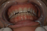
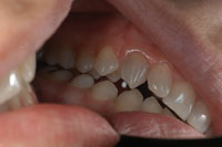
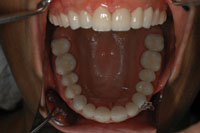
Figure 3. The only teeth touching postsurgically were the last 2 molars (Nos. 2 and 31). |
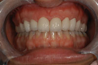
Figure 4. Note separation of patient’s left side in maximum intercuspation (centric occlusion). |
 |
 |
|
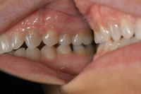
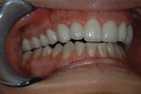
Figure 5. Patient’s right side; only the last teeth are in contact. This is maximum intercuspation. |
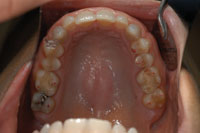
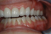
Figure 6. Patient had an otherwise healthy mouth. |
Upon examination the patient presented with a severe malocclusion (Figures 3 to 5) with contact on 2 teeth only in the right posterior region, and a 3-mm slide from centric relation to centric occlusion (all on one tooth). She also had an open bite on all other teeth with impossibility to create contact on any other tooth even by mandibular movement. Upon palpation the right TMJ was very tender, as were the lateral pterygoid, medial pterygoid, and attachment of the temporalis at the coronoid process.
Doppler auscultation revealed significantly augmented blood flow on the right side compared to the left side, with no significant noise upon straight opening; but left excursion revealed a click in the right TMJ. The patient also reported light crepitus in the morning. Opening was limited to 25 mm, and lateral excursions were minimal because of pain. The periodontium was healthy, and the 28 teeth present needed no restoration due to decay or fracture (Figure 6). Soft tissues were normal, as were the glands and saliva. At this time the patient had not yet undergone MRI, but had an appointment for MRI.
DIAGNOSIS
The following diagnosis was made: severe malocclusion iatrogenically induced; disc displacement in the right TMJ with recapture upon opening (Piper III A, Dawson Type II A); capsulitis of the right TMJ; muscle inflammation; headache; limited movements; and inadequate masticatory function.
TREATMENT PLAN
Phase I
The first step was to obtain MRI results to eliminate possible clinical misdiagnosis about the state of the TMJs. We feared, even though no clinical signs pointed there, that the cause of mandibular regression and rapid opening of the bite postsurgically might have been severe damage to the condyles, avascular necrosis, or another significant form of rapid bone degeneration.
The next step was to fabricate, as quickly as possible, an occlusal splint equilibrated to centric relation with absolute anterior guidance.
We then wanted to re-evaluate the patient as soon as the MRI was done. Prognosis after Phase I was reserved.
Phase II
Upon receiving the results of the MRI we could confirm that our clinical observations were correct, and the patient had no condylar degeneration. We proceeded to adjust the splint as we had prescribed. The patient was instructed to wear the splint 24 hours a day for a period of 3 weeks and to come back for re-evaluation.
Discussion of Why the Mandible Regressed Postsurgically
In this case, the conclusion that the joints were healthy led us to question why the mandible had so violently regressed. Relapse, the usual explanation, was simply not valid. We suspect that when the divided parts of the mandible were fixated together, the anterior segment was correctly advanced into proper occlusion, but the posterior segments, instead of having the condyles seated in centric relation, were also advanced. If the surgeon, during fixation, does not positively maintain the con-dyles in centric relation, then there will always be regression postsurgically.
Prognosis After Phase II
At this point, because the condyles were confirmed healthy, and the joints therefore were manageable, the prognosis of relieving the patient’s pain was good, considering that prior to surgery the patient had no TMJ pains.
Results of Phase II
Immediately after insertion of the equilibrated splint the patient felt total relief. As long as the splint was worn, there was no pain. This therapy was continued for 3 months to confirm stability.
Discussion of Phase II Results
Technically, phase II was a complete success, as the patient was comfortable and without pain as long as she wore the appliance. However, it was awkward for the patient to wear a splint at all times. She is an accountant in a large company, and meetings occur daily. Also, she found little pleasure in eating with the splint in place. We had to find a better solution. Because it was demonstrated that a proper occlusion was the solution for this patient (as is very often the case), we offered her 3 options.
PHASE III
The first option was an overdenture that could be made to fit the lower teeth. Such an overdenture should be stable and equilibrated to perfection.
Another option was reconstruction with crowns of the upper, lower, or both arches after studying the possibilities and conveniences with well-made waxups.
The third option was to return to orthodontics and oral surgery to correct the mishap of the first attempt.
After discussion of all possibilities, the patient absolutely refuted the idea of returning to ortho-sugery, even though this solution offered to maintain her dentition in a natural state. She was unable financially at the time to have crowns made, so she opted temporarily for the over-denture on the lower arch.
As we had suspected, however, even though occlusal comfort was attained with the overdenture, the patient was not totally satisfied with the aesthetics, as the lower anteriors were quite long, and just having “something” in the mouth was an annoyance. The patient began to wear the appliance less regularly, and sure enough the pains came back. Within a few months she returned, seeking crowns.
After wax-ups were made (one waxing on the lower only, the other waxing on the upper only), it was determined that an upper rehabilitation was the more aesthetically pleasing option. To make perfectly sure, however, we took an impression of the upper wax-up, poured Protemp 3 Garant (3M ESPE) into it, and reset the impression in the mouth. We left the temporary uncemented, as it was an aesthetic “tryout” to last only a few days.
 |
 |
|
Figure 7. Teeth prepared for zirconia-based crowns (Lava [3M ESPE]). Preps are wide chamfer. |
Figure 8. Zirconia-based crowns by technicians Marc Nantais and Michel Bourque of Laboratoire De Porcelaine Dentaire Longueuil. |
 |
|
Figure 9. Note proper alignment of teeth, sufficient depth of bite to create anterior guidance, and proper cusp tip-to-fossa posterior relationship. |
During these days the patient was comfortable in all aspects. Her occlusion was perfected and comfortable. She had no pains, had no removable appliance to annoy her, and aesthetics were more than pleasing. An appointment was made, and the 14 upper teeth were carefully prepared (Figure 7) to receive zirconia-based crowns. As all details concerning final occlusion had been tested and retested, predictability was very high and prognosis was excellent. Crowns were cemented using RelyX luting cement (Figure 8).
At one year postoperatively, even though the patient has been through episodes of daily stress with her job, and even through very high levels of stress as she and her husband were in the process of adopting a foreign child, she has had no pain and has taken no medication for pain. Control ap-pointments at 3 months, 6 months, and one year postoperatively confirmed occlusal stability and total patient comfort. No adjustment has been needed (Figure 9).
CONCLUSION
 |
 |
|
Figure 10. Cuspid rise right side; no other tooth is in contact. Masseters and temporalis are relaxed. |
Figure 11. Cuspid rise left side; the rise starts immediately from maximum intercuspation. Patient cannot perceive any lateral or protrusive contact on posteriors. |
This case was special. It illustrates most dentists’ misconceptions about what is a good or bad occlusion and the true influence it has. The orthodontist and the surgeon who treated the patient had no real clue as to whether their treatment would bring relief. If they had taken the opportunity to make the patient an equilibrated occlusal splint, they would have found that a proper occlusion was indeed the proper solution. Their prognosis would have been good instead of uncertain.
If the technical aspect of their work had succeeded, the patient might have been relieved of her pains. But no one would have known the true reason. The orthodontist and the surgeon worked on the assumption that an Angle Class I occlusion is a good occlusion. You can’t get any farther from the truth. Angle’s classification is descriptive, not qualitative. What defines a proper occlusion is not how it looks, but how it functions. In a good occlusion when the patient clenches in maximum intercuspation, the condyles are fully seated in the fossae and properly placed in the non-innervated center of the disc. There is no deviation, no slide. Condyles are in centric relation, teeth are in maximum intercuspation. Centric relation = centric occlusion.
From this centric position, all movements should be guided by anterior teeth. No posterior teeth should touch in any excursive movement, lateral or protrusive. These are the 2 first and basic principles of good occlusion (Figures 10 and 11).
Therefore, in the great majority of cases, this is how a rehabilitated occlusion should be; whether the occlusion is corrected by orthodontics, surgery, equilibration, or prosthetics (removable splints, crowns, bridges, implants), the end result should be the same. The case described in this article could and should have been pretested with an equilibrated splint. Care should have been taken by the referring dentist, the orthodontist, and the surgeon to produce the same occlusion with the treatment.
When the patient came to us, that is what we did. We used an equilibrated splint to test and make sure a corrected occlusion was the solution for the patient. We rapidly concluded it was, and then proceeded to correct the dentition to make it correspond to the same principles we planned in the splint. There was no guessing. It was highly predictable that the rehabilitation with crowns would produce the same comfort, and actually even more than the splint.
In this specific case we concluded that a proper occlusion was the solution for the patient. Looking at the models taken before ortho-surgery (Figure 1), we know there was no canine contact and no anterior guidance, and we can suspect that there was also a CR-CO slide. This patient should have had a choice. Ortho-surgery was one of the possible answers to the problem. The patient could have had a simple occlusal equilibration with either composite or porcelain added to the lingual of her upper canines to produce the anterior guidance. Another choice could have been to equilibrate the posteriors and crown from one upper canine to the other to correct both guidance and aesthetics. This patient did not necessarily need to undergo orthodontic therapy and surgery. It was an option. Our personal opinion is that in these cases ortho-surgery is the better technical solution. But the ortho-surgery must be correctly done, and the result has to be excellent. The occlusion produced by the treatment has to be close enough to the goal so that either the orthodontist or the referring general practitioner can equilibrate the dentition to perfection once the ortho-surgery phase is completed and stable.
Most general practitioners don’t have discussions with their orthodontist and/or surgeon concerning the end occlusal pattern. Most of us rely on the ortho-surgery team to “decide.” This should not by any means be the case. Be proactive and learn about occlusion; talk about it with your specialists. We have been doing it for years and have progressively gotten our local orthodontists, surgeons, lab technicians, etc to better understand why patients are not comfortable and how to get them comfortable.
Success in occlusal-related problems resides in pretesting and respecting the principles. Our patient now thinks the world of us. We know we just understand occlusion. So can you.
Dr. Aubé graduated from the University of Montreal dental faculty in 1984. He continued at McGill University in a residence program with emphasis on full-mouth crown and bridge rehabilitation. Anterior guidance was already his way of choice. He completed the full program at Dawson Center and studied TMJ anatomy, physiology, and pathology with Dr. Mark Piper. He also completed a research project with the Montreal Migraine Clinic on the effect of occlusal equilibration on tension-type headaches and migraines. He was invited to present the results at a meeting of the American Academy of Restorative Dentistry, and maintains a private practice in Montreal. He can be reached at centredentaireaube@videotron.ca or (514) 526-3294.