
It is a source of frustration that there appears to be a general lack of knowledge in the dental profession regarding what makes aesthetic composite techniques work easily, without stress, and produce consistently reliable long-term results.1 The failure of our educational systems to instruct the majority of students in the science as well as the art of aesthetic bonding is a disservice to dentistry. One of the primary reasons for this omission is a general lack of understanding of composite technology by the teachers themselves.2
This article is designed to demonstrate many of the outstanding properties found in direct resin bonding techniques and to refute certain misconceptions that exist.3,4 Important concepts include the following:
 |
 |
|
Figure 1. Fractured direct resin veneer—12.5 years post-op after preparation—note long bevels. |
Figure 2. Primer and bonding adhesive in place. Primer on exposed dentin and enamel only. Adhesive on both tooth surface and composite resin. Polymerized. |
 |
 |
| Figure 3. Application of Creative Color Opaque A1 to block out underlying darkness while blending into external shade of microfill. Carried to middle of bevel only. Polymerized. | Figure 4. Initial placement of microfill B1 (used 12.5 years previsously) with 8A titanium coated instrument. |
 |
 |
| Figure 5. Continued sculpting and blending microfill with IPTC short-bladed titanium coated instrument. | Figure 6. Application of microfill complete and polymerized. Note: thorough blending of new material into the old microfill surface. |
 |
 |
| Figure 7. Use of Brasseler ET9 12 fluted carbide bur to start contouring and blending of one material into another. | Figure 8. Use of coarse FlexiDisc to complete blending of one material into another. |
 |
 |
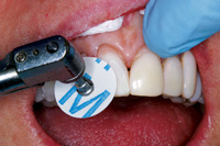
| Figure 9. After use of the coarse disc, note the complete blending of new material into old microfill with no demarcation. | Figure 10. Continuation of polishing through various grits of FlexiDisc—medium, fine, and superfine—to complete the finishing and polishing process. |
(1) Direct resin bonding truly stands the test of time.
(2) An explanation in simple terms of the science of bonding and the clinical significance of understanding it; pinpointing reasons for its failure.5,6
(3) Direct resin bonding is a quality technique, and the aesthetic clinician must be adequately remunerated to perform to perfection.7
(4) Direct resin bonding is not a second-choice alternative. In fact, it frequently outperforms lab-driven techniques.8
(5) Composites are not only dentistry’s most useful materials, but they also conserve the most tooth structure.9
The versatility of these materials is so great that a wide variety of clinical situations are indicated for their use10,11: (1) class I, II, III, IV, and V restorations of all kinds; (2) removal and replication of enamel defects; (3) diastema closure12; (4) incisal reinforcement; (5) tooth lengthening; (6) complete veneering; (7) restorative orthodontics/rearrangement; (8) smile development and widening; (9) development of proper canine rise, incisal guidance, and increase in vertical dimension; (10) gingival replication13; (11) patient and lab mock-ups; (12) temporary partials; (13) full-bonded crowns; (14) resin-retained bridges; (15) repairs of all kinds14 (composite, porcelain, porcelain and metal, exposed dark metal gingival margins, and unsightly metal [gold] margins); (16) restorative dentistry for cleft palate patients15; and (17) masking and increasing value of tetracycline stained dentition,16 the unsightly single discolored tooth, endodontic and implant access areas, and metal posts.
CASE REPORT
A patient came into my office almost 13 years ago desiring a reshaped, brighter, and lighter smile. She had previous grafting, but the recession was so severe that even though a good result was achieved, she still needed some periodontal-enhancing restorative treatment. She also required some gingival replication to create a better smile profile. At that time, we used Renamel Gingafill (Cosmedent), a gingival-colored microfill, to replicate the labial contours and to keep her from having hyper-extended teeth.
 |
 |
| Figure 11. Finishing and polishing of proximal surfaces with 15-µm diamond strips followed by fine/superfine FlexiStrips (shown here). | Figure 12. Invisible repair completed. Things to note: (1) retention of polish long-term; (2) excellent color stability; (3) no demarcation of any kind between old and new microfill; (4) surface texture of veneers exhibits no wear; (5) no recession after 12.5 years—notice health of gingival tissue; and (6) note gingival recession on lower anteriors and compare with upper veneer—what does that tell you? |
 |
 |
|
Figure 13. Smile immediately after direct placement of 8 upper anterior veneers—May 23, 1991. |
Figure 14. Smile 12.5 years post-op demonstrating an invisible repair and total color stability—December 9, 2003. |
 |
 |
| Figure 15. May 23, 1991: immediate post-op. | Figure 16. December 9, 2003: same vibrant smile showing invisible repair. |
 |
 |
| Figure 17. Additional long-term results showing 8 anterior veneers 8 years post-op. Note the exceptional gingival health of all long-term results. | Figure 18. Additional long-term results showing 4 incisors veneered against existing dentition 8 years post-op. |
 |
 |
| Figure 19. Additional long-term results showing anterior veneers 10 years post-op. | Figure 20. Additional long-term results showing 6 anterior veneers 18 years post-op. |
What allows such outstanding long-term results?
(1) The ability to maintain a long-term polish. The proper material selection that accurately simulates the enamel surface. The only material to date that can achieve this long-term is microfill. Why? Because the smaller and more uniform particle size allows you to polish to the most lustrous, enamel-like surface known to dentistry, and it will maintain this polish long term.17
(2) Microfill has a great resistance to wear. It exhibits minimal or no plucking because of the density of the filler and small particle size, and since it can be polished to such a high shine, the material tends to resist wear due to abrasion.
(3) Microfill has excellent tissue compatibility. It shows great margination beneath the free margin of the gingival because of the small particle size.
(4) The outstanding color stability of microfill is probably due to the small particle size and overall density of the material that prevents plucking, pitting, wear, and loss of surface texture.18
(5) Highly polished microfills offer superior aesthetics because their reflective and refractive index mimic tooth structure most accurately. Resin materials are properly pigmented to mimic natural tooth stru
cture, and resin has more lifelike translucency and natural fluorescence that can match both natural dentition and porcelain.
(6) Invisible repairs with color-stable microfill composites are easily accomplished. The use of microfill allows for better and more invisible reparability due to its small particle size. When applied and finished properly, the new restoration blends into the old without demarcation.
Knowledge of the use of proper polishing systems makes margins between the new and old material completely disappear when one truly understands how to use them.
Of course, color stability plays an important role in your restoration. When color doesn’t change, the new and old material blend together as one, without distinction.
Figures 1 through 16 provide a detailed description of the patient’s repair. In addition, Figures 17 through 20 demonstrate other long-term results achieved with microfill composite.
CONCLUSION
All of the qualities I have described are meant to demonstrate how composite restorations can truly stand the test of time. In my personal practice, I have worked with composites for more than 30 years and completely limited my practice for the last 15 years to direct resin bonding. My patient files probably represent in dentistry today one of the largest bodies of actual clinical results working with composite resins.
References
- Mopper KW. The subtleties of anterior direct-bonded class III restorations. Pract Periodont Aesthet Dent. 1990;2(6):17-20. (Now: Practical Procedures and Asesthetic Dentistry.)
- Mopper KW. The value of the hands-on learning experience. J Calif Dent Assoc. 1995;23(4):37-40.
- Meijering AC, Creugers NH, Roeters FJ, et al. Survival of three types of veneer restorations in a clinical trial: a 2.5-year interim evaluation. J Dent. 1998;26(7):563-568.
- Garber DA. Direct composite veneers versus etched porcelain laminate veneers. Dent Clin North Am. 1989;33(2):301-304.
- Duke ES. The science and practice of dental adhesive systems. Compend Contin Educ Dent. 2003;24(6):417-424.
- Mopper KW. Why dentists miss shades frequently. Dent Econ. 1994;84(1):82-83.
- Willhite C. Dramatic smile makeovers using direct resin veneers. Compend Contin Educ Dent. 1997;18(7):646-657.
- Mopper KW. Creative solutions for everyday esthetics. Dental Practice Report. 2000;8(6):46-50.
- ADA Council on Scientific Affairs. Direct and indirect restorative materials. J Am Dent Assoc. 2003;134(4):463-472.
- Mopper KW. Renamel Restorative System Manual. Chicago, Ill: Cosmedent, Inc; 1994.
- Feigenbaum N, Mopper KW. A Complete Guide to Dental Bonding. New Jersey: Johnson & Johnson Dental Products Co; 1984.
- Mopper KW. Diastema closure: A natural for direct bonding procedures. Pract Periodont Aesthet Dent. 1990;2(1):22-28. (Now: Practical Procedures and Asesthetic Dentistry.)
- Trushkowsky RD. Camouflage of recession with a gingival-simulating composite resin. Esthet Dent Update. 1995;6(2):41-42.
- Fahl N Jr. Predictable aesthetic reconstruction of fractured anterior teeth with composite resins: a case report. Pract Periodontics Aesthet Dent. 1996;8(1):17-31.
- Mopper KW. Aesthetic dilemma: restoring the quality of life in a cleft lip, cleft palate patient. Dent Today. 2000;19(6):46-53.
- Mopper KW. Pink opaque for stained dentition. Contemp Esthet Restor Pract. 2000;4(4):40-41.
- Miller MB, Costellanos IR. Microfills. REALITY. 2003;17:576.
- Lu H, Roeder LB, Powers JM. Effect of polishing systems on the surface roughness of microhybrid composites. J Esthet Restor Dent. 2003;15(5):297-303.
- Mopper KW. Porcelain versus composite: What’s the treatment of choice? Synergy 1990;Winter:1-10. (For more information, contact Dental Lab Publications, 205 Liberty Square, East Norwalk, CT 06855.)
- Mopper KW. Dentistry’s most underutilized material—microfill: a key ingredient for successful anterior esthetics. Dental Town. 2003;4(1):20.
Dr. Mopper has been a clinician and a teacher with a practice in Winnetka, Ill, for more than 40 years. He is cofounder and chairman for Cosmedent, and researches new materials and techniques for dentistry. He is also founder and fellow of the American Academy of Cosmetic Dentistry, a fellow of the American Academy of Pediatric Dentistry and the America College of Dentistry, and a member of the America Academy of Aesthetic Dentistry. He can be reached at (847) 441-6080.

