How often have you wondered what you could do to contribute to the lives of your patients? Certainly, many of the things you do as a hygienist each day contribute to the health of your patients, but what if you actually saved their lives? How do you think that would make them feel? How do you think that would make you feel?
THE FACTS ABOUT ORAL CANCER
Oral cancer kills one person every 60 minutes in our country. It kills more people than skin cancer. It kills more people than cervical cancer. It kills our parents, our friends, and our children, and yet most dental professionals don’t take its threat seriously enough to do anything about it. It’s time to take our jobs as healthcare providers seriously, and start focusing on the health of our patients. If we don’t, the statistics show we’re going to have to start focusing on their deaths. It takes just 3 minutes to do something about this devastating health problem.
GET STARTED NOW
So, how do you get started? The first thing to do is get excited about doing this. There are three reasons why I am passionate about doing what I call the “Ritz Carlton” approach to extraoral cancer exams: it saves time, patients love it, and it saves lives.
It Saves Time
I found that once I started implementing this exam on a regular basis, it cut my appointment time by at least 5 to 10 minutes. The reason: patients cut down on their personal chit-chat because by performing the examination you elevated your status to healthcare professional in their mind. They got focused on the real reason they were sitting in your chair—to get healthy.
Patients Love It
Let’s face it, having your face and neck “massaged” feels great! I have never run into a situation where a patient was uncomfortable with the exam. Most of my patients ask me to keep going, and they look forward to this portion of their appointment. Plus, it instantly relaxes people. I know that a lot of patients come to the office from work and are uptight or stressed. This exam helps them focus on the appointment and not outside issues.
It Saves Lives
I have a friend who was lecturing one evening to a group of oral surgeons about oral cancer and early detection. She is not a dental professional, but she has a very personal investment in this topic that I will get into later. During the class, one of the surgeons actually discouraged general dentists and hygienists from utilizing early detection procedures. He felt there was no reason for it because if they found oral cancer the patient was just going to die anyway. In my opinion, this approach to oral cancer is why the mortality rate for it has not significantly dropped in the past 40 years. By the way, my friend who was lecturing is currently watching her mom face each day being fed through a stomach tube, not being able to swallow because her mouth has the consistency of leather after chemotherapy and radiation, and living a life devoid of many simple pleasures for the time she has left.
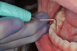
- Palpate and visually inspect lips.
- Check labial vestibules.
- Inspect buccal mucosa.
- Inspect retromolar pads.
- Inspect hard and soft palate.
- Depress patient’s tongue and inspect tonsillar area and pharynx.
- Use a moistened 2 X 2 gauze pad to hold tongue and check lateral borders.
- Palpate tongue and check both dorsal and ventral aspects.
- Visually inspect and palpate floor of mouth.
If anything looks suspicious, inform the patient, have him look in the mirror, and alert the dentist. If the dentist feels the lesion warrants a biopsy, you can refer the patient to an oral surgeon or perform the biopsy in your office. By utilizing a brush biopsy, such as the one from Oral CDX, you can quickly and painlessly evaluate the lesion. The Oral CDX brush biopsy has been tested extensively against traditional scalpel biopsies and has proven to be 100% accurate with no false positives when used according to instructions. A kit provides all necessary items to perform the procedure. The doctor simply brushes the lesion (with no need for anesthetic) until pinpoint bleeding occurs. The auxiliary then transfers the cells from the brush onto a slide, applies a fixative, and allows the slide to dry for 15 minutes. The slide is placed into a transport case and is mailed along with a patient information sheet to the Oral CDX lab. The lab will fax you the results of the biopsy within 7 to 14 days along with a description of the cellular representation.
the test done rather than referring him to the oral surgeon and “hoping” he made the appointment before losing his nerve.
Each year 9,000 to 10,000 people die from oral cancer in the United States, and 30,000 new cases are diagnosed. Ninety-five percent of people affected are over the age of 40, and while the two highest risk factors for oral cancer are the use of alcohol and tobacco, anyone can be affected. Other risk factors may include poorly fitting dentures, chronic irritation from jagged/decayed tooth surfaces, exposure to damaging UV light, and chewing the lips and inside of the mouth. The most common site for oral cancer is the tongue, but it can strike any part of the mouth, including the lips. If the cancer is not treated early, it can spread deep into the lymph glands of the neck.
One of the myths surrounding oral cancer is that it is a death sentence. This is a myth not only believed by dental professionals, but also by patients. The truth is that with early detection, oral cancer is almost always curable. The ADA has recently launched a massive campaign to increase public awareness of oral cancer and early detection. It is our responsibility to follow through with this campaign by partnering with our patients and with companies that offer early detection devices.
CONCLUSION
By conducting a thorough examination, imagine how many lives you could save. Imagine how many lesions could be detected early by taking the time to evaluate for oral cancer and by utilizing the latest technologies in diagnosis. What have you been missing by not really looking? Stop being afraid or reluctant to find oral lesions. It is our responsibility as primary oral healthcare providers to use all our knowledge and clinical skills to save the lives of our patients. One person dies every hour from oral cancer. Research shows that 70% to 80% of the people who die could have been saved through early detection. Let’s not allow one more patient to die unnecessarily because we were unwilling to perform a life-saving exam. It takes just 3 minutes.
Suggested Reading
American Cancer Society. What are the key statistics about oral cavity and oropharyngeal cancer? Available at: http://www.cancer.org. Accessed March 18, 2002.
American Dental Association Dental Newsline. The dentist’s role in spotting oral cancer. Available at: http://www.ada.org. Accessed March 18, 2002.
Cancer News on the Net. Facts about oral cancers; a review by Dr. Greg Bobier. Available at: http://cancernews.com. Accessed March 18, 2002.
Mayo Clinic Health Information. What is oral cancer? Available at: http://www.mayoclinic.com. Accessed March 18, 2002.
Oral Health Information; ADHA Online. Oral cancer facts. Available at: http://www.adha.org. Accessed March 18, 2002.
Ms. Mitchell is a full-time practicing hygienist for Dr. Michael Koczarski in Woodinville, Wash. She is a coach with Hygiene Mastery and codirects the hygiene program for PAC~live. Ms. Mitchell lectures and instructs extensively on the comprehensive assessment of dental wants and needs, treatment planning, and codiagnosis of anterior and posterior aesthetic dentistry. She can be contacted at (425) 486-2200.