Today, restorative dentistry emphasizes minimally invasive approaches to care. This encompasses prevention, remineralization, and when needed, adhesive restorations. These approaches lessen the chance for subsequent adverse outcomes, including the advancement of tooth decay, pulpal involvement, and tooth fracture.
As dentists, our goal is to have the knowledge of various dental products in order to select the best material for any given scenario. In cases where isolation is a problem, resulting in moisture contamination, or there is a high caries rate due to medications, diet, or age; I have personally found the use of resin-modified glass ionomer restorations very useful. Some indications for its use include the following: small Class I, II, III, and V restorations; deciduous teeth restorations, geriatric restorations, pit and fissure sealants, core buildups, root surface restorations, cervical erosion, and abfraction lesions.
CASE PRESENTATION
 |
 |
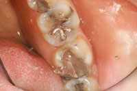
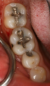
| Preoperative occlusal view of teeth Nos. 13 to 15. | Postoperative occlusal view of teeth Nos. 13 to 15. Note the natural aesthetics achieved when using this minimally invasive dentistry restorative technique. |
A female patient presented to our office for a 6-month hygiene appointment. While receiving her periodic oral examination, we identified a resin sealant on tooth No. 14 (placed many years prior) was breaking down. There was also some staining and incipient decay present on the occlusal surfaces of teeth Nos. 13 and 15.
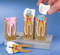
Capturing an image of these teeth on the intraoral camera (RF Systems Lab), we indicated the areas of concern on the flat screen monitor (Pre-op image). Then, using the DemoDent (DemoDent) patient education model, we described to the patient in detail what was occurring in the tooth (Figure 1): “There are 3 layers in a tooth as illustrated in this model (Figure 2). The white is the enamel, the yellow is the dentin, and the pink is the nerve. Your cavity is in the biting surface of the tooth, where food and debris like to collect. When the cavity is in the enamel (the white layer) you usually do not have any pain or sensitivity with it. In fact in most cases, by catching the cavity early, we can clean it out without the need for anesthetic. Once the cavity has gone through the enamel and into the dentin (yellow layer), it spreads much more quickly. Patients may experience some sensitivity to hot, cold, and sweets, depending on how deep it has extended. Once the cavity gets into the nerve (pink layer), patients usually experience constant, throbbing pain. We want to prevent this by stopping the cavity as soon as possible.”
After explaining the situation using the image on the screen and the anatomical model, I found that the patient better understood her dental condition and was very eager to get started.
CLINICAL TREATMENT
 |
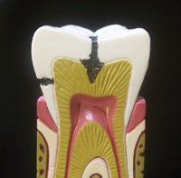 |
| Figure 1. DemoDent patient education model system. | Figure 2. Close-up view of the progression of decay. |
 |
 |
|
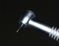
Figure 3. Micro-Preparation Burs (KOMET USA) used for preparation of the teeth. |
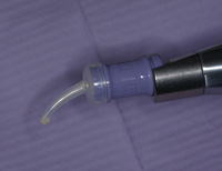
Figure 4. Riva Light Cure RMGI (SDI) was made ready for application. |
Once the patient agreed to treatment, she was scheduled for the restorative part of the procedure. All risks, benefits, and alternatives were discussed with the patient about using a resin-modified glass ionomer restoration (Riva Light Cure [SDI]). A combination of glass ionomer and composite resin, these fillings are a mixture of glass, an organic acid, and resin polymer that harden when light-cured. (The light activates a catalyst in the restoration that causes it to cure in seconds.) This combination of a glass ionomer and resin has excellent aesthetics, high fluoride release, and it chemically bonds to tooth structure. In fact, it has free movement of fluoride, which provides benefits to surrounding and adjacent tooth surfaces. Fluoride is held within the glass ionomer matrix without being bound by the structure. If the level outside the glass matrix is lower, then fluoride ions are released. Conversely, if the fluoride level is higher (ie, topical fluoride) then fluoride will recharge the glass ionomer matrix. In addition, these filling materials are not subject to shrinkage and microleakage, since the bonding mechanism is an acid-base reaction and not a polymerization reaction.
Using Micro-Preparation Burs (KOMET USA) the old resin restoration and caries were removed from tooth No. 14, as well as any staining or decay from teeth Nos. 13 and 15 (Figure 3). Riva Conditioner (SDI) was applied to the teeth for 10 seconds and then rinsed thoroughly with water. Any excess water was removed, being careful not to desiccate the tooth and to keep the dentin slightly moist. The Riva Light Cure RMGI (SDI) capsule was activated and placed in an amalgamator for 10 seconds. Once activated, the capsule was loaded in the dispensing gun and placed into the preparations (Figure 4). Final set was achieved after light-curing for 20 seconds using the Radii Plus (SDI). After light-curing, finishing of the restorations was accomplished under water spray using Q-Finisher burs (KOMET USA). As seen in the postoperative images (Post-op image), the combination of glass ionomer and resin in this restorative material yielded a very aesthetic and functional restoration. In addition, it provides a high fluoride release and bonds well to the tooth structure.
CONCLUSION
After many decades of improvements in oral health, tooth decay is on the rise again. Much of the blame can be placed on today’s high-sugar diet consisting of fast food, soda pop, sport juices, and energy drinks. Another factor that comes to mind is the fact that the baby boomers are living longer. A majority of these patients may be taking medications that are causing severe drying in the mouth that results in a high caries rate, that they are simply challenged in brushing and flossing properly because of dexterity issues.
Resin-modified glass ionomer restorations are ideally suited for use in the treatment of patients who are at high risk for caries. Whatever the situation, it is important for all dentists to select the right restorative dental material for the right case in order to ensure long-term restorative success.
Dr. Nazarian is a graduate of the University of Detroit-Mercy School of Dentistry. Upon graduation, he completed an AEGD residency in San Diego, CA, while serving in the United States Navy Dental Corps. He is a recipient of the Excellence in Dentistry Scholarship and Award. Dr. Nazarian maintains a private practice in Troy, Mich, with an emphasis on comprehensive and restorative care. His articles have been published in many of today’s popular dental publications. He also serves as a clinical consultant for The Dental Advisor, Dental Team Concepts, and Dental Compare. He has conducted lectures and hands-on workshops on aesthetic materials and techniques internationally. He can be reached at (248) 457-0500 or visit the Web site demo–dent.com.
Disclosure: Dr. Nazarian is the creator of the DemoDent patient education model system.