 |
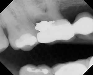
| Figure 1. Panoramic radiograph demonstrating two antroliths in the maxillary sinus. |
 |
| Figure 2. Panoramic radiograph demonstrating a fourth molar in the area of the maxillary right sinus. |
 |
| Figure 3. Panoramic radiograph demonstrating bilateral fourth molars in association with the maxillary sinuses. |
 |
| Figure 4. Panoramic radiograph demonstrating a small fourth molar in area of the maxillary right sinus. |
 |
| Figure 5. Panoramic radiograph demonstrating a third molar in the area of the maxillary right sinus. |
There appears to be controversy concerning the significance of ectopic teeth and antroliths/rhinoliths within the maxillary sinus. Does such a finding necessitate further investigation and the possibility of surgical intervention? The occurrence of ectopic teeth in the maxillary sinus has been reported to be a rare finding.1 Ectopic teeth and antroliths within the maxillary sinus, the intrusion of teeth secondary to trauma, and the intrusion of foreign substances because of dental treatment and trauma have been reported in the literature.2-6 We report on the occurrence of ectopic teeth and antroliths within or adjacent to the maxillary sinus in a population of dental school patients.
METHODS
A dental school patient population was surveyed for the presence of ectopic teeth and antroliths within the maxillary sinuses over a period of 2 years. Panoramic radiographs were surveyed, and approximately 6,000 patients were evaluated for symptomatology. The dental school population was approximately 60% African-American, 15% Hispanic, 15% white, and 10% Asian.
RESULTS
Four patients demonstrated either third or fourth molars within or adjacent to the maxillary sinus (Table). One patient had findings consistent with two rhinoliths/antroliths within the maxillary sinus. The ages of the patients ranged from 14 to 50 years.
Three patients were female and two were male. All patients with positive findings were African-American.
The first case was a 38-year-old black male who was seen for routine dental radiographs. The medical history was noncontributory. The radiographic findings demonstrated dental pathology as well as two antroliths or decalcified teeth within the left maxillary sinus (Figure 1).
The second case involved a 32-year-old black female who was seen for routine dental radiographs. The patient was 7 and a half months pregnant. The panoramic radiograph revealed a fourth molar in the right maxillary sinus (Figure 2).
The third case was a 50-year-old black male who was seen for routine dental radiographs. Two fourth molars were found within or adjacent to the right and left maxillary sinuses (Figure 3). The medical history noted seasonal sinusitis. The radiograph also revealed impacted mandibular left third and fourth molars, and the patient reported previous surgery for removal of mandibular right third and fourth molars. Therefore, the patient had previous knowledge of his impacted molars.
The fourth case was a 42-year-old black female who was seen for routine dental radiographs. The panoramic radiograph demonstrated a small maxillary right fourth molar within or adjacent to the maxillary sinus (Figure 4). The medical history was noncontributory.
The fifth case was a 14-year-old black male who was seen for routine dental radiographs in the process of being evaluated for orthodontic therapy. The panoramic and cephalometric radiographs demonstrated a maxillary right third molar within or adjacent to the sinus (Figure 5). The medical history was noncontributory.
DISCUSSION
In our review of the dental and medical literature, the finding of an ectopic tooth or teeth, or an antrolith (rhinolith) in the maxillary sinus was rare. However, in the experience of radiology faculty in schools of dentistry, these conditions, although relatively rare, are well known. Approximately 3,000 panoramic radiographs from adult patients are taken each year at Howard University College of Dentistry. This evaluation of dental findings within the maxillary sinus was begun in February 1996, and continued for 2 years. Therefore, this evaluation detected an incidence of approximately 1:1,200 (5 in 6,000). Further subdivided, the incidence was 1:1,500 for teeth and 1:6,000 for antroliths.
Much of the literature regarding dental findings within the maxillary sinus reports patients with symptomatic complaints,7-11 while fewer reports note asymptomatic findings.1,4,12 Not surprisingly, asymptomatic findings are seldom as interesting as symptomatic findings. Many lesions of the maxillary sinus are clinically asymptomatic. Lesions that do not block the flow of fluid or gas through the ostium do not usually provoke pain. A foreign body or ectopic tooth blocking flow within a maxillary sinus would tend to cause an increase in pressure, which could result in swelling and pain. However, if the ectopic tooth was not causing any blockage, there would be no increased pressure and, therefore, no symptoms. When lesions of the maxillary sinus encroach upon adjacent structures, they may cause symptoms of the face, eye, nose, or oral cavity.13
Although the tooth-like structures in the first case were consistent with partially decalcified teeth, the appearance of these structures was consistent with antroliths. The discoveries of tooth-like structures or teeth within (or adjacent to) the sinuses were all the result of routine radiographic evaluations. None of these patients presented with symptoms related to findings within the sinus.
Another issue is whether impacted third or fourth (supernumerary) molars visualized radiographically to be within the maxillary sinuses are truly within the sinuses or are merely adjacent to the sinuses. Krenmair et al14 evaluated the incidence, morphology, and clinical implications of septa within the maxillary sinuses. They reported that 16% to 28% of subjects demonstrated septa. The septa compartmentalize or separate the ectopic tooth (or teeth) and the maxillary sinus, and thus the interpretation of whether the tooth is adjacent to or actually within the maxillary sinus is conjectural. It appears that septa were present in all of the cases reported here involving impacted third or fourth molars within (or adjacent to) the maxillary sinuses. Furthermore, none of the cases reported here were associated with patient complaints related to the affected sinus. Therefore, it was our assessment that these patients did not require treatment or referral as a result of the findings. Of course, it is necessary to inform patients of the findings and advise yearly follow-up evaluations. This conservative view differs from that of Bodner et al,15 who advocated removal of asymptomatic impacted third molars within the maxillary sinus. In the first of the three cases presented regarding teeth within the maxillary sinus, they described the diagnostic and surgical issues related to the removal of an asymptomatic third molar. There was no stated reason for removing the asymptomatic ectopic tooth.
Panoramic radiology is not the imaging technique of choice for viewing the maxillary sinus.15 However, panoramic radiographs provide a view of the maxillary sinus and are a popular dental radiographic technique. If further evaluation is necessary, the patient should be referred to a specialist in oral and maxillofacial radiology, and a Water’s view or computed axial tomography (CAT) can be ordered. Bodner et al15 reported that CAT scans utilizing an Elite 2400 Scanner with dental CAT software was superior to other radiographic techniques for evaluation of teeth within the maxillary sinus.
As the absence of symptomatology was also reported in the previous literature,1,15-17 it is the opinion of the authors that an ectopic tooth within (or adjacent to) a maxillary sinus is a relatively benign condition. A few cases in the literature report symptomatology secondary to an ectopic tooth in the maxillary sinus. Jude et al10 reported a case of bilaterally placed ectopic molars in the maxillary right and left sinuses. The ectopic molar on the right side caused an osteomeatal complex obstruction and chronic enlargement of that sinus. In the case presented by Jude et al10 it was necessary to surgically remove the ectopic molars. The ectopic molar within the maxillary right sinus was blocking the flow of fluid, which resulted in swelling. In order to remedy this symptom, the right ectopic tooth was removed. The reason for removing the ectopic tooth from the maxillary left sinus was because of the problematic history for this patient regarding the right side. Hoggins and Brady18 reported a case of a symptomatic ectopic tooth. However, the location was subcondylar rather than within the maxillary sinus. Theaker et al3 reported a case of chronic sinusitis secondary to endodontic paste being expressed into the maxillary sinus. Hara et al2 reported a maxillary incisor intruding into the frontal sinus because of trauma, with the secondary development of rhinorrhea and nasal obstruction.
| Table. Survey Results | ||||||||||||||||||||||||||||||
|
A dentigerous cyst within the maxillary sinus is a different problem. Takagi and Koyama7 reported an impacted second premolar associated with a dentigerous cyst in the maxillary sinus of a 6-year-old child. The child originally reported with swelling of the cheek. The second premolar was located surgically and was moved orthodontically into its proper position. Generally, dentigerous cysts are associated with third molars and are treated by surgical removal of the tooth and cyst. There are a number of reports related to dentigerous cysts within the maxillary sinus.7,8,15 Patients with suspected impactions, or unerupted or missing adult teeth, should be evaluated for dentigerous cysts. Therefore, the primary clinical finding in cases of a dentigerous cyst would be a missing tooth. These cysts can enlarge and impinge upon other structures, resulting in morbidity. Dentigerous cysts present with a well-circumscribed radiolucency surrounding an impacted tooth. The differential diagnosis regarding the fifth case presented in the present study would include a dentigerous cyst. Our suggestion for this 14-year-old patient is to monitor his condition on a yearly basis.
CONCLUSION
In conclusion, our investigation utilizing panoramic radiography demonstrated five cases of ectopic teeth or antroliths in the area of the maxillary sinus in a total of 6,000 patients. No clinical symptoms were reported in any of these cases. As symptomatic ectopic teeth or antroliths within the maxillary sinus were not encountered in our study, symptomatic odontologic findings in the area of the maxillary sinus appear to be an even less frequent event. Thus, in our opinion, the treatment of choice after discovery of asymptomatic odontologic findings in the area of the maxillary sinus is continued observation.
References
1. Elango S, Palaniappan SP. Ectopic tooth in the roof of the maxillary sinus. Ear Nose Throat J. 1991;70:365-366.
2. Hara A, Kusakari J, Shinohara K, et al. Intrusion of an incisor tooth into the contralateral frontal sinus following trauma. J Laryngol Otol. 1993;107: 240-241.
3. Theaker ED, Rushton VE, Corcoran JP, et al. Chronic sinusitis and zinc-containing endodontic obturating pastes. Br Dent J. 1995;179:64-68.
4. Cohen MA, Packota GV, Hall MJ, et al. Large asymptomatic antrolith of the maxillary sinus. Oral Surg Oral Med Oral Pathol. 1991;71:155-157.
5. Karges MA, Eversole LR, Pointdexter BJ, Jr. Antrolith: report of case and review of literature. J Oral Surg. 1971;29:812-814.
6. Manjaly G, Pahor AL. Antral rhinolithiasis and tooth filling. Ear Nose Throat J. 1994;73:676-679.
7. Takagi S, Koyama S. Guided eruption of an impacted second premolar associated with a dentigerous cyst in the maxillary sinus of a 6-year-old child. J Oral Maxilofac Surg. 1998;56:327-329.
8. Vele DD, Sengupta SK, Dubey SP, Dokup MK. Cystic lesions of the paranasal sinuses: report of two unusual cases. J Laryngol Otol. 1996;110:1157-1160.
9. El-Sayed Y. Sinonasal teeth. J Otolaryngol. 1995;24:180-183.
10. Jude R, Horowitz J, Loree T. Ectopic molars that cause osteomeatal complex obstruction: a case report. JADA. 1995;126:1655-1657.
11. Di Felice R, Lombardi T. Ectopic third molar in the maxillary sinus: case report. Aust Dent J. 1995;40:236-237.
12. Ectopic teeth in the maxillary sinus:diagnosis and treatment. Dental Update. 1995;22:146-148.
13. Thunthy KH. Diseases of the maxillary sinus. Gen Dent. 1998;46:160-167.
14. Krenmair G, Ulm C, Lugmayr H. Maxillary sinus septa: incidence, morphology and clinical implications. J Cranio-Maxillofac Surg. 1997;25:261-265.
15. Bodner L, Tovi F, Bar-Ziv J. Teeth in the maxillary sinus – imaging and management. J Laryngol Otol. 1997;111:820-824.
16. Kawana T, Yamamoto H, Akiba M, et al. A case of an ectopic tooth in the maxillary sinus. J Nihon Univ Sch Dent. 1985;27:114-118.
17. Zeskov P. Dubravec L, Sarajlic M. Unusual intracranial localization of an ectopic tooth. [SerboCroatian (Roman)] Neuropsihijatrija. 1976; 24:87-94.
18. Hoggins GS, Brady CL. A chronic sinus of the face because of an ectopic tooth. Br J Oral Surg. 1964;2:37-39.
Dr. Brown is associate professor, Departments of Oral and Maxillofacial Pathology and Oral Diagnosis and Radiology, Howard University College of Dentistry, and clinical associate professor, Department of Otolaryngology, Georgetown University Medical Center. He is past president of the American Academy of Oral Medicine.
Dr. Coleman-Bennett is associate professor, Department of Oral Diagnosis and Radiology, Howard University College of Dentistry. She is board eligible in the specialty of oral and maxillofacial radiology.
Dr. Abramovitch is associate professor, Department of General Dentistry, Dental Branch, University of Texas at Houston Health Science Center. He is board certified in the specialty of oral and maxillofacial radiology.


