When you were in dental hygiene school, did you ever imagine that one day you would actually save a patient’s life? I never did. To me dentistry just wasn’t that critical or dramatic. That was for physicians and emergency medical technicians, not dental hygienists or even dentists.
 What I’ve learned over the last few years, however, has changed my mindset:
- Oral cancer kills more people nationwide than either melanoma or cervical cancer.
- Approximately 30,000 new cases of oral cancer are diagnosed each year in the United States, and 8,000 to 10,000 people die annually of oral cancer.
- Oral and pharyngeal cancers are the only cancers that have not had a significant decrease in mortality rates in the last 50 years. This is because oral cancer is still being diagnosed predominantly in advanced stages.
- More than half of oral cancers have metastasized to a distant site at the time of diagnosis.
- Oral cancer has among the worst 5-year mortality rates of all cancers. Only 50% of patients with oral cancer survive beyond the 5-year mark.
- Studies have reported that dental professionals fail to recognize oral cancer in 69% of cases presented to them and have missed almost twice as many asymptomatic oral cancers as they have found. The innocuous appearance and painless nature of early stage cancerous lesions contributes to this failure to detect.
- Studies reveal that 83% of all oral and pharyngeal cancers are detected not by dental professionals but by medical doctors, particularly anesthesiologists. This can occur as a patient is preparing to have surgery for some other reason and the anesthesiologist detects a tumor as he or she is trying to intubate.
RISK FACTORS
By now, most of us are very familiar with the risk factors associated with oral cancer:
- tobacco use
- 40+ years of age
- men twice as often as women
- African Americans more than Caucasians
- excessive alcohol consumption
- immunocompromised status
- excessive exposure to sunlight (exposed skin/lips).
What may be surprising is that 25% of all oral cancers occur in people who have none of the risk factors. Oral cancer screening examinations have been taught in dental schools across the country for at least the last 20 years, yet for most of us, they are not part of our everyday routines. We now know we must perform this exam for every patient, every time, if we want to save lives. It’s time to hold our profession and ourselves accountable. Early detection of oral and pharyngeal cancers is our responsibility.
WHAT YOU NEED TO KNOW
The purpose of the oral cancer screening examination is not to diagnose but to identify, document, and bring to the doctor’s attention anything that is not “normal.” The doctor will determine with you if a brush biopsy or a referral to a pathologist or oral surgeon is warranted.
WHAT TO LOOK FOR
Two lesions that could be precursors to cancer are leukoplakia (white lesions) and eryth-roplakia (red lesions). Although less common than leukoplakia, erythroplakia and lesions with erythroplakic components have a much greater potential for becoming cancerous. Any white or red lesion that does not resolve itself in 2 weeks should be re-evaluated and considered for biopsy to obtain a definitive diagnosis.
Other possible signs and symptoms that might raise a red flag for the dental professional include the following:
- a lump or thickening in the oral soft tissues
- soreness or a feeling that something is caught in the throat
- difficulty swallowing or chewing
- ear pain
- difficulty opening the jaw or sticking out the tongue
- hoarseness
- numbness of the tongue or other areas of the mouth
- swelling of the jaw that causes dentures to fit poorly or become uncomfortable.
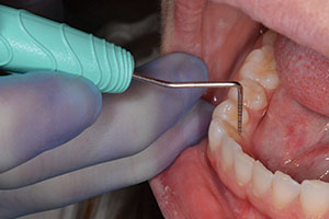
EXAMINATION PROTOCOL
The World Health Organization, the Centers for Disease Control and Prevention, and the National Institutes of Health recommend a standard exam-ination procedure. It requires adequate lighting, a dental mouth mirror, a 2×2-inch gauze square, gloves, eye protection, and a mask. The use of magnification loupes is highly recommended (zeiss.org or orascoptic.com). Magnification increases the ability to detect minor changes. The exam may take approximately 4 to 5 minutes initially. I have found that with practice it takes no more than 2 to 3 minutes to perform a thorough extraoral and intraoral examination.
CONCLUSION
Start today. Pamper your patients by performing the head and neck cancer screening examination for every patient at every visit. Get on the path to early detection of oral cancer and saving lives in the dental office.
Sources
The National Oral Health Information Clearinghouse (a service of the National Institute of Dental and Craniofacial Research, National Institutes of Health). Detecting Oral Cancer: A Guide for Health Care Proessionals. January, 1997. Web site available at: nohic.nidcr.nih.gov/pubs/detect/pages/contents.html.
The Combined Health Information Online Web site is available at: chid.nih.gov.
The American Cancer Society Web site is available at: cancer.org.
World Oncology Network Web site is available at: worldoncology.net/dental.htm
The OralCDx Web site is available at: oralcdx.com.
The Zila Web site is available at: zila.com.
Ms. Gilliam is a practicing dental hygienist, dental hygiene coach, speaker, and author. She is passionate about early detection of oral cancer. She can be reached for consulting or comments at kathryngilliam@yahoo.com.