INTRODUCTION
Digital radiography offers many advantages over the use of x-ray film in pediatric and adolescent dentistry. The author has had the pleasure of utilizing digital radiography for more than 10 years. There are many benefits of this technology, including the ability to have high-quality radiographs, being more efficient, and providing cost savings by using digital radiography over x-ray film. This article will discuss the advantages of digital radiography in working with children and adolescents, which will include improved diagnostic ability, radiography of traumatic injuries, hospital patients, sending radiographs via the Internet to insurance companies and other dentists, patient safety, cost savings, and patient/parent education. Digital radiography has been in use for more than 25 years. During this time, researchers have improved the quality of the radiographic images. The author will discuss the advantages and disadvantages of using digital radiography for children and teens. The author’s discussion of digital radiography in this article will be limited to intraoral radiographs.
Digital radiography is not an experimental modality. The technology is reliable and versatile, which expands the diagnostic and image-sharing possibilities of radiography in dentistry. The utilization of brightness and contrast, task-specific image processing, and sensor-independent archiving are advantages that digital radiography has over conventional film-based imaging.1 Here is a list of some of the uses/advantages of intraoral digital radiography for children and adolescents:
- Detection of caries
- Detection of periodontal disease
- Diagnosis of other pathological conditions
- Traumatic injuries
- Radiographs of disabled children and adolescents
- Developmental and genetic conditions
- Endodontics
- Patient and parent education
- Sending radiographs
- Saving time/money
- Offering “high-tech” dentistry for your patients.2
CASE EXAMPLES
Let’s look at some case histories to illustrate the above uses and advantages of digital radiography.
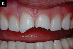
Case 1: Traumatic Injuries
Patient A is a 3-year-old male who fell onto a wooden chair and displaced his maxillary central incisors lingually. The emergency room physician referred the patient to the author, and a digital intraoral radiograph was taken that allowed the evaluation of the extent of the injury (Figures 1a to 1c). The advantage of the digital radiograph was to immediately determine if the patient had moved during the procedure. If a consultation with a pediatric dentist or oral surgeon was indicated, this radiograph could be copied onto photo quality paper and/or emailed to the consulting dentist. If this patient relocated to another dentist or was visiting his family from another locale, the radiograph could be emailed to the dentist of record.3
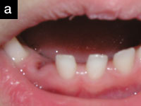
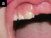
Patient B is a 3-year-old girl who fell against a picnic table and avulsed her mandibular right primary lateral incisor.
 |
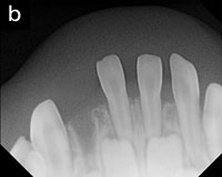 |
| Figure 1a. Avulsed primary incisor. | Figure 1b. Digital radiograph of avulsed tooth. |
 |
| Figure 1c. Dexis intraoral radiograph. |
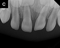
Case 2: Developmental Disturbances to the Dentition
A 6-year-old patient with a history of trauma to the primary incisors at age 2 years exhibited an over-retained discolored primary central incisor and an unerupted permanent central incisor. In this case, the digital radiograph provided the means to detect any possible developmental disturbances (Figures 2a and 2b).
 |
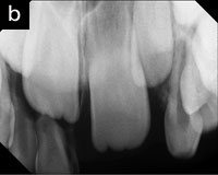 |
| Figures 2a and 2b. Clinical and digital radiographic view of overretained primary central incisor. |
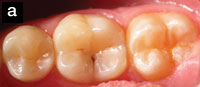
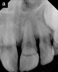
Case 3: Coronal Fracture
This 10-year-old female had a coronal fracture which occurred during a sledding accident. The endodontist e-mailed the radiographs taken during the patient’s several months of treatment and follow-up examination and treatment to the pediatric dentist (Figures 3a and 3b).
 |
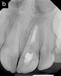 |
| Figure 3a. Digital radiograph showing coronal fracture. | Figure 3b. Postendodontic treatment. |
Case 4: Patient and Parent Education
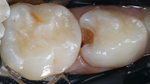
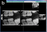
Due to the ability to magnify the image, the patient and parent can be shown any dental disease seen on the radiograph which helps in patient/parent education (Figures 4 a to 4e).
 |
 |
| Figure 4a. Occlusal digital x-ray. | Figure 4c. Bite-wing digital x-ray (Gendex 770 Intraoral x-ray machine). |
 |
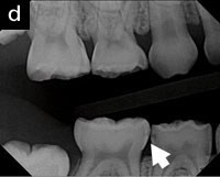 |
| Figure 4b. DEXIS sensor with angled corners. | Figure 4d. Note the caries detected on this. |
 |
| Figure 4e. Note enlargement of digital x-ray (DEXIS) bite-wing digital radiograph (arrow). |
Case 5: Pulpal Necrosis
Ben is an 18-year-old developmentally disabled adult. Due to an injury and subsequent pulp necrosis of the maxillary right central incisor, he required endodontic treatment. Under general anesthesia in an outpatient surgical center, a root canal treatment was performed. Digital radiography was utilized to allow the necessary radiographs to be taken without the need to wait for developing x-ray film. One of the digital radiographs was taken while Ben was under general anesthesia is shown in Figure 5.
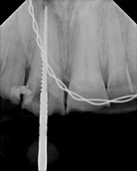 |
| Figure 5. Note the intubation tube and the endodontic file. |
ADVANTAGES OF DIGITAL RADIOGRAPHY
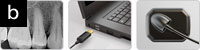
There are many advantages of digital radiographs. Digital radiography allows the dental team to take quality radiographs with ease and efficiency. No chemicals or waiting for x-ray film processing makes the time that the patient needs to be in the dental chair much less. This is especially important for the young or disabled patient.4 The size and shape of the sensor is important for the comfort for the patient. One digital x-ray company has cut the corners off of the sensor design (DEXIS) which aids in the comfort of the patient.
Quality Control and Image Enhancement
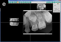
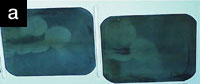
Another advantage is the quality of radiographs attainable. It is very frustrating and often embarrassing to receive poor quality radiographs from colleagues. The radiographic film was underdeveloped and the resulting poor quality is neither readable nor of archival quality. Imagine what our dental team felt about the quality of the dentistry from the dental office that sent us these x-rays. On the other hand, with the software from some digital radiography companies, the dental team can receive e-mailed radiographs and place them into patients’ digital radiographic records (Figures 6a and 6b).
 |
 |
| Figure 6a. Underdeveloped x-ray film. | Figure 6b. Quality control with digital x-rays. |
Yet another advantage of digital radiography is the ability to enhance the radiographic view by enlarging, brightness control, and improvement of the contrast. This makes the digital radiograph more diagnostic to detect fractures, caries, periodontal conditions, tumors, and cysts.5
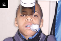
Special Needs Patients
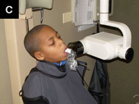
As a pediatric dentist, I have many children and disabled adult patients in my practice. One of the techniques that I utilize and teach to my residents is the use of digital radiography instead of x-ray film. The advantages are the speed of taking the radiograph and the ability to determine the quality of the radiograph immediately. I also enlist the help of the parent/guardian in the taking of the radiographs (Figures 7a and 7b).
 |
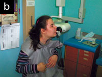 |
| Figure 7a. Mother of special needs child helping with digital x-ray. | Figure 7b. Special needs patient with DEXIS radiography. |
General Anesthesia
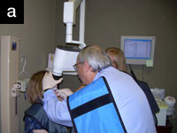
One of the advantages in using digital radiography for patients that are treated while under general anesthesia is the rapidity in taking the radiograph and in the saving of time by determining immediately the quality of the radiograph. This allows the progression of treatment to be accelerated, thus preventing the prolonged time that the patient needs to be under general anesthesia (Figure 8). Also, due to the software that accompanies the digital radiograph program, the radiographs are placed into the sequence previously set up by the dentist.
Save Time and Money
With digital radiography, you will not need to purchase film mounts and take the time required to mount the processed radiographs. You will also save money on the film processor and chemicals that would have been needed to develop traditional x-ray film. The author has been able to train his dental team to take full-mouth digital radiographs in less than 10 minutes without the use of chemicals and film mounts. This saves both time and money. In more than 10 years of using the digital sensors in our multidentist practice, we have only had to replace one sensor. If you are thinking about getting a wireless sensor, you must consider the possibility of the sensor being lost or ruined by accidently leaving it in a dental gown and going through the washing machine—ouch! Some of the digital radiography companies have multiple sizes of sensors; I suggest using the “keep it simple” approach. Another concern you might have is the learning curve in switching from conventional to digital radiography. It took our team of dentists, hygienists, and assistants less than 2 hours to get used to the Rinn type holders for the sensor. There are other holders for the sensor that are even more versatile (Figure 9).6
 |
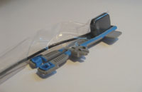 |
| Figure 8. DEXIS digital x-rays with Progeny intraoral x-ray machine used for special needs adult patient under general anesthesia. | Figure 9. DEXIS Digital Sensor and Eezee-Grip Digital Sensor Holder (DENTSPLY Rinn). |
CLOSING COMMENTS
As one can see, there are many advantages to using digital radiography in your practice. Consider making the change to digital if you have not already done so. Both your patients and dental team will appreciate the effort and the investment.
References
1. van der Stelt PF. Filmless imaging: the uses of digital radiography in dental practice. J Am Dent Assoc. 2005;136:1379-1387.
2. Child PL Jr, Christensen GJ. Digital radiography: an improvement? Dent Today. 2010;29:100-102.
3. Magid KS. Digital X-ray file sharing redefines the “solo” practitioner. Dent Today. 2007;26:140, 142-143.
4. Margolis F. Digital radiography in pediatric and special care dentistry. fredmargolis.com/DigitalRadiography.pdf. Accessed August 17, 2011.
5. Christensen GJ. Why switch to digital radiography? J Am Dent Assoc. 2004;135:1437-1439.
6. Dalin J. Dispelling the myths about digital radiography. Dental Economics. 2003. dentistryiq.com/index/display/article-display/188621/ articlesdental-economics/volume-93/issue-9/features/dispelling-the-myths-about-digital-radiography.html. Accessed August 17, 2011.
Dr. Margolis received his BS and DDS from The Ohio State University and his certificate in pediatric dentistry from the University of Illinois College of Dentistry. Dr. Margolis is a clinical instructor at Loyola University’s Oral Health Center and an adjunct clinical assistant professor at the University of Illinois College of Dentistry. He has received Mastership from the Academy of Laser Dentistry and is in full-time private practice of pediatric dentistry in Buffalo Grove, Ill. Dr. Margolis has contributed articles to both lay and professional journals. He is the author of a book, Beautiful Smiles for Special People, which is a course manual for dental personnel treating the disabled patient. He is co-author of a book, Pediatric Laser Dentistry and Atlas, to be published by Quintessence in 2010. He has lectured internationally. He can be reached at kidzdr@comcast.net.
Disclosure: Dr. Margolis receives product from DEXIS for use in his office.