 |
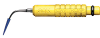
| Yellow Cavitron with blue plastic tip (DENTSPLY). |
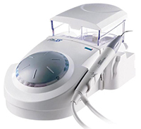 |
| P5 Newtron (ACTEON). |
INTRODUCTION
The use if dental implants is a highly successful and widely accepted procedure. However, the longevity of an implant-supported restoration may be compromised if hygienists provide improper or incomplete maintenance procedures/techniques.
Protocol for the maintenance of dental implants is a topic of much debate and documented research. The difference between periodontal maintenance and dental implant maintenance is striking; however, the products and instruments viable for dental implant care have the safety and efficacy to function for all types of restorative materials and for the natural dentition. The care of dental implants might seem intimidating for the novice; however, a hygienist need not learn an entirely different skill set. The principles are the same, but some techniques and products will differ from that of natural dentition.
The preferred term for those procedures, previously referred to as supportive periodontal therapy or periodontal recall and including the maintenance of implants, is periodontal maintenance.1
The purpose of the article is to provide guidelines for the assessment and safe prophylaxis of the implant patient.
“The Common Threads”
The title of this article, “Common Threads,” points to the similarities between the natural dentition and dental implants.
Specialists in dentistry, including general dentists, orthodontists, and periodontists, employ dental implants. Therefore, the patient, medical history, and the treatment plan that accompany dental implant therapy will confirm or deny what products/instruments hygienists can and should choose.
To provide a safe prophylaxis for the implant patient, including helpful home care aids, may often seem a daunting task for the implant treatment team. However, owing to the work of dedicated researchers and clinicians, useful information abounds. Dentists, hygienists, and dental assistants must dedicate themselves to studying and sharing the most current research and products available to ensure dental implants and the natural dentition remain safe and healthy during professional maintenance visits.
No matter the system, the majority of implants have several components in common. Most are made of a metal known as titanium, a metal widely accepted and approved by the US FDA for certain uses in the human body. Titanium has been selected as the metal of choice for the dental market because of its resistance to attack by bodily fluids, high strength, and the ability to bond with human bone.2
Successfully placed titanium dental implants bond with the jawbone through a process known as osseointegration. The Glossary of Implant Dentistry, written under the auspices of the International Congress of Oral Implantologists, defines osseointegration as: “The direct contact between living bone and a functionally loaded implant surface without interposed soft tissue at the light microscope level.”3 Dr. Per-Ingvar Brånemark’s work, illustrated in research performed on domestic canines, proved that the titanium became so imbedded in the jawbone that it could not be removed in the experimental samples.4
The priority during implant maintenance is to “do no harm” to the bone, the integrated implant, or the surrounding soft tissue. The additional common factor is the screwlike surface of a dental implant known as “threads.” Threads provide immediate stability for the implant during surgical placement, and offer continued stability during osseointegration and for the life span of the implant. The threads are useful landmarks to quickly and efficiently document the status of a dental implant over time. Threads exposed to the oral cavity must be maintained with instruments/products deemed safe and viable for a titanium surface. The aforementioned common factors are of utmost importance during the evaluation and proper prophylaxis for all types of implants.
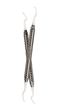 |
| Hu-Friedy instruments. |
Communication and the Common Thread
Recognizing that dental implants support clinical practice in many specialties, the necessity for accurate yet empathetic conversation styles vary with the complexity of the case. To illustrate this point, the implant team member must recognize the diverse applications for implant therapy. The application of mini-implants as a temporary anchoring device in orthodontic procedures requires a less formal communication style than the language necessary (finesse and sensitivity required) for the patient who needs full-mouth rehabilitation due to trauma or disease.
However, the common message for any implant application and the concomitant maintenance of the oral cavity must be explained and understood by the patient. The at-home maintenance expected from the patient, and the absolute commitment to sustained professional prophylaxis of implant restorations, must be incorporated into the treatment plan from conception.
STEPS FOR IMPLANT MAINTENANCE
Common Threads for Maintenance Frequency
The prevalence of tooth loss in the United States for adults age 18 years or older is 9.7%, increasing to 33.1% at age 65 years and older.5 Armed with the knowledge that bacterial pathogens that infect a natural sulcus can be a source of infection for the peri-implant tissue, it is imperative that the bacteria be suppressed and the inflammatory response reduced at regular intervals. It should be suggested that implant patients, especially those who lost their teeth to disease, should be encouraged to commit to a regular 3-month maintenance schedule.
Dental implant maintenance has components similar to that of the periodontally involved patient, coupled with clinically different diagnostic steps. The goal for the hygienist is to incorporate systematic documentation of clinical markers, such as the health status of both the soft tissue and bone supporting the dental implant.
The steps for implant maintenance are the same for all patients and should include visual inspection of the coronal prosthesis and peri-implant tissue, assessment of stability for both the prosthesis and the titanium implant, evaluation of occlusion, assessment of peri-implant tissue, debridement of hard and soft deposits; final polish, radiographic evaluation, and home care instruction.
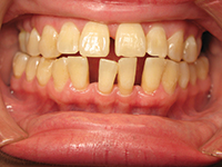
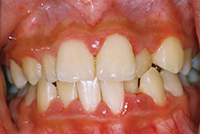
Visual Inspection
The process of deciphering health or disease begins with a visual inspection. Visual inspection provides initial insight for the health of the attached/keratinized tissue, and the free gingiva surrounding both natural dentition and dental implants. Both natural teeth and implants may have the complement of thick-keratinized and stippled-gingival margins. Often, the dental implant site may lose keratinized tissue during the surgical phase of treatment, or it may have been lost previously due to periodontal disease. The lack of keratinized tissue should be the first “cause for pause” to contemplate the need for probing the perimucosal seal surrounding an implant.
Gingival Sulcus Versus Perimucosal Seal
Gingival mucosa surrounding a dental implant differs from that encircling natural dentition in a profound manner. The tissue surrounding an implant lacks the biological seal afforded to natural teeth. In health, natural dentition and jawbone are protected by a sulcus. The sulcus is equipped with a variety of fibers (13 in all) that provide a stable epithelial attachment to the tooth surface. The attachment provides a barrier above the level of the bone, helping to combat bacterial invasion of the jawbone.
The tissue surrounding a dental implant does not include a tight and attached sulcus (as with a natural tooth); rather, it has an area called the peri-implant sulcus, or the perimucosal seal. The adaptation of regenerated gingival epithelium (after surgical placement) to an implant is critical for the development of a perimucosal seal.6 The perimucosal seal has one horizontal fiber within the crevicular epithelium that encircles the body of the implant and prosthetic component. The perimucosal seal provides a barrier against bacterial invasion from the oral cavity but, when compared to a natural sulcus, it is lacking the strength of an attachment. Hence, the debate to “probe or not to probe” begins.
Probing the Implant Site
The topic of probing the perimucosal seal surrounding a dental implant is a spark for much debate. Some clinicians believe that to probe an area that meets healthy visual assessment criteria may cause more harm than the benefit of the potential data provided. Other clinicians believe documenting any change in pocket depth should be measured.7 Moving forward, diverse practice philosophies and continued research will guide hygienists. The agreed protocol for probing is that it should be performed with a plastic probe with minimal force. One point that clinicians seem to agree upon is: after one year of stable probing, the hygienist or dentist should probe only the buccal and lingual surfaces; and the interproximal areas (mesial and distal) can be monitored by radiographic interpretation. A note of importance regarding pocket depths around implants restorations: the interface among the body of the implant, the abutment, and selected restorative design may increase depth of pocket readings. Readings of 5 to 6 mm pockets may be present (due to the implant design), but will be free of bleeding or inflammation. Probing depths alone are not indicative of trouble or disease around an implant site.
 |
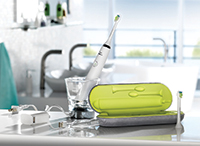 |
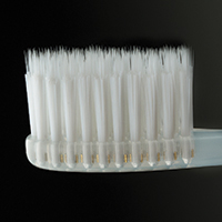
| Nimbus MicroFine (Nimbus Dental). | DiamondClean sonic toothbrush (Philips Sonicare). |
 |
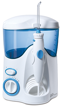 |
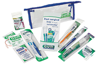
| G∙U∙M Sunstar Soft Picks and interdental brushes. | WaterPik Ultra Water Flosser and Sensonic (Water Pik). |
 |
| Crest PRO-HEALTH alcohol free (Procter and Gamble). |
Assessment for Mobility
Arguably the most important component of an implant maintenance appointment is to detect movement in either the prosthesis or in the body of the implant in the jawbone. Mobility in either location will cause complications for the longevity of the implant and can jeopardize the stability of the peri-implant tissue.8 Complaints of pain expressed by the patient should be a red flag for further investigation.
Prosthetic Component—To check the coronal stability of the prosthesis, the use of a stiff instrument (such as a mirror handle) placed on the buccal and lingual surface will help the hygienist detect any mobility problems.
Should the implant restoration include 2 or more implants, similar to a traditional fixed bridge, the hygienist should place an instrument under the pontic and pull gently upward (in an occlusal direction) while feeling for any movement. Should movement be detected, the question to be asked becomes, “Is it simply the prosthetic component or is specific data needed to ascertain the stability of the implant within the jawbone?” Radiographic evaluation is usually the best method to determine if the movement involves more than a coronal prosthetic component. It is important to note that mobility is unacceptable, potentially damaging, and must be addressed immediately by the dentist.
Occlusal Assessment—Problems with the occlusion (bite forces) have been touted by some clinicians and researchers to be the number one reason for implant failure. Malocclusion or uneven occlusal forces acting on natural teeth can cause loss of form, function, and health. However, the natural dentition has the benefit of periodontal ligaments which can, for a time, provide shock absorption to protect damage to the bone and soft tissue. For the dental implant, there is no forgiveness at the bone-titanium junction with any malocclusion. Occlusal trauma, gone undetected or untreated, will eventually cause osseointegration to fail.9 Therefore, the occlusion should be evaluated by the dentist every 3 months for the first year, followed by at least biannual evaluations for the life of the implant. Occlusal examinations should be part of every implant maintenance appointment.
Peri-Implant Tissue Assessment—The tissues surrounding dental implants are more susceptible to inflammation and bacterial infection than those surrounding the natural dentition. The hygienist should be attentive to the differences in structure of the peri-implant attachment. The tissue is more fragile, has the complement of one singular horizontal fiber, and does not mechanically attach to the body of the implant. The factors noted make the peri-implant tissue less able to withstand a bacterial assault. Inflammation and infection can alter the healthy status of the peri-implant tissue and should be inspected closely. Patients with both natural dentition and an implant prosthesis are at particular risk for peri-mucositis and peri-implantitis, especially in the presence of periodontitis in the natural dentition. The sulci around natural teeth may provide anaerobic sites for periodontal pathogens that can cross contaminate peri-implant tissue. Two categories with respect to inflammation or disease are: peri-implant mucositis and peri-implantitis.
SIGNS OF DISEASE
• Red, purple, cyanotic (a bluish discoloration of the gingiva and mucous membranes resulting from inadequate oxygenation of the blood)
• Shiny, loss of stippling, fibrotic
• Enlarged, cratered
• Bleeding on probing
• Exudates or suppuration.
PERI-IMPLANT MUCOSITIS (Reversible)
Similar to Gingivitis
• Main symptom is inflammation without evidence of bone loss
• Caused by insufficient oral hygiene and/or poor prosthetic design or occlusion
• Often caused by loose abutment (enables bacterial infiltration)
• Can lead to peri-implantitis.
PERI-IMPLANTITIS
• Recognized by radiographic or clinical evidence of bone loss
• Gingival expression may mimic a periodontal abscess, suppuration and BOP may occur
• Can cause rapid bone loss
• Treatment involves infection control and possible modification of exposed implant surface; resective or regenerative procedures may also be indicated.
• The status of osseointegration can be labeled healthy, ailing, or failing. The thread count that remains successfully integrated with the jawbone denotes the viability of an implant. If more than 50% of the threads do not remain integrated, the implant is failing.
Debridement of Hard and Soft Deposits
Debridement of an implant prosthesis and the surface of exposed titanium must be performed with safe instruments/materials. The importance of safety and efficacy of products used to maintain health around the titanium body of an implant cannot be overemphasized. The safety and suggested materials for maintenance of dental implants is ever-changing. All members of the implant team must be continually evaluating the products/materials used in the implant environment. The instruments and products listed are examples deemed safe for use around implants. (These are displayed for learning purposes only.) Discussions with manufacturers should include the acquisition of white papers to prove the safety and efficacy of use around titanium.
Cavitron and Piezo Units
The use of a Cavitron (DENTSPLY) or Piezo (SATELEC) unit has been widely accepted in the literature to enhance the breakup of biofilm and for the removal of hard deposits. Hygienists must first ensure that any instrument selected is safe for use on a titanium surface. Published in vivo studies have examined the effects of ultrasonic scalers covered with plastic sheaths and ultrasonic scalers with carbon tips versus metal scalers. The conclusion drawn: carbon- and plastic-tipped ultrasonics maintained smooth implant surfaces while traditional metal tips resulted in damaged implant surfaces.10
Scalers and Curettes
At present, the use of scalers and curettes made of plastic or acrylic resin are most favorable for use around the peri-implant site. No matter the instrument of choice, subgingival scaling around dental implants is a clinical skill that deserves continued education and practice. Hard deposits clinging to titanium are not as tenacious as those found on natural dentition; this is because the surface of titanium does not promote a tight bond for calculus. Short working strokes with light pressure are usually sufficient to remove most hard deposits. One trick of the trade for the removal of hard deposits is to use the air syringe to dry the exposed body of the implant; following this step, the deposit will often flake off without the aid of much assistance from a manual scaler.
Final Polish
Polishing the titanium portion of the implant prosthesis may not always be necessary, but if performed, it can be accomplished with rubber cups and nonabrasive polishing paste (or tin oxide).11
Nonabrasive polishing gels may include over-the-counter gel toothpaste but should not be labeled as “whitening.” As an alternative to professional polishing paste labeled as “implant specific,” the hygienist may use a slurry of water and tin oxide. (Tin oxide has been used for polishing in the dental field for decades.)
Radiographic Assessment
The literature varies concerning the need for radiographic follow-up after stable initial integration is achieved.
Examples of varying protocol are:
• Initial placement: 3 months, 6 months, 12 months, every 2 years.12
• Initial placement: 6 months, 12 months, and every 2 years if no pathology present.
• Initial placement: every 6 months if pathology present.13
Radiographic interpretation will aid the hygienist in documenting the number of threads that remain successfully integrated. The status of osseointegration can be labeled healthy, ailing, or failing. If an implant is ailing (the loss of more than 3 threads), a clinician may step in with resective procedures to stop the inflammatory response; or the use of debridement and oral antibiotics may be helpful.
The density of the bone, and any change in thread count exposure, must be monitored carefully until an infection is under control. If more than 50% of the threads do not remain integrated, the implant may receive a failing status. What does this mean for the implant? Rarely are implants removed unless mobility and total bone loss occur. Attempts to remediate bone loss and reclaim health and stability are of utmost importance. Procedures to eliminate disease and re-establish bone are well-documented and are often a successful means to maintain a failing implant. The removal of an implant would be the last resort; it is only recommended after attempts to remediate a poor situation are unsuccessful.
Common Threads for Home Care Aids
Implant maintenance empowers the hygienist to build a rapport complete with trust and integrity with the patient. The success of implant dentistry depends upon the hygienist and patient working in concert to plan a home care strategy that will complement the prosthesis and the ability of the patient. The communication skills of the hygienist, coupled with specific home care aids, will help ensure patient compliance.
The common factor for all home-care aids is that the products must be made of materials that will not scratch the surface of the titanium implant or the porcelain surface of the prosthesis. The products safe for use on dental implants are also viable for cosmetic dentistry maintenance materials.
A resource card for your implant patient is a useful tool for the hygienist, the referring dentist, and the patient. For each case, a small card (laminated and small enough to fit into a wallet) should be provided to the patient with relevant information on it (name of the implant company, length and width of each implant, corresponding tooth area, and the type of cement or screw used to fix the prosthetic portion onto the body of the implant). Sometimes the cards will also list the name of the dental laboratory that fabricated the case.
Manual Toothbrushes—Soft or extra soft manual toothbrushes, if used correctly, may provide satisfactory breakup of the biofilm surrounding the dental implant. The dental literature supports the use of soft or extra soft brushes as a means to clean the surface of a titanium implant safely.
Sonic and Electric Brushes—Research (supported by Philips Sonicare) reports that sonic and electric toothbrushes are safe and successful for the removal of soft deposits from a titanium implant surface. Both types of brushes can provide added security for those patients with limited dexterity.
Results from Philips Sonicare further stated that the Sonicare DiamondClean standard and compact brush heads resulted in significantly less dentin abrasion than manual toothbrush (P < .05). There was no significant difference between Sonicare DiamondClean standard and compact brush heads. The conclusion formed from the in vitro study; the Sonicare DiamondClean standard and compact brush heads were found to cause about 50% less dentin wear than a manual toothbrush.14 The study also provided research that all sonic and electric toothbrushes are safe for use around dental implants. The study demonstrated that the use of Sonicare Elite (or other power toothbrushes) does not affect implant retention strength in vitro for up to 2 years of simulated clinical brushing with toothpaste. This suggests that the exerted vibration from using a power toothbrush has no effect on implant longevity, hence corroborating that power toothbrushes are safe to use with dental implants.15
Home Irrigation Systems
Irrigation units are helpful in debriding an implant prosthesis of soft debris and food particles. The units should be filled with solutions safe for use around implants, and they should be used on a very low setting. The safest solution is water, but most importantly, a solution free of alcohol is best. The water stream should be aimed horizontally between the interdental spaces.
Floss, Interdental Picks, and Brushes
Floss—Different shapes of floss such as flat tape, round string, or Super Floss are safe for use around dental implants. However, caution must be used in selection. Floss with pumice, or whitening products placed on or into the floss matrix, should not be used.
Interdental Brushes—Interdental picks and brushes are helpful for reaching between contacts and under prosthetic designs with closed contacts. Similar to natural dentition restored with fixed bridge work, the brushes and picks provide the patient a much needed aid in homecare. All parts of the picks or interdental brushes must be made of plastic or coated with plastic.
Mouthrinses and Fluoride—The plethora of mouthrinses available around the globe can blur a patient’s ability to make a safe selection, so a helpful hint from the hygienist is in order.
One factor must be clear: the safest rinse or premade solution is one free of both alcohol and whitening solution. Rinses containing alcohol can be used with the understanding that it may exacerbate the symptoms of dry mouth and must be monitored daily.
Fluoride solutions (both in-office and at-home rinses) must be free of acidulated phosphate. Neutral sodium fluoride has been proven safe for use in patients with mixed dentition and in implant restorative cases.
CLOSING COMMENTS
With more than 400,000 implants now being placed yearly, implant dentistry is here to stay. The hygienist need not learn an entirely new system of documentation, nor discover new and different home care aids. Yet, differences in managing dental implants versus natural dentition do exist. Therefore, a constant review of new literature and materials/techniques should be a part of the hygienist’s daily practice responsibilities.
References
- American Academy of Periodontology. Supportive treatment. In: Proceedings of the World Workshop in Clinical Periodontics. Chicago, IL: American Academy of Periodontology; 1989:IX-24.
- Titanium in dental implants. titanium.com/markets/medical/dental-implants. Accessed August 15, 2012.
- Jalbout Z, Tabourian G; International Congress of Oral Implantologists; New York University College of Dentistry. Glossary of Implant Dentistry. Upper Montclair, NJ: ICOI; 2004:40.
- Ring ME. A thousand years of dental implants: a definitive history—part 1. Compend Contin Educ Dent. 1995;16:1060-1064.
- US Department of Health and Human Services. Oral Health in America: A Report of the Surgeon General. Rockville, MD: US Department of Health and Human Services, National Institute of Dental and Craniofacial Research, National Institutes of Health; 2000.
- Silverstein L, Garg A, Callan D, et al. The key to success: maintaining the long-term health of implants. Dent Today. 1998;17:104-111.
- Sison SG. Implant maintenance and the dental hygienist. Access. May-June 2003;suppl 1:1-12. adha.org/downloads/sup_implant.pdf. Accessed August 2, 2012.
- McKinney RV Jr, Steflick DE, Koth DL, et al. The scientific basis for dental implant therapy. J Dent Educ. 1988;52:696-705.
- Eskow RN, Smith VS. Preventive periimplant protocol. Compend Contin Educ Dent. 1999;20:137-154.
- Kawashima H, Sato S, Kishida M, et al. Treatment of titanium dental implants with three piezoelectric ultrasonic scalers: an in vivo study. J Periodontol. 2007;78:1689-1694.
- Matarasso S, Quaremba G, Coraggio F, et al. Maintenance of implants: an in vitro study of titanium implant surface modifications subsequent to the application of different prophylaxis procedures. Clin Oral Implants Res. 1996;7:64-72.
- Mombelli A, Lang NP. The diagnosis and treatment of peri-implantitis. Periodontol 2000. 1998;17:63-76.
- Ward ST, Czuszak CA, Thompson AL, et al. Assessment and maintenance of dental implants: clinical and knowledge-seeking practices of dental hygienists. J Dent Hyg. 2012;86:104-110.
- Moore M, Putt M, Jain V, et al. In vitro assessment of dentin wear resulting from the use of the Philips Sonicare DiamondClean power toothbrush. The Science Behind Sonicare [brochure]. Philips Oral Healthcare, Inc., 2011. Data on file.
- Castellon R, Fernunson MA, Garcia-Godoy F, et al. Effect of power toothbrushes on retention strength of implant crowns and abutments under simulated clinical conditions. The Science Behind Sonicare [brochure]. Philips Oral Healthcare, Inc., 2011. Data on file.
Further Reading and Information
There are many professional associations willing and able to help hygienists and other team members keep abreast of changes in dental implant therapy and maintenance: American Academy of Periodontology (perio.org), American Academy of Implant Dentistry (aaid-implant.org), The Academy of Osseointegration (osseo.org), and the International Congress of Oral Implantologists (icoi.org).
Ms. Wadsworth owns Lisa C. Wadsworth, Inc, a company focused on consulting and personal coaching for the dental community. With more than 3 decades of experience in the implant and periodontal arena, she began her career as a dental assistant, progressed to certification in implant surgical assisting, and lastly a degree in dental hygiene. Ms. Wadsworth has received Fellowship status with the Association of Dental Implant Auxiliaries, served as a dental hygiene educator for OraPharma, and is recognized by Philips Sonicare as a key opinion leader. She is a member of the Speaking Consulting Network and lectures on a national scale. Topics include implant dentistry, periodontal protocols, professional development, and ergonomics. As a speaker, she has honored by Dentistry Today as a Leader in Continuing Education since 2007. Ms. Wadsworth has served as a contributing editor for Modern Hygienist from 2005 to 2007. She has been published in such journals as RDH magazine, Dental Assisting Digest, Dental Practice Report, and Implant News and Views. She can be reached at (215) 262-6168 or via the Web site lisawadsworth.com.
Disclosure: Ms. Wadsworth is a key opinion leader for Philips Sonicare. She receives financial compensation for opinions about products as well as lecture content.