INTRODUCTION
Generalized tooth wear, attrition, and erosion can significantly decrease an individual’s vertical dimension of occlusion (VDO). Considered the 3-D space between fixed and movable occluding anatomic components when either resting or functioning, VDO is the cornerstone for ensuring a healthy and functional occlusion, as well as appropriate restoration proportions and ideal smile design for long-term service and patient satisfaction.1,2
Therefore, it is imperative that a patient’s VDO be re-established when full-mouth rehabilitations are undertaken to treat generalized tooth wear.3-7 VDO significantly affects restorative outcomes and influences patient comfort, aesthetics, restoration anatomy and morphology, material selection, and treatment longevity (ie, the likelihood of withstanding mastication forces).8
 |
| Figure 1. Preoperative, full-facial view of a male patient with severe attrition, erosion, and tooth wear. |
 |
| Figure 2. Pre-op view of the patient’s natural smile. |
The ideal VDO varies among individuals; is affected by stress, posture, disease, medication, airway, and other factors; and can be determined in a number of ways, including the use of cephalometry, diagnostic wax-ups or set-ups, and/or phonetic sounds.2,6,7,9,10 Regardless, according to Dr. Bob Lee’s minimally invasive bio-rejuvenation (Lee coined it bioesthetics, but the author prefers to use the term bio-rejuvenation) dentistry principles, determining and restoring a patient’s VDO should be predicated on the joints seating in their most superior, anterior, and medial position (ie, anterior proprioceptive guidance), which would enable proper tooth form, appropriate occlusion with maximal intercuspal position, and reduced masseter and temporalis elevating muscle activity.11-13 When the joints (ie, condyle/disc complex) are healthy and functioning properly, a physiologically correct occlusal relationship is observed with little to no attrition, anterior teeth occlude properly, posterior teeth are non-interfering, and the inferior lateral pterygoid muscle releases during elevator muscle contraction and maximal intercuspal position.12,14
In the absence of a healthy and comfortable joint relationship, it is necessary to stabilize, rehabilitate, and adapt the patient’s occlusion. Additive interim restorations, in addition to deprogrammers and other orthotic devices, have been used to stabilize and simultaneously alter an individual’s occlusion.15,16 In particular, a Condylar Centering Orthotic (C2O) device is a custom-made, clear acrylic splint that is worn in the mouth 24/7 until joint stabilization is achieved (ie, an average of 6 to 12 weeks); weekly splint adjustments are necessary to ensure proper jaw positioning.
Additive restorations (ie, direct composite), on the other hand, historically have been intended to enhance aesthetics for the short term and to enable dentists and patients to evaluate the anticipated therapeutic results of definitive, yet more invasive and costly, indirect partial- or full-coverage restorative treatments.15-17 However, whereas traditional full-mouth rehabilitation with indirect restorations commits patients to a lifetime of expensive re-restorations, additive adhesive composite resin reconstruction is predictable, minimally invasive, and cost effective for many patients with generalized wear and loss of VDO.12,18 It can also be considered a “long-term” solution if the following principles of biologic occlusion are met: (1) a verified, fully seated joint; (2) biologic tooth morphology; and (3) proper form of the occluded dentition (ie, an interarch relationship).
Fortunately, recent advancements in the material characteristics (eg, strength, wear resistance and durability, and lower polymerization shrinkage), optical properties (eg, more shades and opacities), and handling (eg, thixotropic, easier to polish with higher gloss/luster) of direct composites lend to their use for the long-term restoration of VDO in cases of generalized wear, according to the principles of minimally invasive bio-rejuvenation dentistry.19-21 Among the materials available is an injectable nanohybrid composite (G-ænial Universal Flo [GC America]) that demonstrates high strength, excellent wear resistance, high gloss retention, precise handling, and lifelike aesthetics.22-24 The author has found this to be an ideal injectable composite resin material when undertaking the bio-rejuvenation techniques in his practice, and the protocol for its use will be described in the following case presentation.
CASE REPORT
Diagnosis and Treatment Planning
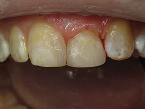
A 49-year-old male patient presented with severe wear and erosion of all of his teeth, resulting in a decreased VDO and mid-facial collapse (Figures 1 to 3). The patient was self-conscious about his facial and smile appearance, and he was also aware of, and concerned about, having already destroyed much of his natural tooth structure. He was reluctant to further “grind down” even more of his natural teeth to accommodate indirect full-coverage crown restorations.
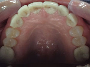
Diagnostic records were obtained (ie, a proper series of photographs, radiographs, full-arch/full-mouth VPS impressions, a centric relation [CR] bite registration, a face-bow transfer, and models), and a comprehensive examination was performed. The examination revealed generalized severe wear of the teeth, resulting in decreased VDO (Figures 4 and 5). Tooth No. 12 had previously been restored with an oversized, full-coverage crown restoration. The patient’s periodontal health was excellent, and there were no systemic or medical issues contraindicating treatment.
 |
 |
| Figure 3. Pre-op, retracted, close-up view of the patient’s severely worn dentition. | Figure 4. Pre-op occlusal view of the patient’s maxillary arch, revealing severe wear and attrition. |
 |
 |
| Figure 5. Pre-op occlusal view of the patient’s mandibular arch, revealing severe wear and attrition. | Figure 6. Pre-op models were mounted and analyzed to determine the ideal vertical dimension of occlusion (VDO). |
 |
 |
| Figure 7. The patient’s joints were s tabilized using a Condylar Centering Orthotic device (C2O). |
Figure 8. View of the centric relation mounted models at the restored VDO. |
 |
 |
| Figure 9. Occlusal view of the diagnostic wax-up. | Figure 10. Right-lateral view of the diagnostic wax-up. |
 |
 |
| Figure 11. Lumaloc Composite Transfer Stints were used to ensure the accuracy of the composite resin placement. | Figure 12. Postoperative view of the completed composite restorations (G-ænial Universal Flo [GC America]) on the mandibular arch. |
 |
 |
| Figure 13. The accuracy of the restorations was confirmed using the validator. | Figure 14. Post-op view of the restored maxillary and mandibular teeth. |
 |
 |
| Figure 15. Post-placement close-up view of the maxillary central incisors following application of the OPTIGLAZE (GC America). | Figure 16. Left-lateral view of the lithium disilicate crown restorations on teeth Nos. 12 and 13. |
 |
 |
| Figure 17. The occlusion was verified based on mandibular and maxillary centric contact points. | Figure 18. View of the mandibular centric contact points. |
An examination of the temporomandibular joint (TMJ) demonstrated the need to stabilize the joints prior to determining and restoring the patient’s VDO. Thorough model analysis was completed using mounted study models on an AD2 Semi-Adjustable articulator (Advanced Dental Designs) (Figure 6). It was explained to the patient that by first stabilizing his jaw joints and muscles with the condyles seated in the CR position, it would be possible to restore his worn teeth to an aesthetic, functional, and comfortable position in a minimally invasive and cost-effective manner that would simultaneously enhance his overall facial appearance.12 However, the patient was informed that despite the enhanced durability and wear-resistance of direct composite resins, he should expect the need for ongoing maintenance and repairs in the years ahead. Given the minimally invasive nature of this treatment plan, this was acceptable to him.
The bio-rejuvenation treatment planned for the patient would therefore first involve stabilizing his condyles, subsequently opening his VDO, and then doing minimally invasive bio-rejuvenation dentistry with direct composite restorations. Teeth Nos. 2 to 11, Nos. 14 and 15, and Nos. 18 to 31 would be treated using a combination of universal injectable composite (G-ænial Universal Flo) and characterization glazes (OPTIGLAZE Color [GC America]). The composite selected for this case demonstrates physical and material characteristics similar to compactable composites, with requisite strength and wear resistance, ability to easily polish, and enamel-like properties.
Tooth No. 12, which had previously been treated with an oversized, full-coverage crown restoration, along with tooth No. 13, was treatment planned for High Density Micronization (HDM) lithium disilicate full-coverage restorations (GC Initial LiSi Press [GC America]). Since it would have been difficult to properly increase the anterio-posterior width of tooth No. 13 with composite, both teeth Nos. 12 and 13 were restored with crowns to create unity and resolve size discrepancy issues.
 |
| Figure 19. View of the maxillary centric contact points. |
 |
 |
| Figure 20. Post-op full-facial view of the patient after completion of minimally invasive bio-rejuvenation dentistry. |
Figure 21. Post-op natural smile view of the patient’s bio-rejuvenation dentistry. |
Condylar Stabilization
The patient’s joints were stabilized and his occlusion analyzed using a C2O (Figure 7). The C2O is a permissive anterior-guided orthotic that eliminates any eccentric interferences, allows the condyles to fully seat, and eliminates muscle hyperactivity. This treatment phase involved weekly orthotic adjustment appointments to ensure proper jaw positioning. (TMJ physical therapy utilizing the C2O is taught at the Texas Center for Occlusal Studies and Minimally Invasive Dentistry [txcos.com].)
Determining VDO
To make a definitive diagnosis and treatment plan based on the now stabilized joints, an open CR bite record was taken at a VDO that would enable restoration to proper tooth form, horizontal overjet, overbite, and centric contacts (Figure 8). Alginate impressions were taken, after which models were poured and mounted on a semi-adjustable articulator with a face-bow transfer.2,25 Aesthetically and functionally, the patient had lost VDO, and analyzing the mounted models confirmed it would be possible to restore teeth Nos. 2 to 11, Nos. 14 and 15, and Nos. 18 to 31 with an additive, bio-rejuvenation approach.
A complete diagnostic wax-up was then fabricated by the dental laboratory team, taking into consideration the correct incisal edge/length, emergence profile, and other morphologic characteristics (eg, biologic principles) for the patient’s rejuvenated teeth (Figures 9 and 10). The wax-up, along with clear vinyl polysiloxane (VPS) (Lumaloc [Ultradent Products]) stints (Figure 11) for accurately transferring the wax-up to the mouth using an injectable composite resin, as well as the acrylic validators for verifying accuracy of the completed restorations, were returned to the dental office.
Minimally Invasive Bio-Rejuvenation
In this case, the goal of minimally invasive bio-rejuvenation treatment was to establish an ideal VDO that would correspond with and support the patient’s improved joint/muscle relationship. First, the lower incisors would be restored. They were micro-abraded, with no enamel prepared/removed. The teeth were acid-etched for 20 seconds and rinsed thoroughly and dried, after which a universal adhesive (Peak Universal Adhesive [Ultradent Products) was applied, air thinned, and light cured.
Next, with the matrix stint (Lumaloc) in place in the mouth, the teeth were restored using shade A1 of the injectable composite (Figure 12). The composite was then light cured through the clear stint, after which it was removed. Once the initial replacement layer had been placed and cured, the accuracy of the restorations was confirmed with the validator (Figure 13). Once the teeth were restored with the A1 shade, the teeth were polished to completion. Then the rest of the mouth was restored in the following order: maxillary incisors (occlusion verified); maxillary and mandibular first molars (occlusion verified); maxillary and mandibular premolars (note: teeth Nos. 12 and 13 were provisionalized with Tempsmart Provisional crowns, and the occlusion was verified); maxillary and mandibular cuspids (occlusion verified); and, finally, maxillary and mandibular second molars (Figure 14).
Lifelike characterizations were imparted by cutting back 0.3 mm of the facial composite (Figure 14) and then using a variety of light-cured color characterization glazes (Figure 15). The final enamel layer was created by applying the JE shade of the same injectable composite over the now characterized dentin A-1 layer. The restorations were then polished to completion, thus enhancing mammelons, translucencies, and other characterizations created by the utilization of the OPTIGLAZE Color.
Full-arch polyether impressions (Impregum Soft [3M]) were taken for the fabrication of the lithium disilicate full-coverage crown restorations by the dental lab team for teeth Nos. 12 and 13. These restorations were seated at a subsequent appointment (Figure 16).
In this case, the patient was restored to a fully functioning occlusion (Figures 17 to 19) at a comfortable VDO, which simultaneously enhanced his facial and smile appearance (Figures 20 and 21). By restoring his occlusion to a stable condylar position in CR at a proper VDO, a position at which his mastication muscles were relaxed and functioning comfortably, his facial muscles and smile also became more relaxed, with minimal pressure.12 This optimal occlusion is consistent with Dr. LD Pankey’s recommendations that condyles should be fully seated in the fossa, and that all posterior teeth should touch simultaneously and evenly, with the anterior teeth touching lightly.26
CLOSING COMMENTS
As demonstrated in this article, even the most severe cases of tooth wear with a loss of VDO can be restored with minimally invasive direct composite resin bio-rejuvenation. Essential to achieving this objective is working from a stable condylar position (ie, by first stabilizing the joint), building the correct VDO, and creating true anterior proprioceptive guidance. The latter 2 are predicated on creating the proper overjet/overbite and the appropriate form of inter-arch occlusal relationship. Because preservation of natural tooth structure is of the utmost importance, clinicians can use materials like advanced injectable composites (eg, G-ænial Universal Flo) that can be precisely placed, demonstrate high wear resistance, and be polished easily.
By using the clinical protocol and materials demonstrated herein, the properly trained clinician can achieve the treatment objectives of a full-mouth bio-rejuvenation case in a minimally invasive and cost-effective manner for patients.
References
- Chapman RJ, Mehta N. The craniofacial complex and vertical dimension: proper positioning improves speech, breathing, eating, and appearance. Inside Dentistry. 2013;9:54-78.
- Rivera-Morales WC, Mohl ND. Restoration of the vertical dimension of occlusion in the severely worn dentition. Dent Clin North Am. 1992;36:651-664.
- Boucher CO. Complete denture prosthodontics—the state of the art. J Prosthet Dent. 1975;34:372-383.
- Lucchini JP, Lavigne J, Spirgi M, et al. The centric relation. IV. Variations in condylar positions according to the methods of measuring centric relation and to the patient’s clinical type [in French]. SSO Schweiz Monatsschr Zahnheilkd. 1978;88:1-12.
- Ash MM Jr, Ramfjord SP. Reflections on the Michigan splint and other intraocclusal devices. J Mich Dent Assoc. 1998;80:32-46.
- Gattozzi JG, Nicol BR, Somes GW, et al. Variations in mandibular rest positions with and without dentures in place. J Prosthet Dent. 1976;36:159-163.
- Pound E. Controlling anomalies of vertical dimension and speech. J Prosthet Dent. 1976;36:124-135.
- Kois JC, Phillips KM. Occlusal vertical dimension: alteration concerns. Compend Contin Educ Dent. 1997;18:1169-1177.
- Rivera-Morales WC, Mohl ND. Relationship of occlusal vertical dimension to the health of the masticatory system. J Prosthet Dent. 1991;65:547-553.
- Ferrario VF, Sforza C, Serrao G, et al. Three-dimensional assessment of the reliability of a postural face-bow transfer. J Prosthet Dent. 2002;87:210-215.
- Lee RL. Esthetics and its relationship to function. In: Rufenacht CR, ed. Fundamentals of Esthetics. Chicago, IL: Quintessence Publishing; 1990:137-208.
- Stewart H. Minimally invasive bio-rejuvenation dentistry: a conservative approach to full-mouth rehabilitation. Dent Today. 2017;36:94-98.
- Williamson EH, Lundquist DO. Anterior guidance: its effect on electromyographic activity of the temporal and masseter muscles. J Prosthet Dent. 1983;49:816-823.
- Dawson PE. Functional Occlusion: From TMJ to Smile Design. St. Louis, MO: Mosby Elsevier; 2007:45-55.
- Nanda A, Jain V, Manak K, et al. An alternative adhesive based technique of raising the occlusal vertical dimension. Indian J Dent Res. 2014;25:505-508.
- Nanda A, Jain V, Srivastava A. An electromyographic study to assess the minimal time duration for using the splint to raise the vertical dimension in patients with generalized attrition of teeth. Indian J Dent Res. 2011;22:303-308.
- Burrow MF. Attrition and erosion: restorative planning and performance. Ann R Australas Coll Dent Surg. 2012;21:97-100.
- Stewart H. Conservative full mouth rehabilitation using the principles of bioesthetic dentistry. Contemporary Esthetics. 2006;10:46-56.
- Palmer KM. Use of additive dentistry decreases risk by minimizing reduction. Compend Contin Educ Dent. 2012;33:346-352.
- Sensi LG, Strassler HE, Webley W. Direct composite resins. Inside Dentistry. 2007;3:76.
- Fortin D, Vargas MA. The spectrum of composites: new techniques and materials. J Am Dent Assoc. 2000;131(suppl):26S-30S.
- Sumino N, Tsubota K, Takamizawa T, et al. Comparison of the wear and flexural characteristics of flowable resin composites for posterior lesions. Acta Odontol Scand. 2013;71:820-827.
- Kitasako Y, Sadr A, Burrow MF, et al. Thirty-six month clinical evaluation of a highly filled flowable composite for direct posterior restorations. Aust Dent J. 2016;61:366-373.
- Sachdeva P, Goswami M, Singh D. Comparative evaluation of shear bond strength and nanoleakage of conventional and self-adhering flowable composites to primary teeth dentin. Contemp Clin Dent. 2016;7:326-331.
- Dawson PE. New definition for relating occlusion to varying conditions of the temporomandibular joint. J Prosthet Dent. 1995;74:619-627.
- Mahan PE, Wilkinson TM, Gibbs CH, et al. Superior and inferior bellies of the lateral pterygoid muscle EMG activity at basic jaw positions. J Prosthet Dent. 1983;50:710-718.
Dr. Stewart maintains a private practice in Flower Mound, Texas, and is CEO, director, and clinical instructor for the Texas Center for Occlusal Studies and Minimally Invasive Dentistry (txcos.com). A frequently published author, he lectures extensively nationally and internationally about advanced minimally invasive restorative and cosmetic dentistry, occlusion, and composite resin techniques, with an emphasis on biologic and systems-focused treatments. A mentor at the Schuster Center for Professional Development in Scottsdale, Ariz, Dr. Stewart can be reached via email at hal.stewart@verizon.net.
Disclosure: Dr. Stewart has received honorarium in the past for speaking engagements sponsored by Ultradent Products and GC America.
Related Articles
The Wonderful World of Teeth Whitening
Restoring a Class IV Fracture Using a Compactable, Universal Nanohybrid Composite
Universal Nanohybrid Composite Comes in Four Shades