INTRODUCTION
In aesthetic dentistry, material science has played a key role in the development of restorations with a more lifelike appearance. Despite the progress shown in the development and application of more minimally invasive dentistry (MID) with lab-fabricated restorations, direct composite resin still plays a vital role in conservative anterior and posterior restorations. During the last few decades, there has been a tremendous interest in recreating the natural appearance of a dentition while using conservative aesthetic- and restorative-dentistry treatment protocols. With current advancements in the physical and aesthetic properties of composite resins, direct resin restorations have developed into a viable long-term restorative solution1 (Figures 1 and 2).
Philosophy of MID in Cosmetic Restorative Treatment
In correcting dental problems, one must understand the importance of approaching treatment planning with the appropriate material and clinical perspectives. With advances in material science, the ability to remineralize and heal demineralized tooth structure not only reduces the incidence of future caries, but it also allows for less tooth removal during the restorative treatment.2,3
 |
 |
| Figure 1. Minimally invasive dentistry can correct failing alloy restorations with recurrent caries. | Figure 2. Direct resin restorations have developed into a viable long-term restorative solution. |
 |
 |
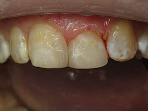
| Figure 3. Caries emerging along the margin of existing porcelain restorations will continue to be a prevalent problem in patients. A minimally invasive prep along the lateral incisor was done to remove marginal decay. | Figure 4. An injectable composite resin (such as G-ænial Universal Flo [GC America]) can be an ideal dental material to use for minimally invasive repairs. |
Adhesion dentistry allows for a conservative cavity design to be used instead of the original G.V. Black preparation designs. So that the restoration could not be dislodged, G.V. Black’s classification for cavity preparation was based upon the need to include resistance and retention form in the prep design. Additional tooth removal to create divergent internal axial walls and retentive grooves were requirements of G.V. Black’s cavity design.2 Presently, the cavity preparation design should be based on the preservation of natural dentition. Still, with the progress of dental adhesives, the additional tooth removal—once required to create mechanical retention and resistance—is no longer needed. Therefore, MID is now a realistic treatment goal with adhesive resins.
Injectable Composite Resins: A Real Game Changer
In the last several years, there has been much progress within the arena of minimally invasive dental materials. Injectable composite resin is truly an outside-of-the-box material designed for fast application and polishing with high gloss retention. In comparison to conventional flowable composite resins, injectable composite resins can be used for long-term restorative purposes. Due to the delivery system used with these flowable composite resins, injectable composites can be used in hard-to-reach areas of the mouth. This gives the clinician another option to confidently restore all restorative classifications with direct resin. These injectable composite resins are a real game changer for minimally invasive restorative dentistry.
The idea behind an injectable composite resin (such as G-ænial Universal Flo [GC America]) is that this material combines the user-friendly aspects of flowable resins with the strength, shades, and ease of handling and polishing that is similar to sculptable state-of-the-art nanohybrid composite resins. Injectable composite resins can be used for both the anterior and posterior teeth (Figures 3 and 4) and results in excellent aesthetics. The unique chemistry of G-ænial Universal Flo features 200 nm strontium glass fillers homogeneously dispersed for high flexural strength, high wear resistance, and low flexural modulus.4 In this material, new silane coupling technology creates a stronger adhesion between the glass fillers and resin matrix, thereby increasing flexural strength of the resin and sustained high surface luster.
 |
 |
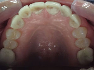
| Figure 5. A 16-year-old patient presented with cosmetic dental challenges, including multiple diastemas, undersized teeth, and a gummy smile. | Figure 6. The retracted view shows a healthy dentition with diastemas. |
 |
 |
| Figure 7. A diode laser (NV Microlaser [DenMat]) was used to remove gingival tissue as a part of the aesthetic corrections implemented in this case. | Figure 8. An adhesive resin (G-Premio BOND [GC America]) was placed and cured prior to sculpting the composite resin veneers (G-ænial Sculpt [GC America]). A matrix was used along the lingual aspect to establish the lingual and incisal contours. |
 |
 |
| Figure 9. Composite resin veneers were first completed on both central incisors. Particular attention was given to create central incisors that would be mirror images of each other. | Figure 10. A silicone matrix was used to develop proximal, lingual, and incisal contours of the lateral incisors and canines. |
 |
 |
| Figure 11. After establishing correct incisal edge position, the composite resin veneers were sculpted to completion. | Figure 12. Completed composite resin veneers along teeth Nos. 6 to 11. In closing multiple diastemas, it is important to develop correct tooth proportion to meet aesthetic needs. |
 |
| Figure 13. With proper diagnosis, treatment planning, and treatment execution, beautiful aesthetic treatment can be achieved using minimally invasive techniques. |
Universal Nanotechnology Composite Resins
The continued trend in direct restorative resins is for a material to be a universal composite resin for both anterior and posterior teeth. It is important that the material exhibit natural aesthetics that allow the clinician to easily achieve natural-looking dentition in all clinical situations. For an aesthetic material to be successful, it must reflect and refract light in a way that is close to that of natural tooth structure. When the composite resin closely matches the optical properties of the surrounding tooth/teeth, a chameleon effect is achieved that renders the restoration invisible. A new nanohybrid composite resin is showing great success because it reflects, refracts, and absorbs light in a similar manner to natural dentition, thus making it simple to achieve the highest level of aesthetics.5
G-ænial Sculpt (GC America) is a strong, high-density material with uniform dispersion that was developed to have improved wear resistance and high gloss polishability for a long-lasting mirror-like surface finish. The new high performance filler features complete 360° silanization of the nanoparticles to strengthen the adhesion between the nanosized (200 nm) glass particles to the resin matrix, thereby increasing the flexural strength and durability of the composite resin. This unique chemistry also gives the material a harder, ceramic-like surface and long-term wear resistance, along with outstanding handling characteristics during clinical placement. In the author’s opinion and clinical experience, G-ænial Sculpt is a true blend of strength and beauty in direct composite resin.
CASE REPORTS
Case 1: Anterior Restorative Application
A 16-year-old female patient presented with cosmetic challenges in her aesthetic zone that included multiple diastemas, undersized teeth, and a gummy smile (Figure 5). It was determined, after careful evaluation, that the gummy smile was due to an altered passive eruption problem. In addition, the undersized nature of her teeth created a facial proportion issue (Figure 6). It was decided that closing the diastemas would increase the proportion of her anterior teeth, thus creating the better facial proportion and balance that would be needed to create an aesthetically appealing smile for this patient. To close the diastemas in this young dentition, a minimal invasive procedure was recommended; no-prep composite resin veneers (teeth Nos. 6 to 11) would be done, without any tooth preparation, to correct this cosmetic dental problem.
 |
 |
| Figure 14. A 43-year-old patient presented with old and failing Class II alloy restorations. | Figure 15. After proper isolation using a rubber dam, the failing alloy restorations and associated dental caries were removed using a caries detection stain (Caries Detector [Zest Dental Solutions]). |
 |
 |
| Figure 16. Using a 7th generation bonding adhesive (G-Premio BOND), a polymer chain union can be created between the composite resin and the resin-modified glass ionomer base (Fuji IILC [GC America]). | Figure 17. An injectable composite resin (G–ænial Universal Flo) was placed in the proximal areas to seal the box. |
 |
 |
| Figure 18. The second premolar was restored in the same way using an anatomic matrix (Composi-Tight [Garrison Dental Systems]) to recreate the correct interproximal contours. | Figure 19. A 15-fluted Q-Finisher bur (No. H274Q-018 [Komet USA]) was used to develop the initial occlusal anatomy and to finish and blend the margins. |
 |
| Figure 20. With modern direct composite resin materials, consistent and predictable clinical success can be achieved for both anterior and posterior teeth. |
Clinical Protocol
After sounding the bone with a periodontal probe, a diode laser (NV Microlaser [DenMat]) was first used to remove excess gingival tissue (Figure 7). Next, the enamel surface was cleaned using a microetcher (MicroEtcher II [Danville Materials]) to remove all dental pellicle from the tooth surfaces. Then, a multipurpose, all-in-one adhesive resin (G-Premio BOND [GC America]) was used as the bonding resin. G-Premio BOND was placed for 10 seconds and then air thinned for 5 to 10 seconds. The adhesive was light cured for 10 seconds (Figure 8). It should be noted that this versatile adhesive resin can be used with a self-etch or total-etch protocol. The result is a very thin (3 µm) adhesive layer with high shear bond strength (35 mPa). Due to the resulting thin adhesive layer, G-Premio BOND can be used for both direct and indirect restorative dentistry.
Using the latest generation of nanohybrid composite resin (G-ænial Sculpt), a simple sequence of composite resin layering was done to blend the composite resin to the existing tooth, creating a polychromatic effect. Using a lingual matrix, a universal shade A-1 was placed along the lingual. The reason for this was to mimic the lingual enamel coloration and the incisal halo. Then placement of subtle characterizations with tints (Kolor-Plus [Kerr]) was done to create subtle incisal characterization. Finally, using a freehand sculpting technique, a youthful translucent shade (G-ænial Sculpt WT [GC America]) was applied over the facial surface and light cured (Sapphire Plus Plasma Arc Curing Light [DenMat]). To consistently and predictably create a new smile with composite resin veneers, a technical sequence needs to be developed and methodically applied. The author places and sculpts the 2 central incisors first so that good symmetry is attained (Figure 9). Then the lateral incisor and canine on either side is sculpted to completion. Through this sequence, all diastemas were fully closed with correct aesthetic proportion in mind (Figures 10 and 11).
Primary and secondary anatomy was sculpted with a medium-grit diamond chamfer bur (No. 850FG-016 [Komet USA]). Then aesthetic contours were refined using aluminum oxide finishing discs (Sof-Lex finishing discs [3M]), anatomical finishing burs (No. H50A-FG-010 [Komet USA]), and Q-Finisher (Nos. H50AQ and H274QFG-018 [Komet USA]) composite resin finishing burs. Next, the microdiamond-infused polishing wheels (Footsies prepolishing 94028M.RA.130 and high-shine 94028F.RA.130 wheels [Komet USA]) created a beautiful high polish for these nanohybrid composite resin restorations (Figure 12). Finally, microdiamond polishing paste (0.5 µm) (Ultradent Diamond Polish Mint [Ultradent Products]) was used to bring out the mirror-like surface luster of this nanohybrid composite resin.
By applying the proper technique and state-of-the-art dental materials, beautiful results can be attained in correcting cosmetic dental problems using minimally invasive practices (Figure 13).
Case 2: Posterior Restorative Application
A 43-year-old female patient presented with old and failing Class II amalgam restorations. Radiographically, caries were evident in association with the failing alloy restorations (Figure 14).
Clinical Protocol
After proper isolation with a rubber dam (Rubber Dam [Hygenic Corp]), the existing alloys were removed (Figure 15). Then all of this patient’s caries were removed using a caries detection stain (Caries Detector [Zest Dental Solutions]). After complete caries removal, the remaining tooth structure was assessed as being strong enough to perform a minimally invasive procedure using nanohybrid composite resin materials.
Initially, a tofflemire matrix system was used so that the anatomical contours of the first premolar could be recreated. A chlorohexidine gluconate (Cavity Cleanser [BISCO Dental Products]) swab was used to remove any surface bacteria from the prepared site. Next, a polyacrylic acid scrub (Dentin Conditioner [GC America]) was done for 20 seconds to condition the dentin surface. A resin-modified glass ionomer (RMGI) (Fuji II LC [GC America]) was placed and light cured to cover the affected and unaffected dentin.6,7,8 After the RMGI base was fully cured, the final preparation was completed using a diamond bur to refine the internal form of the preparation. Marginal beveling (45° taper) was completed along the cavosurface of the final tooth preparation.9
Anatomic wedges were then placed around the tofflemire matrix to seal the gingival box. The latest generation of adhesive resin was used to further seal the dentin in the cavity preparation. Using a 7th generation adhesive (G-Premio BOND), a polymer chain union can be created between the composite resin and RMGI (Figure 16). Following proper placement of the adhesive resin, an injectable composite resin (G-ænial Universal Flo) was placed in the proximal area to seal the box (Figure 17). This formed an intimate union linking the composite resin and the RMGI. Incremental build-up of composite resin was then used to anatomically reconstruct the tooth.
Using a simplified technique of anatomically sculpting composite resin, the clinician can restore the tooth with ease. With a universal composite resin system (G-ænial Sculpt, shade A-1), a natural result can be attained with a single shade of nanohybrid resin.
The final finishing, using composite resin finishing burs (Composite Resin Finishing and Polishing Kit [Komet USA]), was done to contour, develop, and refine accurate margins. A 15-fluted Q-Finisher bur (No. H274Q-018 [Komet USA]) was used to develop the initial occlusal anatomy; then a 30-fluted finishing bur (No. H274UF-018 [Komet USA]) was used to refine the occlusal surface smoothness. Next, the Footsies finishing wheels, followed by a microdiamond polishing paste, were also used to create a lifelike finish and long-lasting surface luster for the first premolar restoration, just as described in the first case. After that, the second premolar was restored using the same clinical protocol. Note that in case 2, an anatomic matrix (Composi-Tight [Garrison Dental Systems]) was used for the second premolar restoration to create anatomic interproximal contours and contacts (Figures 18 to 20).
CLOSING COMMENTS
Using modern and recently improved dental materials, consistent and predictable clinical success can be achieved for both anterior and posterior teeth. As demonstrated in the clinical cases presented in this article, we can now provide to our patients optimal aesthetic and restorative solutions with MID using state-of-the-art direct composite resins.
References
- Kugel G. Materials continue to expand dentistry’s options. Compend Contin Educ Dent. 2012;33:80.
- Mount GJ. Minimal intervention dentistry: rationale of cavity design. Oper Dent. 2003;28:92-99.
- Mount GJ, Ngo H. Minimal intervention: advanced lesions. Quintessence Int. 2000;31:621-629.
- Christensen GJ. New flowable resins: good, bad or just hype? Clinicians Report. 2013;6:1-3.
- Ferracane JL. Resin composites—state of the art. Dent Mater. 2011;27:29-38.
- Tay FR, Sidhu SK, Watson TF, et al. Water-dependent interfacial transition zone in resin-modified glass-ionomer cement/dentin interfaces. J Dent Res. 2004;83:644-649.
- Davidson CL. Glass-ionomer bases under posterior composites. J Esthet Dent. 1994;6:223-224.
- Lin A, McIntyre NS, Davidson RD. Studies on the adhesion of glass-ionomer cements to dentin. J Dent Res. 1992;71:1836-1841.
- Okuda WH. Simplified posterior aesthetics using microhybrid composite resins: techniques for success. Pract Proced Aesthet Dent. 2004;16:135-140.
Dr. Okuda practices in Honolulu, Hawaii. He is the past national president (2002 to 2003), board-accredited member, and board-accredited Fellow of the American Academy of Cosmetic Dentistry (AACD). He has been an international speaker for more than 20 years, and has authored numerous articles on cosmetic and restorative dentistry topics. Dr. Okuda is a Fellow of both the International College of Dentists and the International Congress of Oral Implantologists. He is also the Esthetic Dentistry Expert to the National Dental Expert Advisory Board of the AGD and the esthetic columnist for General Dentistry. Since 2007, Dr. Okuda has been listed in Dentistry Today’s Leaders in CE. In 2007, Contemporary Esthetics presented the coveted “National Cosmetic Practice of the Year” award to Dr. Okuda’s practice. He is also the co-founder of the Give Back a Smile program: a national charitable foundation of the AACD which helps survivors of domestic violence throughout the nation to restore their smiles and lives. He may be reached at okudacosmeticdentistry.com.
Disclosure: Dr. Okuda received honorarium support from GC America for writing this article.
Related Articles
Minimally Invasive Bio-Rejuvenation Dentistry: A Conservative Approach to Full-Mouth Rehabilitation
Direct Composite Resin Restorations for Today’s Practice
Direct Composite Restorations to Mask Intrinsic Staining: An Eighteen-Year Follow-Up